. 2021 Jun;64(6):722-733.
doi: 10.1165/rcmb.2020-0387OC.

Antifibrotics Modify B-Cell-induced Fibroblast Migration and Activation in Patients with Idiopathic Pulmonary Fibrosis

Affiliations

Antifibrotics Modify B-Cell-induced Fibroblast Migration and Activation in Patients with Idiopathic Pulmonary Fibrosis

Mohamed F Ali et al. Am J Respir Cell Mol Biol. 2021 Jun.

Abstract

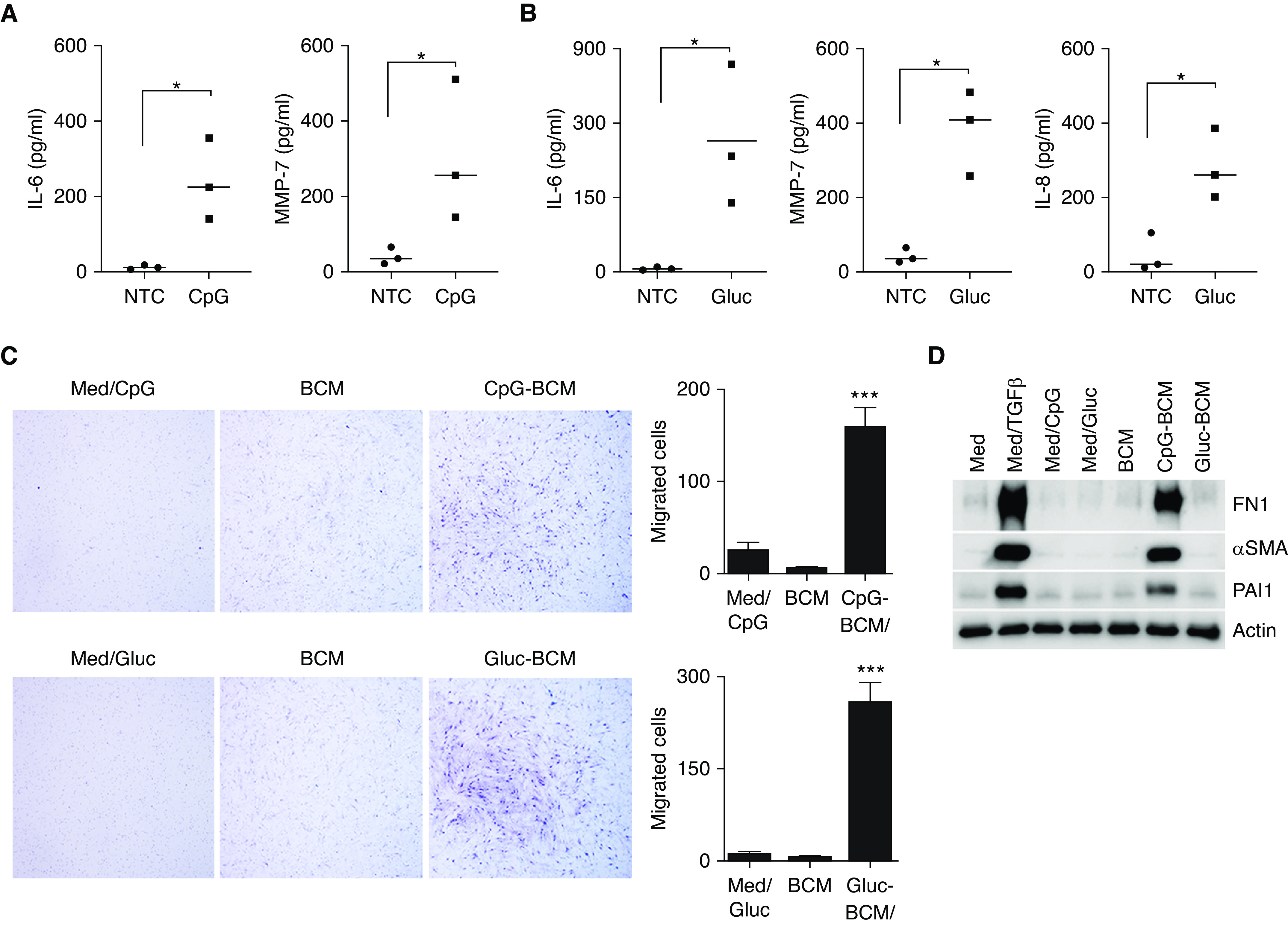
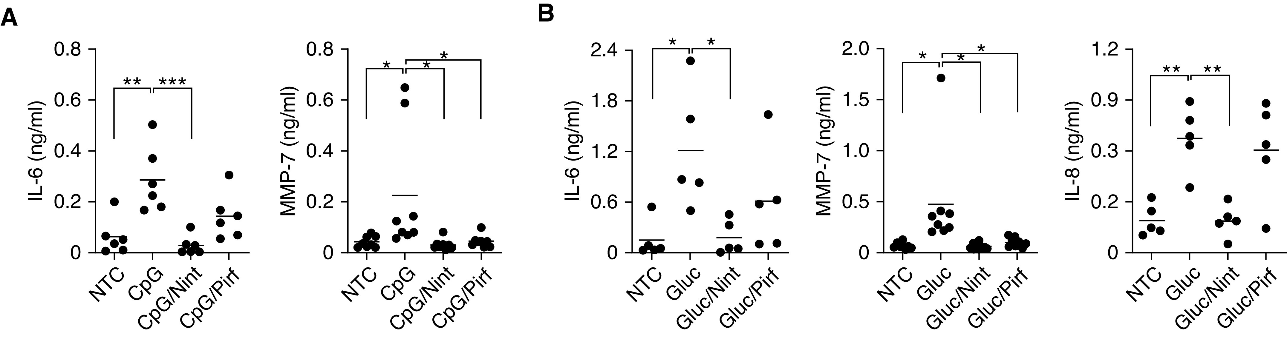
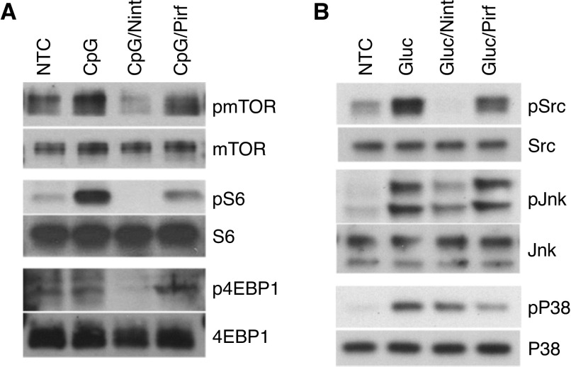
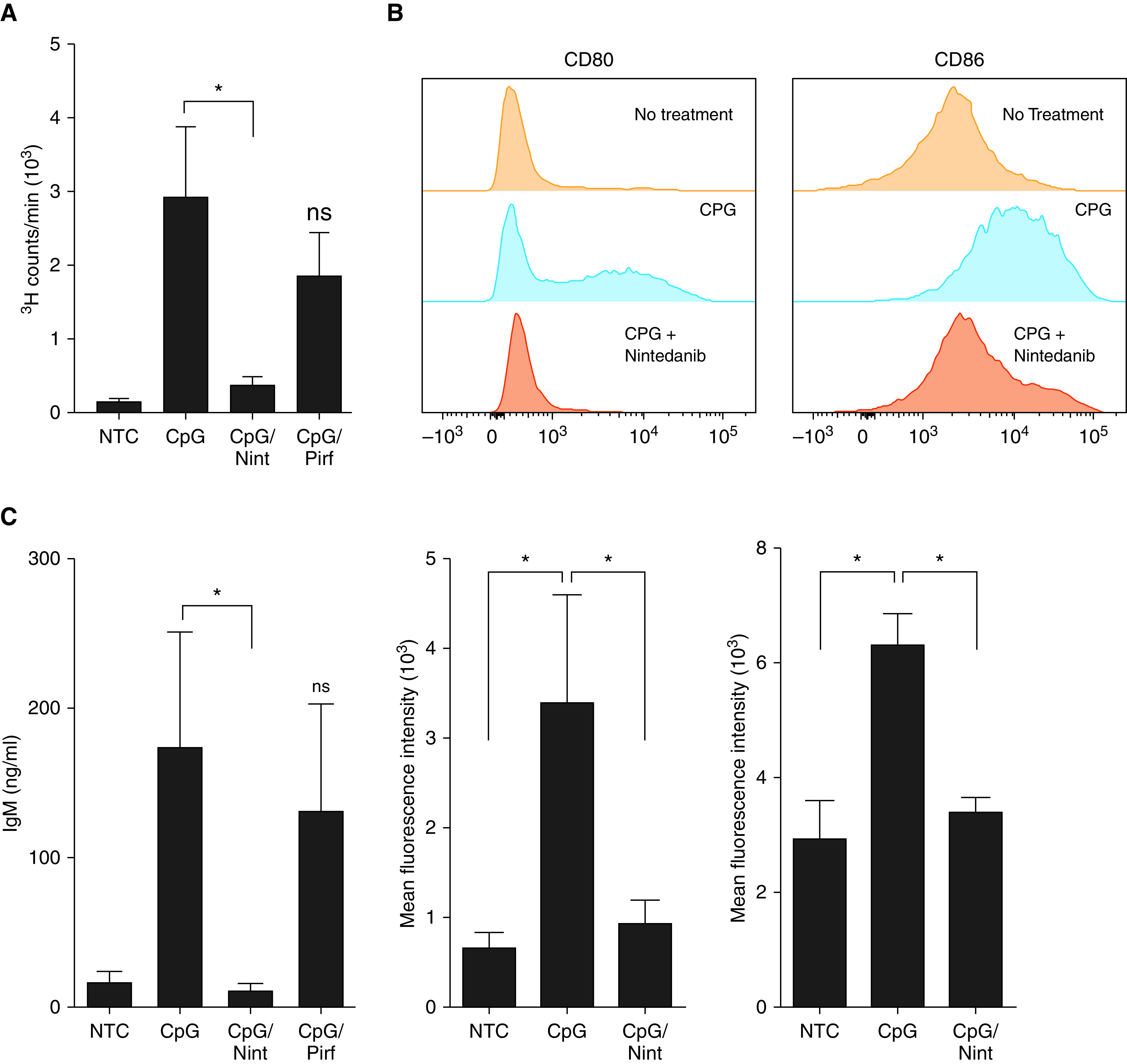
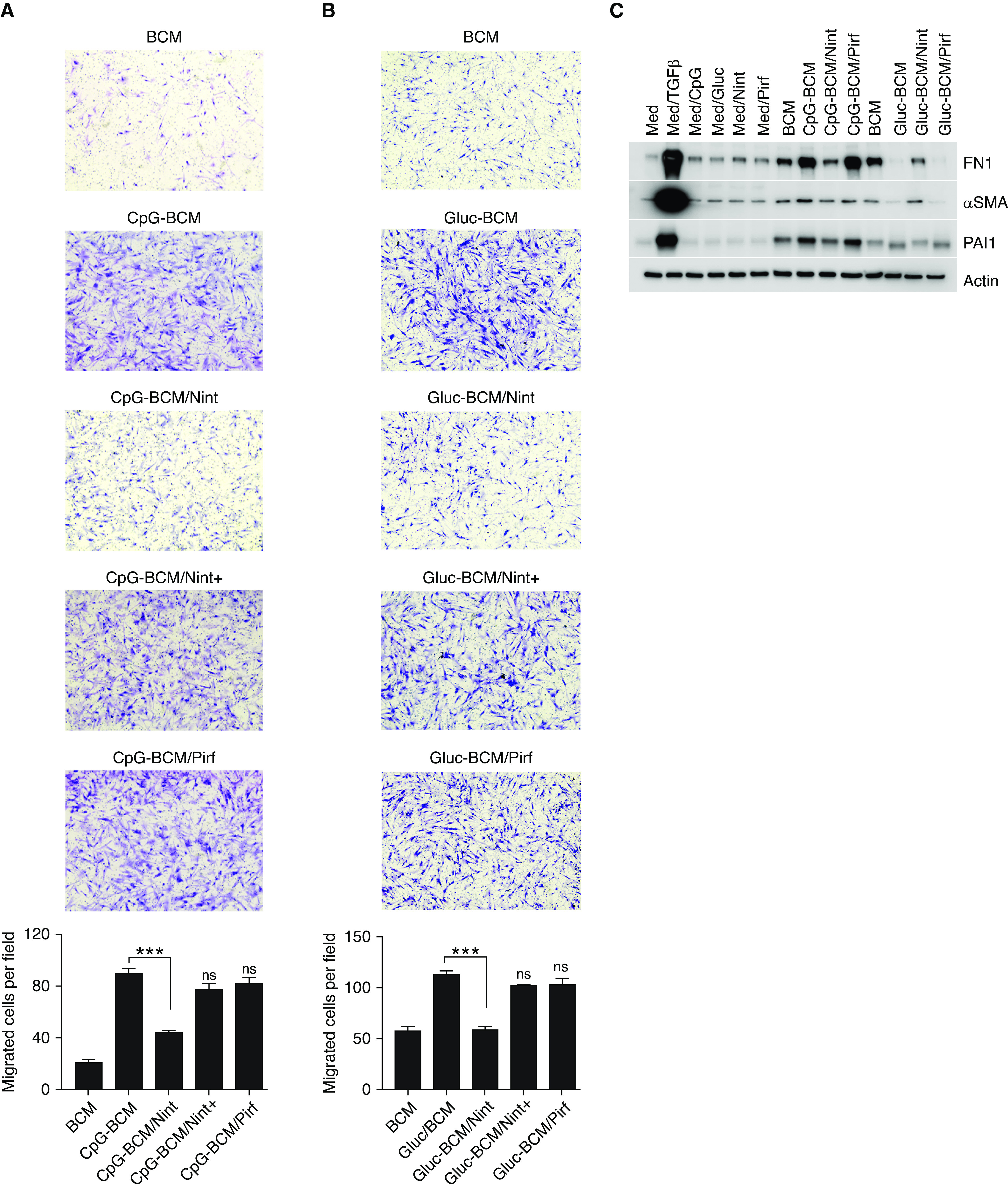
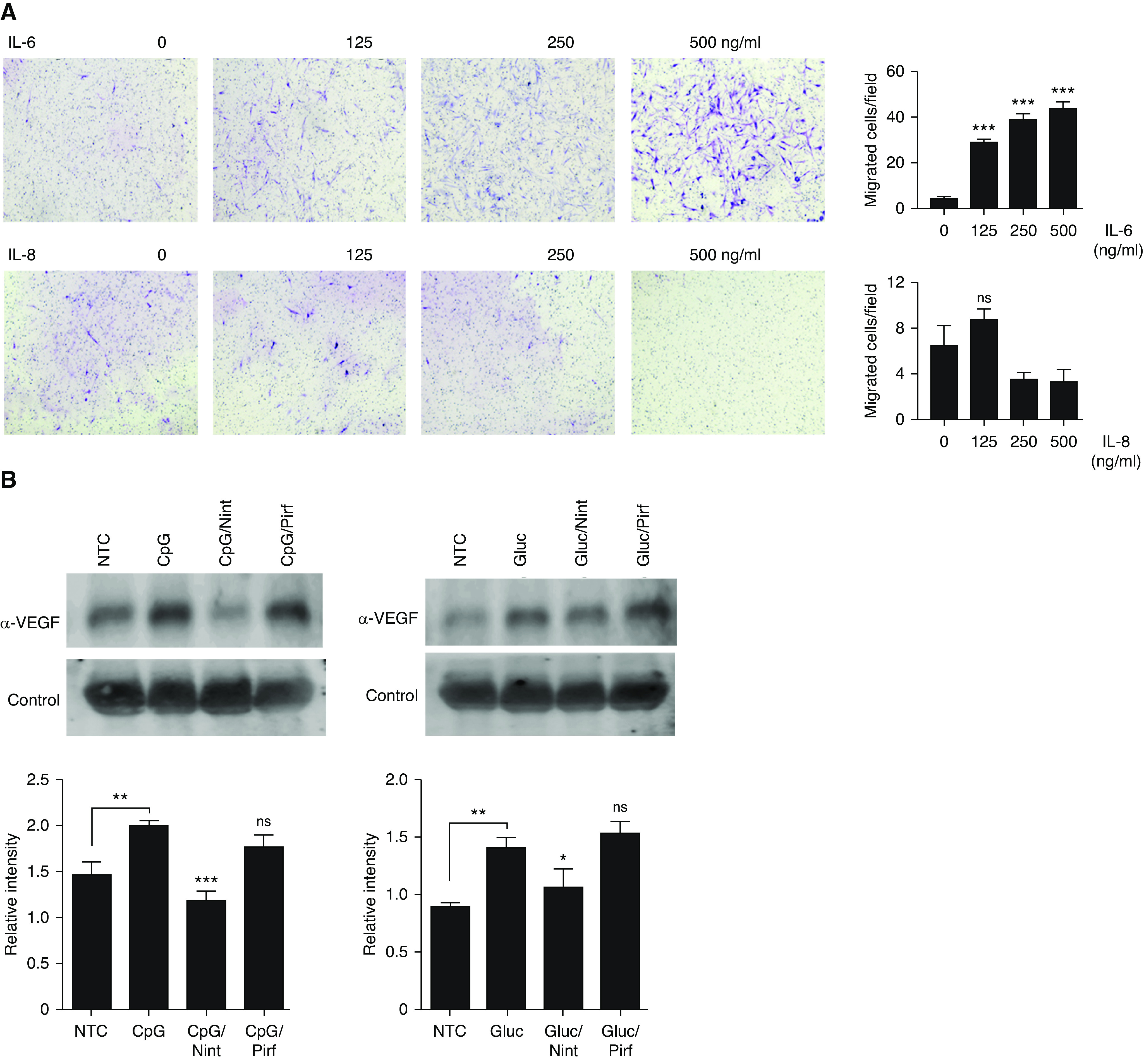
B-cell activation is increasingly linked to numerous fibrotic lung diseases, and it is well known that aggregates of lymphocytes form in the lung of many of these patients. Activation of B-cells by pattern recognition receptors (PRRs) drives the release of inflammatory cytokines, chemokines, and metalloproteases important in the pathophysiology of pulmonary fibrosis. However, the specific mechanisms of B-cell activation in patients with idiopathic pulmonary fibrosis (IPF) are poorly understood. Herein, we have demonstrated that B-cell activation by microbial antigens contributes to the inflammatory and profibrotic milieu seen in patients with IPF. B-cell stimulation by CpG and β-glucan via PRRs resulted in activation of mTOR-dependent and independent pathways. Moreover, we showed that the B-cell-secreted inflammatory milieu is specific to the inducing antigen and causes differential fibroblast migration and activation. B-cell responses to infectious agents and subsequent B-cell-mediated fibroblast activation are modifiable by antifibrotics, but each seems to exert a specific and different effect. These results suggest that, upon PRR activation by microbial antigens, B-cells can contribute to the inflammatory and fibrotic changes seen in patients with IPF, and antifibrotics are able to at least partially reverse these responses.

Keywords: B-lymphocytes; antifibrotics; fibrosis; idiopathic pulmonary fibrosis; pattern recognition receptor.


Figures

Figure 1.
Figure 1.
B-cells from patients with idiopathic pulmonary fibrosis (IPF) respond to microbial antigens by releasing a proinflammatory and profibrotic milieu that induced fibroblast migration and activation. (A and B) Peripheral B-cells from patients with IPF were either left unstimulated or were stimulated with CpG (A) or β-glucan (Gluc) (B), as indicated. IL-6, MMP-7, and IL-8 were measured in the cell supernatant by ELISA. (C) Images of quiescent IPF fibroblasts stained with Wright-Giemsa migrating toward B-cell–conditioned media (BCM). BCM was obtained from B-cell supernatant from unstimulated cells or B-cells stimulated with CpG (CpG-BCM) or Gluc (Gluc-BCM) for 48 hours. CpG and Gluc in regular media were used as controls. Migrated IPF fibroblasts were manually counted using ImageJ software. (D) Cell lysate of IPF fibroblasts stimulated with TGFβ, CpG, Gluc, CpG-BCM, or Gluc-BCM (as indicated) and immunoblotted for FN1 (fibronectin), αSMA (α-smooth muscle actin), PAI1 (plasminogen activator inhibitor-1), or β-actin. Data are representative of three independent experiments. *P < 0.05 and ***P < 0.001. Med = media; MMP-7 = matrix metallopeptidase-7; NTC = unstimulated B-cells; TGFβ = transforming growth factor β.
Figure 2.
Figure 2.
B-cell aggregates are present in fibrotic areas of the lungs of patients with pulmonary fibrosis and show mTOR activation. (A) Hematoxylin and eosin stain of lung tissue from three patients with clinical diagnosis of pulmonary fibrosis (1a–3a) showing pathologic features of usual interstitial pneumonia (UIP). CD20 stain (1b–3b) demonstrates B-cell aggregates (brown) neighboring fibroblastic foci (arrows). High-resolution computed tomography imaging of lung parenchyma of the respective patients demonstrate different patterns of fibrotic changes (1c–3c). (B) FFPE from patients with pathological findings of UIP underwent immunofluorescent staining for CD20 (red) and p-S6 (green). Scale bars, A, 500 µm; B, 500 µm;. FFPE = formalin-fixed, paraffin-embedded tissue.
Figure 3.
Figure 3.
Antifibrotics alter the inflammatory milieu of activated B-cells from patients with IPF. (A and B) B-cells were either left unstimulated or stimulated with CpG (A) or Gluc (B) in the presence of 1 μM of Nint and 100 μM of Pirf, as indicated. IL-6, IL-8, and MMP-7 were measured by ELISA in the cell supernatants of peripheral B-cells from patients with IPF. *P < 0.05, **P < 0.01, and ***P < 0.001. Nint = nintedanib; ns = not significant (P > 0.05); Pirf = pirfenidone.
Figure 4.
Figure 4.
Nint interferes with mTOR and Src kinase signaling pathways, whereas Pirf affects p38 activation and has no effect on mTOR. (A) Phosphorylation of mTOR, S6, and 4EBP1 was detected by IB in the total cell lysates of CpG-stimulated B-cells in the presence of 1 μM Nint or 100 μM of Pirf, as indicated. Total mTOR, S6K, S6, and 4EBP1 were used as controls. (B) Phosphorylation of Src, JNK, and p38 was measured in the total cell lysates after Gluc stimulation of B-cells in the presence or absence of Nint and Pirf, as indicated. Total Src, JNK, and p38 were used as controls. B-cells were isolated from patients with IPF. Data are representative of at least three independent experiments.
Figure 5.
Figure 5.
Nint impairs B-cell proliferation and activation. (A) 3H-thymidine incorporation was assessed in NTC and cells stimulated with CpG. Some cells were pretreated with 1 μM Nint or 100 μM Pirf for 1 hour before CpG stimulation (CpG/Nint and CpG/Pirf, respectively), as indicated. (B) Representative histograms of CD80 and CD86 on peripheral human B-cells. CD80 and CD86 were assessed in NTC, stimulated cells with CpG, and CpG-stimulated cells pretreated with nintenadib, as previously indicated. (C) IgM was measured by ELISA in the cell supernatants of B-cells of NTC, stimulated cells with CpG, and CpG-stimulated cells pretreated with 1 μM Nint or 100 μM Pirf for 1 hour before stimulation for a total of 5 days, as indicated. Data are representative of a typical experimental run of at least three separate experimental preparations, which were performed with different B-cell preparations. *P < 0.05. ns = not significant (P < 0.05).
Figure 6.
Figure 6.
Treatment of B-cells with Nint impairs B-cell–driven fibroblast migration and activation. (A and B) Images of quiescent IPF fibroblasts stained with Wright-Giemsa stain migrating toward BCM, CPG-BCM, and Glu-BCM. Some BCMs were prepared by preincubating B-cells for 1 hour with 1 μM Nint or 100 μM Pirf before stimulation with CpG or β-glucan (CpG-BCM/Nint, CpG-BCM/Pirf, Gluc-BCM/Nint, and Gluc-BCM/Pirf), as indicated. Controls include regular medium (Med) and medium containing CpG, Gluc, Pirf, and Nint (Med/CpG, Med/Gluc, Med/Pirf and Med/Nint, respectively) and are shown in Figure E2. Nint was also added to the BCM only when the Transwell assay is set up to control for any Nint carryover (CpG-BCM/Nint+ and Gluc-BCM/Nint+). Each membrane was imaged and divided into four fields, migrated cells were counted in each field, and means are plotted as migrated cells per field. (C) Cell lysate of IPF fibroblasts stimulated with different BCMs (as indicated) and immunoblotted for FN1, αSMA, PAI1, or β-actin, as indicated. BCM was obtained as previously indicated (A and B). TGFβ was used as positive control. Data are representative of three separate experiments. ***P < 0.001.
Figure 7.
Figure 7.
IL-6 and VEGFA participate in B-cell–mediated fibroblast migration and activation. (A) Images of quiescent IPF fibroblasts stained with Wright-Giemsa stain migrating toward different concentrations of recombinant IL-6 and IL-8 (A) or stimulated with different preparations of BCM, as indicated. Some fibroblasts were preincubated with 5 μM axitinib for 1 hour before the addition of the BCM, as indicated. (B) VEGFA secretion was measured by IB in the cell supernatant of NTC or stimulated cells with CpG and β-glucan for 24 hours as indicated. Some cells were pretreated with Nint and Pirf, as indicated. Nonspecific protein was used as loading control. Band intensities were quantified using ImageJ software version 1.48 (National Institutes of Health), and the relative intensities were calculated by normalizing the intensity of the target protein to that of the loading control. (C) Images of quiescent IPF fibroblasts stained with Wright-Giemsa stain migrating toward different concentrations of recombinant VEGFA or stimulated with different preparations of BCM, as indicated. (D) Cell lysates from IPF fibroblasts stimulated with different concentrations of IL-6 and VEGFA and immunoblotted for FN1, αSMA, PAI1, or β-actin, as indicated. Data are representative of three separate experiments. *P < 0.05, **P < 0.01, and ***P < 0.001. VEGFA = vascular endothelial growth factor A.

Comment in

References

    1. Raghu G, Collard HR, Egan JJ, Martinez FJ, Behr J, Brown KK, et al. ATS/ERS/JRS/ALAT Committee on Idiopathic Pulmonary Fibrosis. An official ATS/ERS/JRS/ALAT statement: idiopathic pulmonary fibrosis: evidence-based guidelines for diagnosis and management. Am J Respir Crit Care Med. 2011;183:788–824. - PMC - PubMed
    1. Richeldi L, du Bois RM, Raghu G, Azuma A, Brown KK, Costabel U, et al. INPULSIS Trial Investigators. Efficacy and safety of nintedanib in idiopathic pulmonary fibrosis. N Engl J Med. 2014;370:2071–2082. - PubMed
    1. King TE, Jr, Bradford WZ, Castro-Bernardini S, Fagan EA, Glaspole I, Glassberg MK, et al. ASCEND Study Group. A phase 3 trial of pirfenidone in patients with idiopathic pulmonary fibrosis. N Engl J Med. 2014;370:2083–2092. - PubMed
    1. Seibold MA, Wise AL, Speer MC, Steele MP, Brown KK, Loyd JE, et al. A common MUC5B promoter polymorphism and pulmonary fibrosis. N Engl J Med. 2011;364:1503–1512. - PMC - PubMed
    1. Noth I, Zhang Y, Ma SF, Flores C, Barber M, Huang Y, et al. Genetic variants associated with idiopathic pulmonary fibrosis susceptibility and mortality: a genome-wide association study. Lancet Respir Med. 2013;1:309–317. - PMC - PubMed

Publication types

MeSH terms