. 2021 May:126:109-118.
doi: 10.1016/j.actbio.2021.03.001. Epub 2021 Mar 6.

Angiogenic hydrogels for dental pulp revascularization

Affiliations

Angiogenic hydrogels for dental pulp revascularization

Zain Siddiqui et al. Acta Biomater. 2021 May.

Abstract

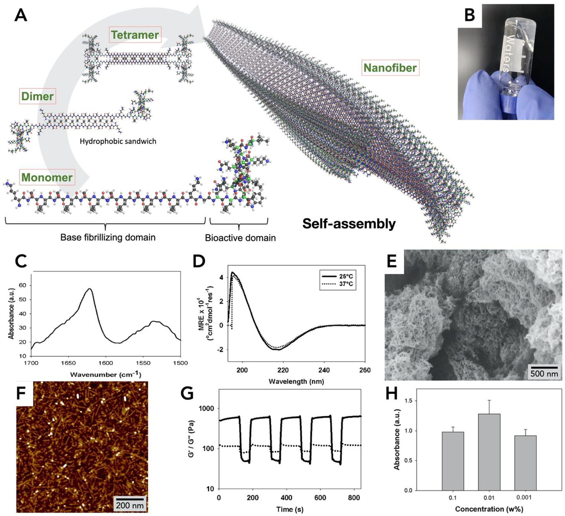
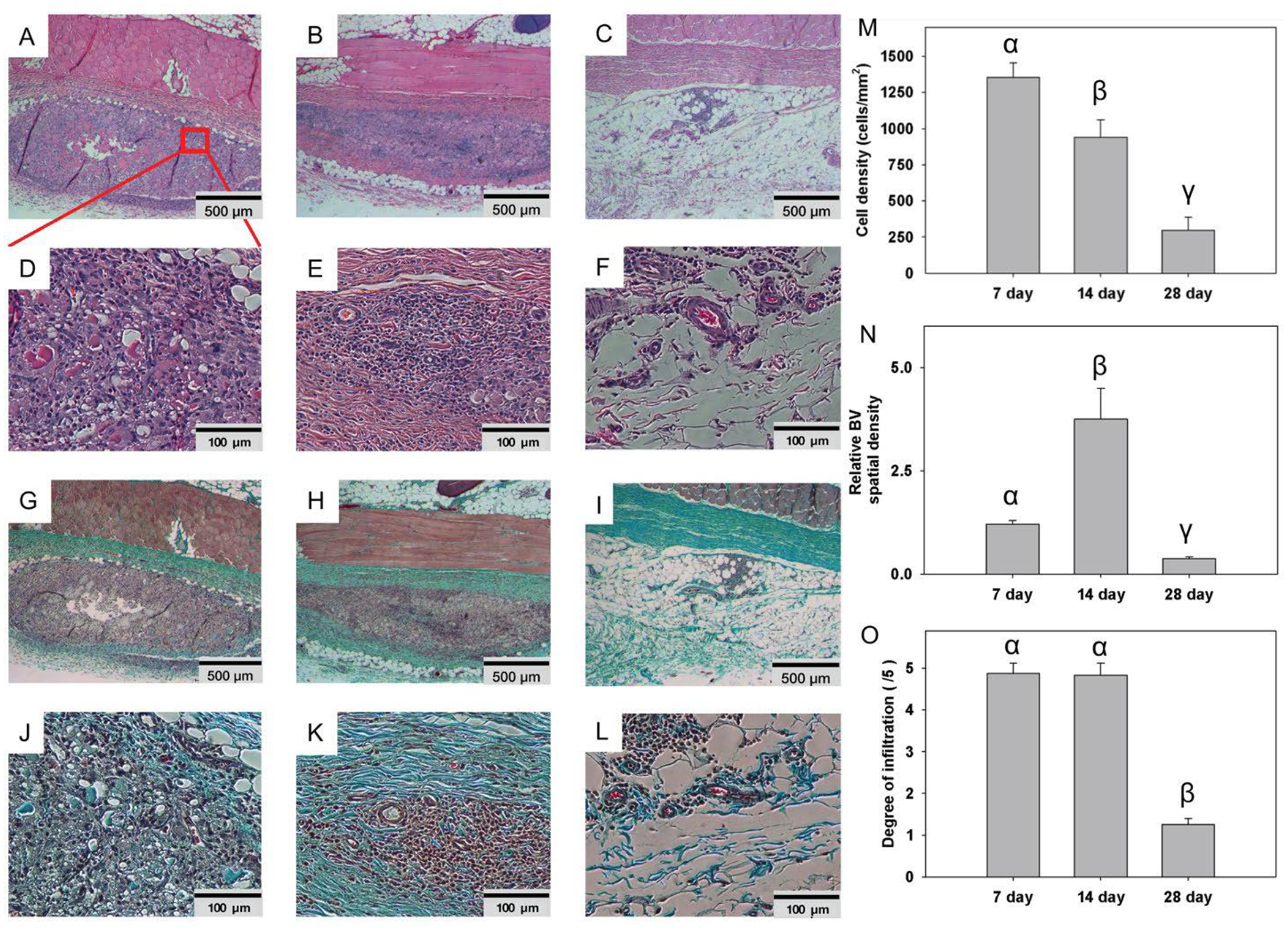
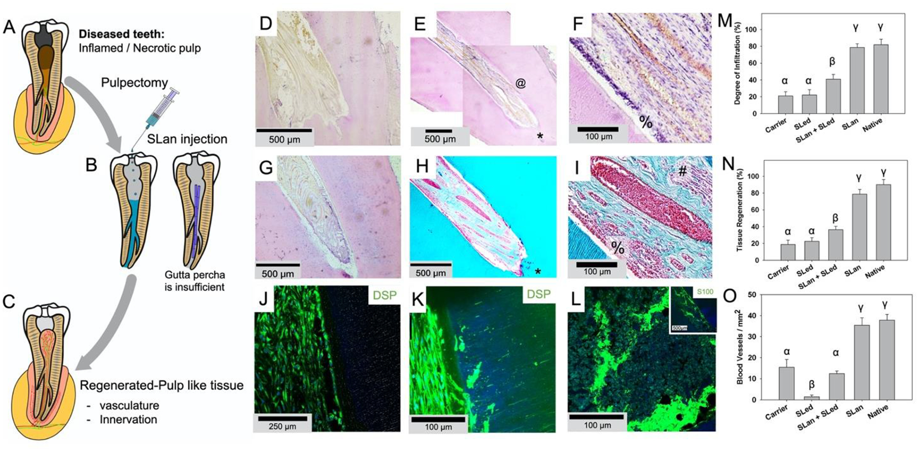
Angiogenesis is critical for tissue healing and regeneration. Promoting angiogenesis in materials implanted within dental pulp after pulpectomy is a major clinical challenge in endodontics. We demonstrate the ability of acellular self-assembling peptide hydrogels to create extracellular matrix mimetic architectures that guide in vivo development of neovasculature and tissue deposition. The hydrogels possess facile injectability, as well as sequence-level functionalizability. We explore the therapeutic utility of an angiogenic hydrogel to regenerate vascularized pulp-like soft tissue in a large animal (canine) orthotopic model. The regenerated soft tissue recapitulates key features of native pulp, such as blood vessels, neural filaments, and an odontoblast-like layer next to dentinal tubules. Our study establishes angiogenic peptide hydrogels as potent scaffolds for promoting soft tissue regeneration in vivo. STATEMENT OF SIGNIFICANCE: A major challenge to endodontic tissue engineering is the lack of in situ angiogenesis within intracanal implants, especially after complete removal of the dental pulp. The lack of a robust vasculature in implants limit integration of matrices with the host tissue and regeneration of soft tissue. We demonstrate the development of an acellular material that promotes tissue revascularization in vivo without added growth factors, in a preclinical canine model of pulp-like soft-tissue regeneration. Such acellular biomaterials would facilitate pulp revascularization approaches in large animal models, and translation into human clinical trials.

Keywords: Acellular scaffolds; Angiogenesis; Pulp revascularization; Self-assembly; Tissue regeneration.


Conflict of interest statement

Declaration of Competing Interest The authors declare that they have no known competing financial interests or personal relationships that could have appeared to influence the work reported in this paper. .A.K. has equity interests in commercialization ventures to translate these and related technologies.

Figures

Fig. 1.
Fig. 1.. Biophysical properties of SLan hydrogel.
(A) Self-assembly of SLan. In aqueous solution dimers and tetramers form assemble into a β-sheet based nanofibers. (B) 1% SLan hydrogel, (C) FTIR spectrum shows β-sheet secondary structure (1624 cm−1 peak) confirmed by (D) Circular dichroism spectra (~195 nm maxima and ~217 nm minima. (E) Scanning electron micrograph of critical-point-dried SLan hydrogel. (F) Individual SLan nanofibers observed in atomic force microscopy. (G) 1% SLan hydrogels are thixotropic. At low strain (1%), G’>G” (indicating solid-like elastic properties), at high strain (100%), G’<G” (signifying liquefaction of the hydrogel and showing dominance of viscous modulus). The switch in rheological properties is fast and reversible (representative plot shown). (H) In vitro cytocompatibility (n=4, p<0.05, CCK8 dye absorbance) of HEPG2 cells shows that SLan did not exhibit cytotoxicity.
Fig. 2.
Fig. 2.. Dorsal subcutaneous implants of SLan in rats.
Implants (n=4) showed rapid infiltration of cells - H&E images of 200 μL bolus implants in Wistar rats as early as Day-7 (A: 500 μm, region magnified in D: 100 μm), completely into the center of implants by Day 14 (B: 500 μm, E: 100 μm), and fully degraded by Day 28 (C: 500 μm, F: 100 μm). Similarly, extracellular matrix (ECM) deposition (collagen, blue) was noted in Masson’s Trichrome staining, with infiltration of blood vessels as early as 7 days (G: 500 μm, J: 100 μm), Day 14 (H: 500 μm, K: 100 μm), with complete resorption of superfluous collagen and vasculature by 28 days (I: 500 μm, L: 100 μm). (M) Cell density analysis of 7,14, and 28 day SLan rat subcutaneous samples. (N) Blood vessel density analysis of 7,14, and 28 day SLan rat subcutaneous samples. (O) Degree of infiltration analysis of 7, 14, and 28 day SLan rat subcutaneous samples.
Fig. 3.
Fig. 3.. Regeneration of vascularized soft tissue in canine root canals.
(A) Caries and trauma may lead to the inflammation and necrosis of the pulp (A). (B) After pulpectomy, implantation of injectable angiogenic SLan hydrogels help regenerate (C) vascularized pulp-like soft tissue in 28 days, unlike inert materials such as gutta percha. In a canine pulpectomy model, disorganized blood clots form for over-instrumentation carrier filled (sucrose-HBSS) control (D). H&E staining of tooth roots of SLan filled teeth showed rapid infiltration of cells and tissue (E), and within crevices in the canal space (@), along with an odontoblast-like layer in apposition to the dentin wall (F - %). In contrast, control dentinogenic SLed hydrogels lead to disorganized tissue (G). Trichrome staining of SLan implants reveals blood vessels (H, I) with collagen deposition (blue); and an odontoblast-like layer (I - %) which stains with dental sialoprotein (DSP) (J) with cytoplasmic protrusions into dentinal tubules (K). S100+ Nerve bundles (Trichrome I-#) were regenerated along the length of the canal (L and inset). (M) Degree of infiltration, (N) degree of tissue regeneration, and (O) densities of blood vessels were similar for SLan and native teeth but significantly greater than controls. (n=8 for SLan, n=4 for all other groups; values are reported as mean ± standard deviation; different Greek letters indicate statistical significance between groups p<0.05).

References

    1. Augustin HG, Koh GY, Organotypic vasculature: From descriptive heterogeneity to functional pathophysiology, Science 357(6353) (2017) eaal2379; doi: 10.1126/science.aal2379. - DOI - PubMed
    1. Carmeliet P, Jain RK, Molecular mechanisms and clinical applications of angiogenesis, Nature 473(7347) (2011) 298–307. - PMC - PubMed
    1. Sarkar B, Nguyen PK, Gao W, Dondapati A, Siddiqui Z, Kumar VA, Angiogenic Self-Assembling Peptide Scaffolds for Functional Tissue Regeneration, Biomacromolecules 19(9) (2018) 3597–3611. - PubMed
    1. Mastrullo V, Cathery W, Velliou E, Madeddu P, Campagnolo P, Angiogenesis in Tissue Engineering: As Nature Intended?, Front Bioeng Biotechnol 8 (2020) 188. - PMC - PubMed
    1. Kumar VA, Liu Q, Wickremasinghe NC, Shi S, Cornwright TT, Deng Y, Azares A, Moore AN, Acevedo-Jake AM, Agudo NR, Pan S, Woodside DG, Vanderslice P, Willerson JT, Dixon RA, Hartgerink JD, Treatment of hind limb ischemia using angiogenic peptide nanofibers, Biomaterials 98 (2016) 113–9. - PMC - PubMed

Publication types

LinkOut - more resources