. 2021 Mar 10;19(3):e3000709.
doi: 10.1371/journal.pbio.3000709. eCollection 2021 Mar.

Blunted diurnal firing in lateral habenula projections to dorsal raphe nucleus and delayed photoentrainment in stress-susceptible mice

Affiliations

Blunted diurnal firing in lateral habenula projections to dorsal raphe nucleus and delayed photoentrainment in stress-susceptible mice

He Liu et al. PLoS Biol. .

Abstract

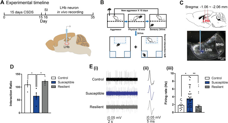
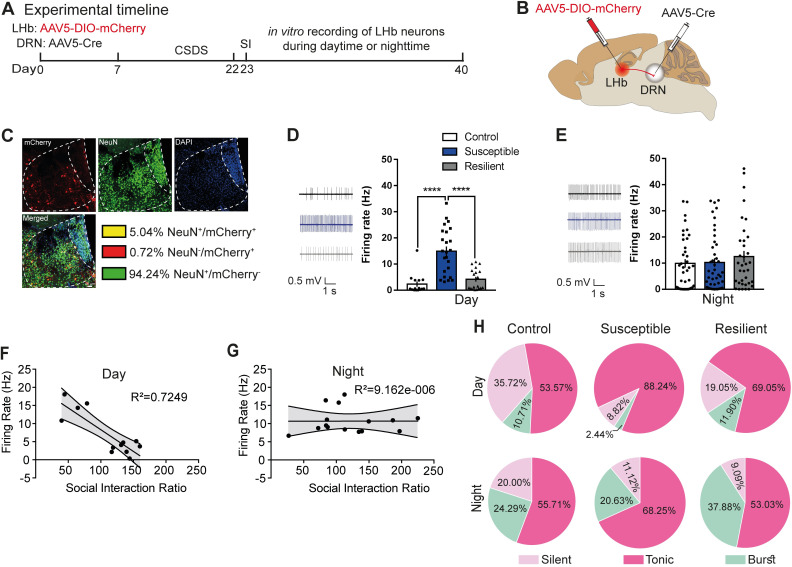
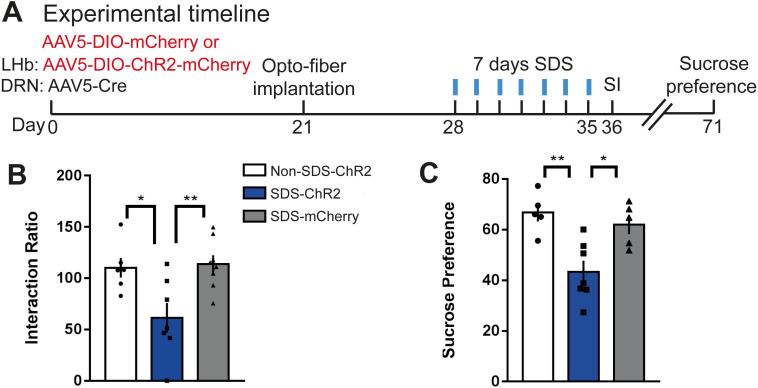
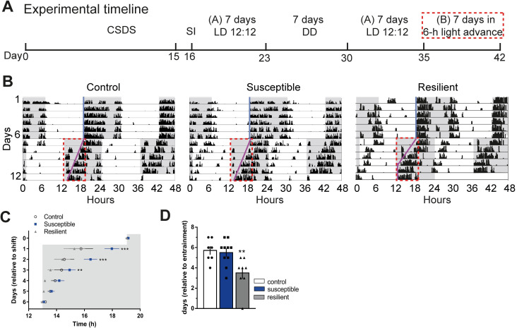
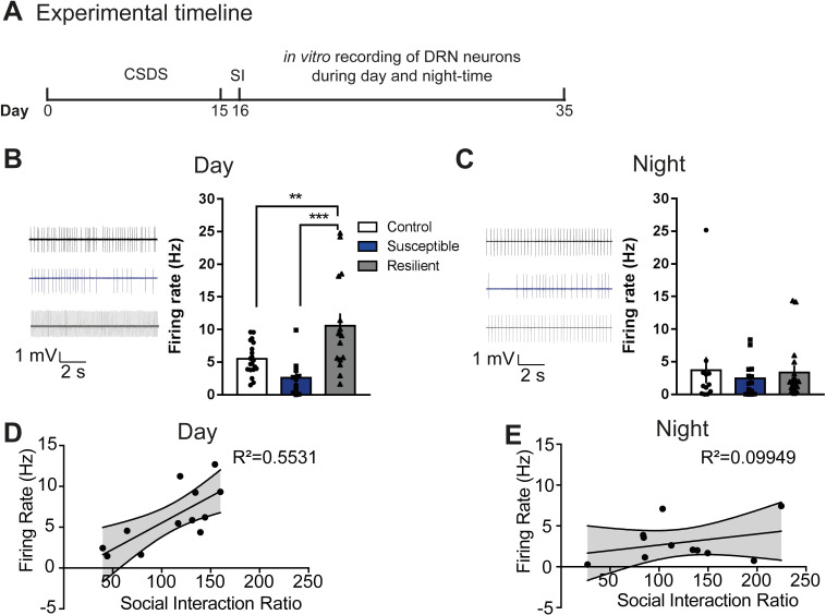
Daily rhythms are disrupted in patients with mood disorders. The lateral habenula (LHb) and dorsal raphe nucleus (DRN) contribute to circadian timekeeping and regulate mood. Thus, pathophysiology in these nuclei may be responsible for aberrations in daily rhythms during mood disorders. Using the 15-day chronic social defeat stress (CSDS) paradigm and in vitro slice electrophysiology, we measured the effects of stress on diurnal rhythms in firing of LHb cells projecting to the DRN (cellsLHb→DRN) and unlabeled DRN cells. We also performed optogenetic experiments to investigate if increased firing in cellsLHb→DRN during exposure to a weak 7-day social defeat stress (SDS) paradigm induces stress-susceptibility. Last, we investigated whether exposure to CSDS affected the ability of mice to photoentrain to a new light-dark (LD) cycle. The cellsLHb→DRN and unlabeled DRN cells of stress-susceptible mice express greater blunted diurnal firing compared to stress-näive (control) and stress-resilient mice. Daytime optogenetic activation of cellsLHb→DRN during SDS induces stress-susceptibility which shows the direct correlation between increased activity in this circuit and putative mood disorders. Finally, we found that stress-susceptible mice are slower, while stress-resilient mice are faster, at photoentraining to a new LD cycle. Our findings suggest that exposure to strong stressors induces blunted daily rhythms in firing in cellsLHb→DRN, DRN cells and decreases the initial rate of photoentrainment in susceptible-mice. In contrast, resilient-mice may undergo homeostatic adaptations that maintain daily rhythms in firing in cellsLHb→DRN and also show rapid photoentrainment to a new LD cycle.


Conflict of interest statement

The authors have declared that no competing interests exist.

Figures

Fig 1
Fig 1. Increased daytime firing in LHb cells of susceptible mice.
(A) Experimental timeline of 15 days CSDS paradigm, SI, and in vivo single-unit recording in the daytime. (B) Detailed schematic of the CSDS paradigm using C57BL/6J male mice and CD1 retired male breeders (aggressor) and SI test. (C) Schematic showing anatomical site of LHb (top) and example site of glass electrode recording in the LHb (bottom). (D) SI data showing that in the presence of a CD1 social target (nonaggressor), susceptible mice display decreased SI ratio (F2,9 = 7.295, P < 0.05, N = 4 mice/group). (E) (i) Example traces, (ii) typical action potentials of recorded LHb cells of control (top), susceptible (middle), and resilient mice (bottom) after SI test. (iii) Increased firing rate in susceptible mice (F2,73 = 7.195, P = 0.0014; N = 4 mice/group, n = 13–37 cells/group). Error bars: mean ± SEM. The raw data can be found in S1 Data. CSDS, chronic social defeat stress; LHb, lateral habenula; MHb, medial habenula; SI, social interaction.
Fig 2
Fig 2. Blunted rhythms in spontaneous firing in cellsLHb→DRN of susceptible mice.
(A) Experimental timeline of viral surgeries to label cellsLHb-DRN, CSDS, SI test, and in vitro slice electrophysiology. (B) Schematic showing surgeries for viral injections to specifically label cellsLHb-DRN. (C) Confocal images showing coexpression of mCherry, NeuN, and DAPI within LHb; and quantified data showing mCherry-expressing neurons (5% ± 0.1% LHb neurons project to DRN). (D) Sample traces of daytime in vitro spontaneous activity in cellsLHb-DRN of mice. Increased daytime spontaneous activity in cellsLHb-DRN in susceptible mice (right) (F2,55 = 22.68, P < 0.0001; n = 15–24 cells from 7 to 11 mice/group). (E) Sample traces of nighttime in vitro spontaneous activity in cellsLHb-DRN of mice. No difference in nighttime spontaneous firing in cellsLHb-DRN in control, susceptible, and resilient mice (right). (F, G) Significant negative correlation between spontaneous activity in cellsLHb-DRN and SI ratio during the day (R2 = 0.7249; P < 0.05) but not the night. (H) Pie charts illustrating the percentage of cells expressing: silent, tonic, or burst firing in the daytime (top) and nighttime (bottom) in labelled cellsLHb-DRN. Susceptible mice exhibit notable increased daytime tonic firing and decrease in silent patterns (χ2 = 6.947, P < 0.05, chi-squared test; n = 28–42 cells from 7 to 11 mice/group). At night, there is no difference in firing patterns among the phenotypes. There is significant increase in burst firing at night compared to day (P < 0.05). Error bars: mean ± SEM. The raw data can be found in S2 Data. CSDS, chronic social defeat stress; DRN, dorsal raphe nucleus; LHb, lateral habenula; SI, social interaction.
Fig 3
Fig 3. Diurnal rhythms in Ih and Kv+- currrents in cellsLHb→DRN of susceptible mice.
(A) Daytime representative traces (left) and statistical data of Ih-currents in response to incremental steps in voltage injections (right). Resilient and susceptible mice display significant increase in Ih-current compared to control (F2,252 = 23.29, P < 0.0001; n = 10 to 14 cells from 7 to11 mice per group). (B) Nighttime representative traces (left) and statistical data of Ih-currents in response to incremental steps in voltage injections (right). There was no difference in current between any of the phenotypes. (C) Daytime representative traces (left) and statistical data of isolated Kv+ channel–mediated currents (right). Susceptible mice display significant increase in peak (F2,216 = 9.94, P < 0.0001; n = 6 to 11 cells from 7 to 8 mice per group; top) and sustained (F2,216 = 24.97, P < 0.0001; n = 6 to 11 cells from 7 to 8 mice per group; bottom) phases of Kv+-currents. (D) Nighttime representative traces (left) and statistical data of isolated Kv+ channel–mediated currents (right). There is no difference in peak and sustained phases of Kv+-currents among the 3 phenotypes. Error bars: mean ± SEM. The raw data can be found in S3 Data.
Fig 4
Fig 4. Daytime optical activation of cellsLHb→DRN during weak social stress induces susceptibility.
(A) Experimental timeline of viral surgeries, optofiber implantation, repeated high frequency optical stimulation patterns, and behavioural tests. (B) In the presence of a CD1 social target (nonaggressor), SDS-ChR2 mice display increased stress-susceptibility as measured by decreased SI ratio compared with non-SDS-ChR2 and SDS-mCherry mice, (F2,18 = 6.905, P < 0.01; N = 6–8 mice/group). (C) The SDS-ChR2 mice display anhedonia as measured by reduction in 1% sucrose intake over 3 h in the sucrose preference test (F2,14 = 9.805, P < 0.01; N = 5–7 mice/group; right). Error bars: mean ± SEM. The raw data can be found in S4 Data. DRN, dorsal raphe nucleus; LHb, lateral habenula; SDS, social defeat stress; SI, social interaction.
Fig 5
Fig 5. Susceptible or resilient mice exhibit different rates of photoentrainment.
(A) Experimental timeline (A, normal light cycle: Lights ON: 07:00; Lights OFF: 19:00; B, new light cycle following 6 h advance: Lights ON: 13:00; Lights OFF: 01:00), red box denotes 6 h lights advance in new LD cycle. (B) Representative double-plotted actograms of activity before and after 6 h shift in LD cycle. Grey background indicating dark phase of the LD cycle. Light-blue and pink lines representing extended regression line derived by onset of activity under normal and new LD cycles, respectively. Dotted red lines outlining the data of activity onsets shown in (c). (C) Susceptible mice display significantly slower initial rate of photoentrainment in the new LD cycle (F2,25 = 10.81, P < 0.001) (D) Resilient mice took significantly less number of days to reentrained in the new LD cycle (F2,22 = 6.587, P < 0.05). N = 7–10 mice/group. Error bars: mean ± SEM. The raw data can be found in S5 Data. CSDS, chronic social defeat stress; DD, complete darkness; LD, light–dark; SI, social interaction.
Fig 6
Fig 6. Blunted rhythms in spontaneous firing in DRN cells of susceptible mice.
(A) Experimental timeline of CSDS paradigm, SI test, and in vitro recordings. (B, C) Sample traces of spontaneous firing of DRN cells in day (B) and in night (C) in control (top), susceptible (middle), and resilient mice (bottom). (B) Susceptible mice exhibit decreased daytime spontaneous firing in DRN cells, while resilient mice exhibit increased daytime spontaneous firing (F2,43 = 9.793, P < 0.001; n = 11–20 cells from 7 to 11 mice/group), (C) No difference in nighttime spontaneous firing in DRN cells. (D, E) Significant positive correlation between spontaneous activity in DRN cells and SI ratio during the day (R2 = 0.5531; P < 0.05) but not the night. Error bars: mean ± SEM. The raw data can be found in S6 Data. CSDS, chronic social defeat stress; DRN, dorsal raphe nucleus; SI, social interaction.

References

    1. Cho JR, Treweek JB, Robinson JE, Xiao C, Bremner LR, Greenbaum A, et al.. Dorsal Raphe Dopamine Neurons Modulate Arousal and Promote Wakefulness by Salient Stimuli. Neuron. 2017;94 (6):1205, e8–19. 10.1016/j.neuron.2017.05.020 - DOI - PubMed
    1. Zhao H, Zhang BL, Yang SJ, Rusak B. The role of lateral habenula-dorsal raphe nucleus circuits in higher brain functions and psychiatric illness. Behav Brain Res. 2015;277:89–98. 10.1016/j.bbr.2014.09.016 - DOI - PubMed
    1. Salaberry NL, Hamm H, Felder-Schmittbuhl MP, Mendoza J. A suprachiasmatic-independent circadian clock(s) in the habenula is affected by Per gene mutations and housing light conditions in mice. Brain Struct Funct. 2019;224 (1):19–31. 10.1007/s00429-018-1756-4 - DOI - PubMed
    1. Bano-Otalora B, Piggins HD. Contributions of the lateral habenula to circadian timekeeping. Pharmacol Biochem Behav. 2017;162:46–54. 10.1016/j.pbb.2017.06.007 - DOI - PubMed
    1. Souetre E, Salvati E, Belugou JL, Pringuey D, Candito M, Krebs B, et al.. Circadian rhythms in depression and recovery: evidence for blunted amplitude as the main chronobiological abnormality. Psychiatry Res. 1989;28 (3):263–78. 10.1016/0165-1781(89)90207-2 - DOI - PubMed

Publication types