. 2021 Mar 10;16(3):e0248348.
doi: 10.1371/journal.pone.0248348. eCollection 2021.

Establishment of a well-characterized SARS-CoV-2 lentiviral pseudovirus neutralization assay using 293T cells with stable expression of ACE2 and TMPRSS2

Affiliations

Establishment of a well-characterized SARS-CoV-2 lentiviral pseudovirus neutralization assay using 293T cells with stable expression of ACE2 and TMPRSS2

Sabari Nath Neerukonda et al. PLoS One. .

Abstract

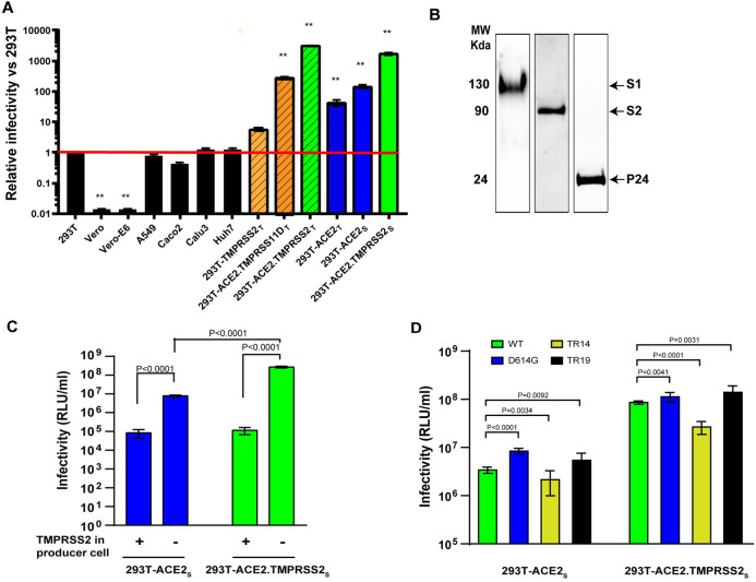
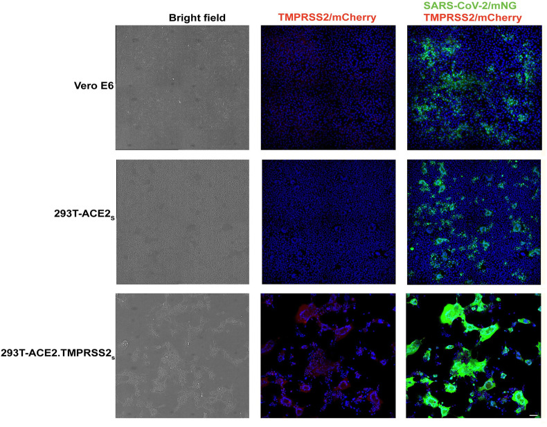
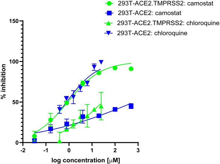
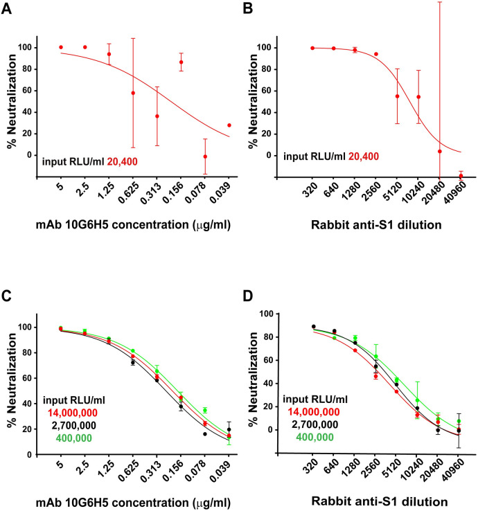
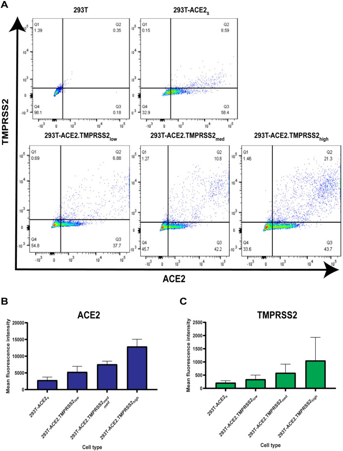
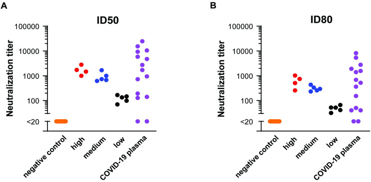
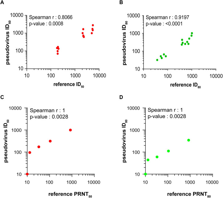
Pseudoviruses are useful surrogates for highly pathogenic viruses because of their safety, genetic stability, and scalability for screening assays. Many different pseudovirus platforms exist, each with different advantages and limitations. Here we report our efforts to optimize and characterize an HIV-based lentiviral pseudovirus assay for screening neutralizing antibodies for SARS-CoV-2 using a stable 293T cell line expressing human angiotensin converting enzyme 2 (ACE2) and transmembrane serine protease 2 (TMPRSS2). We assessed different target cells, established conditions that generate readouts over at least a two-log range, and confirmed consistent neutralization titers over a range of pseudovirus input. Using reference sera and plasma panels, we evaluated assay precision and showed that our neutralization titers correlate well with results reported in other assays. Overall, our lentiviral assay is relatively simple, scalable, and suitable for a variety of SARS-CoV-2 entry and neutralization screening assays.


Conflict of interest statement

The authors have declared that no competing interests exist.

Figures

Fig 1
Fig 1. SARS-CoV-2 lentiviral pseudovirus infectivity under various conditions.
(A) Relative infectivity of SARS-CoV-2 pseudoviruses in various target cells. The Y-axis shows relative infectivity compared to background in 293T cells. Subscript ‘t’ or ‘s’ refers to transient or stable expression, respectively. Red line indicates background level (1 x 10⁴ relative units of luciferase activity/milliliter or RLU/ml). (B) Western blots probed for spike S1/S2 subunits and HIV p24 incorporated into pseudoviruses. Lanes represent different blots (C) Infectivity on 293T-ACE2s and 293T-ACE2.TMPRSS2s of pseudoviruses primed with or without TMPRSS2 during pseudovirus production. (D) Infectivity on 293T-ACE2s and 293T-ACE2.TMPRSS2s of pseudoviruses bearing full-length, wildtype S glycoprotein (WT), an S glycoprotein with the D614G substitution, or S glycoproteins with C-terminal truncations of 14 (TR14) and 19 (TR19) amino acids. Data are shown as means and standard deviations from 3–4 independent experiments (panel A, C and D). The tests for two-group comparison were analyzed using GraphPad Prism software. P values of 0.05 were considered statistically significant. **: P<0.0001, compared to the infectivity in 293T.
Fig 2
Fig 2. Infection of Vero E6, 293T-ACE2s and 293T-ACE2.TMPRSS2s with replicating SARS-CoV-2-mNG virus.
Cells inoculated with 100 PFU/ml of virus (MOI 0.1) were fixed and imaged at 24 hours post infection by confocal microscopy. The left column shows bright field images (black and white), with the 293-ACE2.TMPRSS2 cells (bottom) showing a high degree of cytopathic effect and syncytium formation, resulting in fewer cells. The center column shows merged images of TMPRSS2/mCherry and Hoechst dye (blue) with only the 293-ACE2.TMPRSS2 cells (bottom) showing positive signals in red. The right column shows merged images of SARS-CoV-2/mNG (green), TMPRSS2/mCherry (red), and Hoechst dye (blue). Compared to Vero (top) and 293-ACE2 (middle), the 293T-ACE2.TMPRSS2 (bottom) cells show stronger SARS-CoV-2 nNG signals in green. Scale indicates 50um.
Fig 3
Fig 3. pH-dependent and -independent pathways of cell entry of SARS-CoV-2 S pseudoviruses.
Camostat mesylate inhibits TMPRSS2 activity and chloroquine inhibits endosomal acidification required for cathepsin activity. Cells were pretreated with camostat mesylate or chloroquine for 2 h prior to pseudovirus infection in the presence of inhibitor for a period of 48 h. X-axis indicates log concentration of inhibitor. Y axis indicates percentage inhibition compared to pseudovirus infection without inhibitor treatment. Results shown are representative of three independent experiments.
Fig 4
Fig 4. Optimization of pseudovirus inoculum for neutralization assays.
Neutralizations were performed in 293T-ACE2.TMPRSS2s cells with mAb 10G6H5 (A and C) and a rabbit serum against the S1 subunit (B and D) using various pseudovirus inoculums. X-axis indicates mAb concentration (A and C) and serum dilution (B and D) used. Y axis indicates percentage inhibition compared to pseudovirus infection without mAb or serum treatment.
Fig 5
Fig 5. ACE2 and TMPRSS2 levels at the cell surface.
(A) Cell surface expression of ACE2 and TMPRSS2 on various cell types. (B) Mean fluorescence intensity (MFI) of ACE2 on various cell types compared to 293T cells X-axis indicates MFI and Y axis indicates various cell types analyzed. (C) Mean fluorescence intensity (MFI) of TMPRSS2 on various cell types compared to 293T cells. X-axis indicates MFI and Y axis indicates various cell types analyzed.
Fig 6
Fig 6. Neutralization titers of serum panels.
ID50 (A) and ID80 (B) titers. ID50 = inhibitory dilution leading to 50% neutralization compared to control. ID80 = inhibitory dilution leading to 80% neutralization compared to control. X-axes indicate sample type. Y axes indicate pseudovirus neutralization titers. Each dot represents an independent sample.
Fig 7
Fig 7. Correlation of neutralization titers between different neutralization assays.
Spearman correlation of ID50 (A and C) or ID80 (B and D) values comparing titers generated in the present study to (A and B) the focused concordance samples and (C and D) an NIBSC reference standards with reported titers generated by a plaque reduction neutralization (PRNT) assay.

References

    1. Zhou P, Yang XL, Wang XG, Hu B, Zhang L, Zhang W, et al.. A pneumonia outbreak associated with a new coronavirus of probable bat origin. Nature. 2020;579(7798):270–3. 10.1038/s41586-020-2012-7 - DOI - PMC - PubMed
    1. Gorbalenya AE, Baker SC, Baric RS, de Groot RJ, Drosten C, Gulyaeva AA, et al.. The species Severe acute respiratory syndrome-related coronavirus: classifying 2019-nCoV and naming it SARS-CoV-2. Nature Microbiology. 2020;5(4):536–44. 10.1038/s41564-020-0695-z - DOI - PMC - PubMed
    1. Wu F, Zhao S, Yu B, Chen Y-M, Wang W, Song Z-G, et al.. A new coronavirus associated with human respiratory disease in China. Nature. 2020;579(7798):265–9. 10.1038/s41586-020-2008-3 - DOI - PMC - PubMed
    1. Zhu N, Zhang D, Wang W, Li X, Yang B, Song J, et al.. A Novel Coronavirus from Patients with Pneumonia in China, 2019. New England Journal of Medicine. 2020;382(8):727–33. - PMC - PubMed
    1. Poland GA, Ovsyannikova IG, Crooke SN, Kennedy RB. SARS-CoV-2 Vaccine Development: Current Status. Mayo Clinic Proceedings. 2020;95(10):2172–88. 10.1016/j.mayocp.2020.07.021 - DOI - PMC - PubMed

Publication types

MeSH terms

Substances