. 2021 Oct 7;15(10):1720-1736.
doi: 10.1093/ecco-jcc/jjab044.

Overexpression of Vitamin D Receptor in Intestinal Epithelia Protects Against Colitis via Upregulating Tight Junction Protein Claudin 15

Affiliations

Overexpression of Vitamin D Receptor in Intestinal Epithelia Protects Against Colitis via Upregulating Tight Junction Protein Claudin 15

Ishita Chatterjee et al. J Crohns Colitis. .

Erratum in

  • Correction.
    [No authors listed] [No authors listed] J Crohns Colitis. 2023 Jan 27;17(1):149. doi: 10.1093/ecco-jcc/jjac104. J Crohns Colitis. 2023. PMID: 35971821 Free PMC article. No abstract available.

Abstract

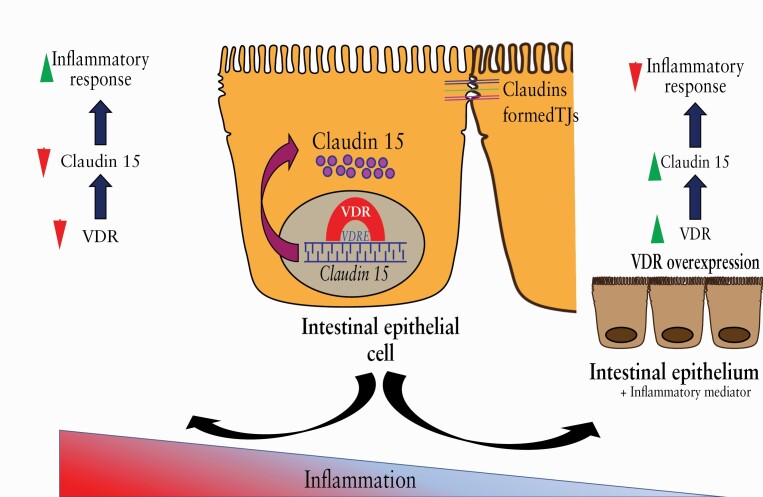
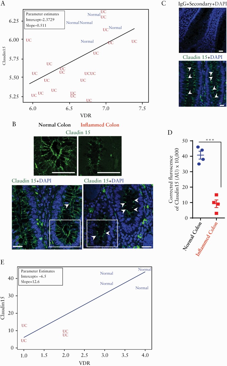
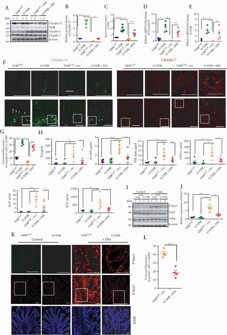
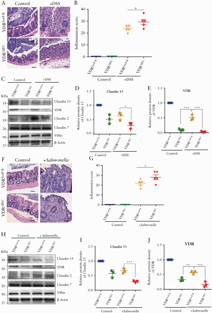
Background and aims: Dysfunction of the vitamin D receptor [VDR] contributes to the aetiology of IBD by regulating autophagy, immune response, and mucosal permeability. VDR directly controls the paracellular tight junction protein Claudin-2. Claudin-2 and Claudin-15 are unique in maintaining paracellular permeability. Interestingly, claudin-15 mRNA was downregulated in patients with ulcerative colitis. However, the exact mechanism of Claudin-15 regulation in colitis is still unknown. Here, we investigated the protective role of VDR against intestinal inflammation via upregulating Claudin-15.

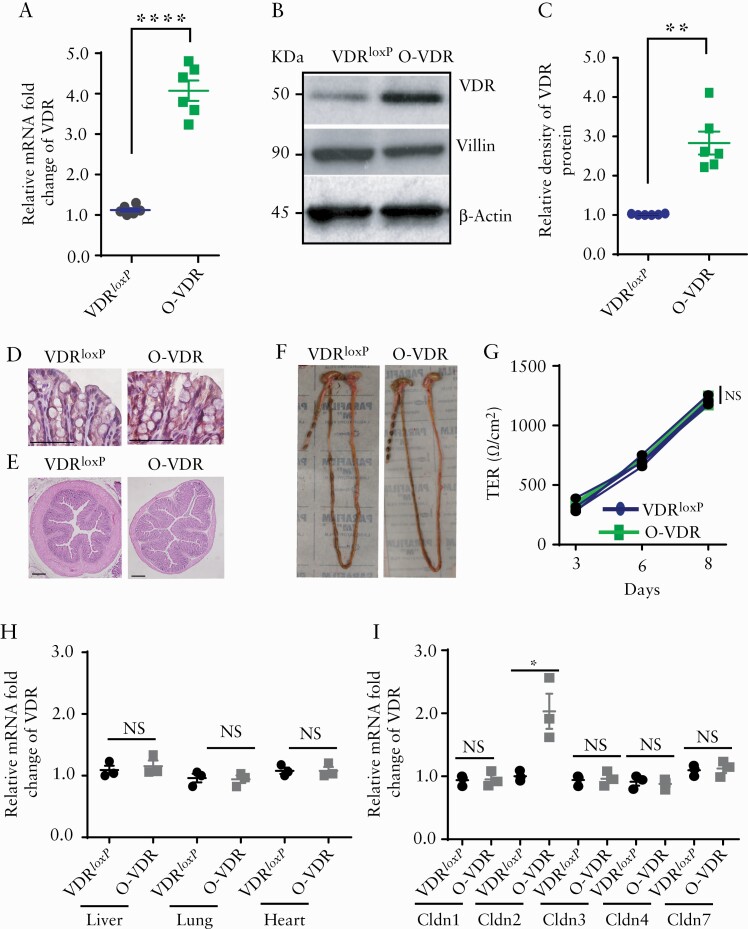
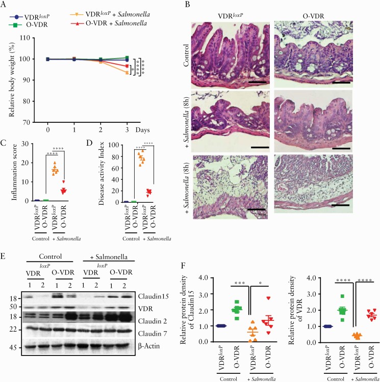
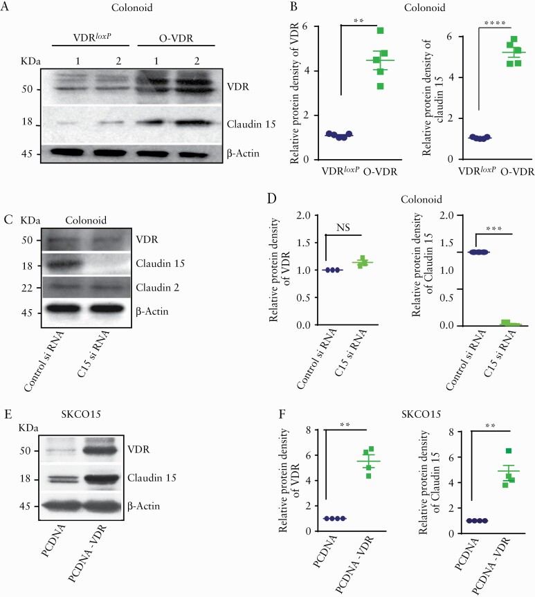
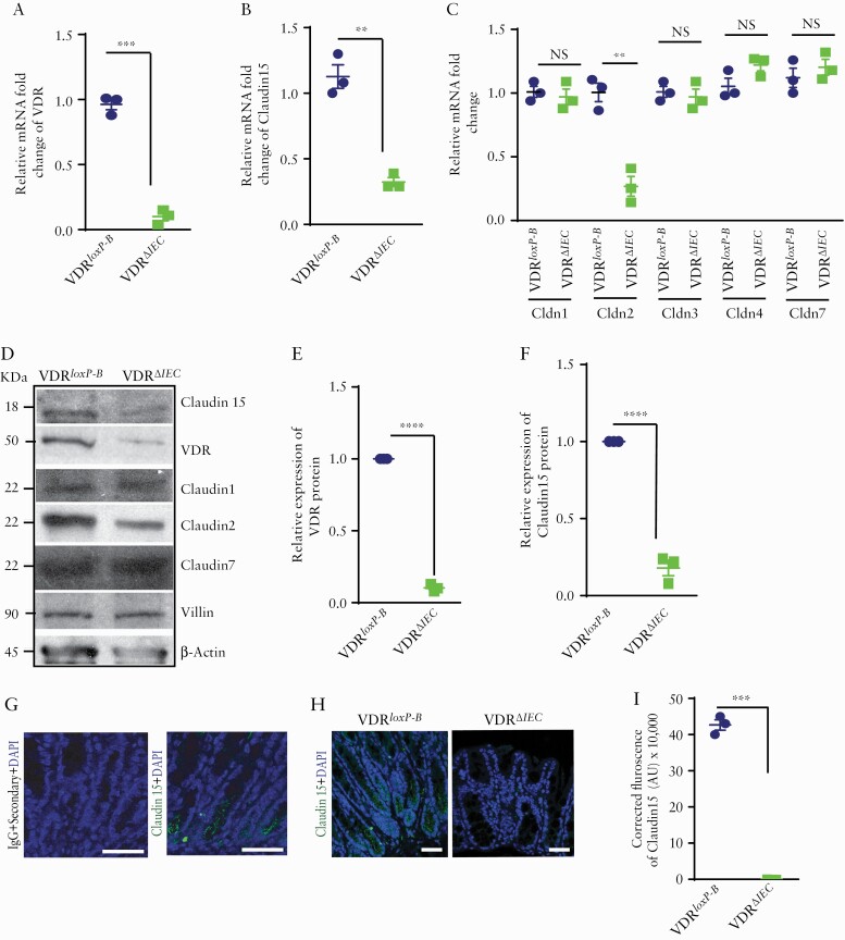
Methods: We analysed the correlation of Claudin-15 with the reduction of VDR in human colitis. We generated intestinal epithelial overexpression of VDR [O-VDR] mice to study the gain of function of VDR in colitis. Intestinal epithelial VDR knockout [VDR∆IEC] mice were used for the loss of function study. Colonoids and SKCO15 cells were used as in vitro models.

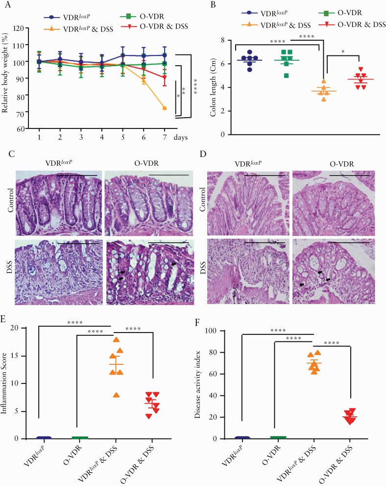
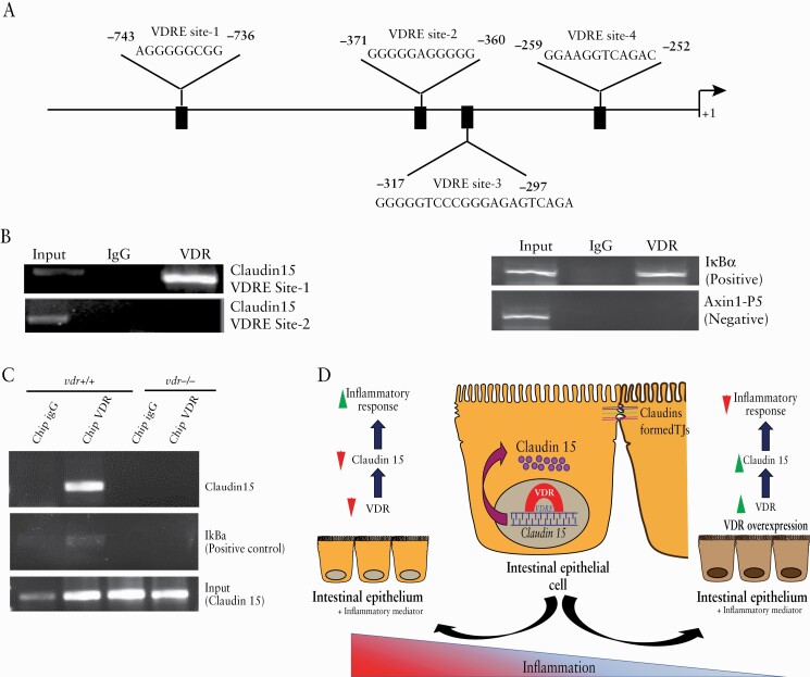
Results: Reduced Claudin-15 was significantly correlated with decreased VDR along the colonic epithelium of human IBD. O-VDR mice showed decreased susceptibility to chemically and bacterially induced colitis and marked increased Claudin-15 expression [both mRNA and protein] in the colon. Correspondingly, colonic Claudin-15 was reduced in VDR∆IEC mice, which were susceptible to colitis. Overexpression of intestinal epithelial VDR and vitamin D treatment resulted in a significantly increased Claudin-15. ChIP assays identified the direct binding of VDR to the claudin-15 promoter, suggesting that claudin-15 is a target gene of VDR.

Conclusion: We demonstrated the mechanism of VDR upregulation of Claudin-15 to protect against colitis. This might enlighten the mechanism of barrier dysfunction in IBD and potential therapeutic strategies to inhibit inflammation.

Keywords: Salmonella; Claudin; Crohn’s disease; IBD; VDR; colonoids; inflammation; tight junction; ulcerative colitis.


Figures

Graphical Abstract
Graphical Abstract
Figure 1.
Figure 1.
Significantly coordinated expression of reduced Claudin-15 and VDR in patients with ulcerative colitis [UC]. [A] Significantly coordinated expression of VDR and Claudin-15 in UC patients. We performed a regression analysis and a scatter plot of VDR against Claudin-15. Values for healthy controls were in blue colour and values for patients were in red colour. GEO database GDS3119 Normal, n = 5; UC, n = 21; the coefficient is 0.51096 with p = 0.0092 in linear regression model. The graph shows VDR and Claudin-15 expression are positively correlated and UC patients are differentiated from normal controls. [B] Immunofluorescence staining detected Claudin-15 protein [green colour] in inflamed and adjacent normal colonic epithelium in biopsy specimens obtained from patients with UC. The nucleus is stained blue with DAPI. Scale bar = 50 µm. Data were analysed by unpaired t test, ***p ≤ 0.001. [C]The specificity of the Claudin-15 immunostaining was verified in the human colon tissue section using IgG [+only secondary antibody] as control [upper panel, n = 4] and a similar section was stained with anti-Claudin-15 antibody [lower panel, n = 4]. [D] Quantification of green fluorescence indicated decreased Claudin-15 expression in the inflamed colonic epithelium of patients with UC. In each figure, values for normal colon indicated by blue colour and for inflamed colon by red colour. [D] Significantly coordinated expression of VDR and Claudin-15 as detected by immunofluorescence in biopsy samples of ulcerative colitis patients and normal controls. Blue indicates normal colon and red indicates UC patients sample. Normal, n = 4; UC, n = 4; the coefficient is 12.6 with p = 0.0059 in linear regression model. VDR, vitamin D receptor.
Figure 2.
Figure 2.
O-VDR mouse model showed intestinal VDR overexpression. An increase in VDR expression in the O-VDR mouse colon was indicated by [A] mRNA, [B] western blot images, and [C] densitometry quantification of the blots. [D] IHC staining with anti-VDR antibody indicated augmented VDR expression in O-VDR mouse colonic epithelium. [E] O-VDR mice exhibited normal colon histopathology. [F] Morphology and normal colon length. [G] TEER of mouse colonoid-derived monolayers remained unchanged between the O-VDR and VDRloxP groups. [H] mRNA expression of VDR in the liver, lung, and heart remained unaltered in OVDR mice as compared with VDRloxP. [I] Increased mRNA expression of claudin-2 mRNA in OVDR mouse colon was detected; however, expression of Claudin- 1, 3, 4, and 7 did not change in OVDR mice as compared with VDRloxP mice. In each figure, values for VDRloxP are indicated by blue colour and for O-VDR are indicated by green colour. n = 3 to 6 mice per group. Data were analysed by unpaired t test for Figure 2A, C, H, and I and two-way ANOVA for 2G. NS = not significant, * p < 0.05, **p ≤ 0.01, and ***p ≤ 0.001. VDR, vitamin D receptor; IHC, immunohistochemical; TEER, transepithelial electrical resistance; ANOVA, analysis of variance.
Figure 3.
Figure 3.
O-VDR mice are protected from acute colitis. Mice 8‐10 weeks old were treated with dextran sodium sulphate [DSS]: 5% [W/V] in the drinking water for 7 days. [A] Weight loss during the course of treatment. The body weight of the O-VDR group was significantly higher than that of the DSS-treated control group. Body weights were analysed by generalised linear mixed models. [B] Colon length in control and O-VDR mice after DSS treatment. Representative images of H&E-stained colonic epithelium sections from [C] the distal colon and [D] the proximal colon. [E] Decreased inflammation score in O-VDR mice compared with VDRloxP following DSS treatment; control mice did not show inflammation and/or any injury. [F] The disease activity index was significantly altered in DSS-treated mice. In each figure, values for VDRloxP are indicated by blue colour and for O-VDR are indicated by green colour, VDRloxP+DSS are indicated in orange, and OVDR + DSS are indicated in red colour. Scale bar = 200 µm, n = 6 [3 male and 3 female] mice per group. Data were analysed for 3B, 3E, and 3F by unpaired t test or one-way ANOVA. *p ≤ 0.05, **p ≤ 0.01, and ****p ≤ 0.0001. VDR, vitamin D receptor; H&E, haematoxylin and eosin; ANOVA, analysis of variance.
Figure 4.
Figure 4.
O-VDR mice retained Claudin-15 expression following acute DSS colitis, thus protecting the host from inflammation. [A] Western blot analysis of mouse colonic tissue indicated decreased VDR and Claudin-15 protein expression following acute DSS colitis; however, Claudin-15 expression was restored in O-VDR mice. Densitometric analysis of [B] Claudin-15 and [C] VDR by western blot. Relative mRNA expression of [D] Claudin-15 and [E] VDR in mouse colonic epithelium. [F] The expression and distribution of Claudin-15 in mouse colonic epithelium is shown by green fluorescence. Nuclear staining is revealed by blue DAPI. [G] The intensity of the immunofluorescence staining of Claudin-15. [H] Cytokines were inhibited in DSS-treated O-VDR mice. [I] Western blot analysis indicating protein levels of p-stat 3, stat3, villin, and actin in VDRloxP and O-VDR mice. [J] Densitometric analysis of p-stat3 expression. [K] Immunofluorescence staining indicating p-stat3 expression and distribution in mouse colonic epithelium. Control VDRloxP and O-VDR mice did not show detectable p-stat3. [L] Quantification of the red fluorescence of p-stat3, n = 3–6. Data were analysed by unpaired t test or one-way ANOVA, *p ≤ 0.05, **p ≤ 0.01, ***p ≤ 0.001, and ****p ≤ 0.0001. Scale bar = 50 µm, n = 3–6. . VDR, vitamin D receptor; ANOVA, analysis of variance.
Figure 5.
Figure 5.
OVDR mice were protected from Salmonella-induced colitis. Mice 8‐10 weeks old were treated with Salmonella typhimurum. [A] VDR over-expressing OVDR mice were protected from Salmonella-induced colitis. Mice (eight to ten week-old) were infected with Salmonella. Body weights of the mice were analysed by generalised linear mixed models. [B] Representative images of H&E of mice caecum without [top panel] and with 8 h [middle panel] and with 4 days [lowermost panel] of Salmonella infection. [C] Decreased inflammation [in caecum] in O-VDR mice was noted as compared with VDRloxP after 8 h of salmonella treatment. [D] The disease activity index was significantly altered in Salmonella infection. [E] Western blot images showing OVDR mice retained VDR and Claudin-15 protein expression in the colon, compared with the VDRloxP group. [F] Densitometric quantification of the western blots. In each figure, values for VDRloxP are indicated by blue colour and for O-VDR are indicated by green colour, VDRloxP+ Salmonella is indicated in orange and OVDR + Salmonella are indicated in red colour. n = 6 for each group. All data in C, D, and F were analysed by unpaired t test or one-way ANOVA. *p ≤ 0.05, **p ≤ 0.01, ***p ≤ 0.001, and ****p ≤ 0.0001. VDR, vitamin D receptor; H&E, haematoxylin and eosin; ANOVA, analysis of variance.
Figure 6.
Figure 6.
VDR regulates Claudin-15 expression in vitro. [A] Western blot image shows higher VDR and Claudin-15 expression in mouse colonoids. [B] Densitometric quantification of the western blots. [C] Protein expression after Claudin-15 siRNA treatment in mouse colonoids. [D] Densitometric quantification of the western blots after silencing of Claudin-15 by siRNA. [E] Western blot images showing increased VDR and Claudin-15 expression in SKCO15 cells following transfection with hVDR. [F] Quantification of the blot. In each figure, values for the control group are indicated by blue colour and for the experimental group are indicated by green colour; n = 3–6 for each group. Data were analysed by unpaired t test or ANOVA, NS p > 0.05, *p ≤ 0.05, **p ≤ 0.01, ***p ≤ 0.001, ****p ≤ 0.0001. VDR, vitamin D receptor; ANOVA, analysis of variance; NS, non-significant.
Figure 7.
Figure 7.
Deletion of VDR reduced Claudin-15 expression in vivo. Reduced VDR and Claudin-15 were detected in VDRΔIEC mice as indicated by [A] VDR mRNA and [B] claudin-15 mRNA. However, mRNA of [C] Claudin-1, 3, 4, 7 was unaltered with exception of Claudin-2. [D] Western blot image showing decreased expressions of VDR, Claudin-15, and Claudin-2 in mouse colon. Densitometric quantification of the western blots of [E] VDR and [F] Claudin-15. [G] No staining was detected in the mice colon tissue section by IgG [+only secondary antibody] whereas similar section stained with Claudin-15 antibody indicated staining in the mice colon as indicated by specific green colour. [H] Representative images of ICC images indicating a lower level of Claudin-15 expression in mousee colon. [I] Quantification of the ICC staining. Scale bar = 50 µm. In each figure, values for the control group are indicated by blue colour and for the experimental group are indicated by light green colour; n = 3 for each group. VDRloxp-B: Belgium VDRloxp/loxp mice used in generating VDRΔIEC mice. Data were analysed by unpaired t test or one-way ANOVA, NS p > 0.05, **p ≤ 0.01, ***p ≤ 0.001, and ****p ≤ 0.0001. VDR, vitamin D receptor; ICC, immunocytochemistry; ANOVA, analysis of variance; NS, non-significant.
Figure 8.
Figure 8.
Reduced Claudin-15 expression due to deletion of VDR in the intestinal epithelium in the experimental colitis. [A] Representative images of H&E of mice with or without DSS treatment. [B] Increased inflammation in VDRΔIEC mice was noted as compared with VDRloxP-B. [C] Representative images of western blot of Claudin-15, Claudin-2, Claudin-7, and VDR after DSS treatment. Densitometric analysis of Claudin -15 [D] and VDR [E] in VDRΔIEC mousee colon in DSS colitis. [F] Representative images of H&E of mouse caecum 4 days post Salmonella infection. [G] Increased inflammation in VDRΔIEC mice was noted as compared with VDRloxP-B after Salmonella infection. [H] Representative images of western blot of Claudin-15, Claudin-2, Claudin-7, and VDR following Salmonella treatment. Densitometric analysis of Claudin -15 [I] and VDR [J] in the colon of VDRΔIEC mice in Salmonella-induced colitis. Scale bar = 50 µm, n = 3–6. Data were analysed by unpaired t test or one-way ANOVA, *p ≤ 0.05, **p ≤ 0.01, and ****p ≤ 0.001. VDR, vitamin D receptor; H&E, haematoxylin and eosin; ANOVA, analysis of variance.
Figure 9.
Figure 9.
VDR regulates Claudin-15 expression in colonic epithelium. [A] Putative VDRE sites in the Claudin-15 promoter. [B] ChIP assay using quantitative PCR followed by SDS-PAGE electrophoresis showed direct binding of VDR to the Claudin-15 promoter. The assay was verified for a primer of a VDRE site on IkBα as a positive control and a primer for a non-VDRE site of Axin1 as a negative control. [C] SDS-PAGE analysis following the ChIP assay verified the binding of VDR to the Claudin-15 promoter, n = 3–6. [D] A working model of intestinal epithelial VDR in maintaining tight junctions and protecting the host from colitis. In colonic epithelial cells, VDR binds directly to the VDRE region of the Claudin-15 promoter, thus increasing the expression of Claudin-15. Different types of tight junction proteins, including Claudin-15, form tight junctions [TJs] in epithelial cells. Chronic inflammation is known to damage these TJs. However, overexpression of VDR protects against inflammation by enhancing the expression of the tight junction protein Claudin-15. VDR, vitamin D receptor; PCR, polymerase chain reaction.

Comment in

  • Letter to the Editor.
    ECCO Governing Board. ECCO Governing Board. J Crohns Colitis. 2022 Nov 23;16(11):1792-1793. doi: 10.1093/ecco-jcc/jjab225. J Crohns Colitis. 2022. PMID: 35073577 No abstract available.

References

    1. Lloyd-Price J, Arze C, Ananthakrishnan AN, et al. ; IBDMDB Investigators. Multi-omics of the gut microbial ecosystem in inflammatory bowel diseases. Nature 2019;569:655–62. - PMC - PubMed
    1. Mineta K, Yamamoto Y, Yamazaki Y, et al. Predicted expansion of the claudin multigene family. FEBS Lett 2011;585:606–12. - PubMed
    1. Edelblum KL, Turner JR. The tight junction in inflammatory disease: communication breakdown. Curr Opin Pharmacol 2009;9:715–20. - PMC - PubMed
    1. Darsigny M, Babeu JP, Dupuis AA, et al. Loss of hepatocyte-nuclear-factor-4alpha affects colonic ion transport and causes chronic inflammation resembling inflammatory bowel disease in mice. PLoS One 2009;4:e7609. - PMC - PubMed
    1. Liu W, Chen Y, Golan MA, et al. Intestinal epithelial vitamin D receptor signaling inhibits experimental colitis. J Clin Invest 2013;123:3983–96. - PMC - PubMed