Review
. 2021 Jun;86(1):32-56.
doi: 10.1111/prd.12361. Epub 2021 Mar 10.

The dental plaque biofilm matrix

Affiliations
Review

The dental plaque biofilm matrix

Nicholas S Jakubovics et al. Periodontol 2000. 2021 Jun.

Abstract

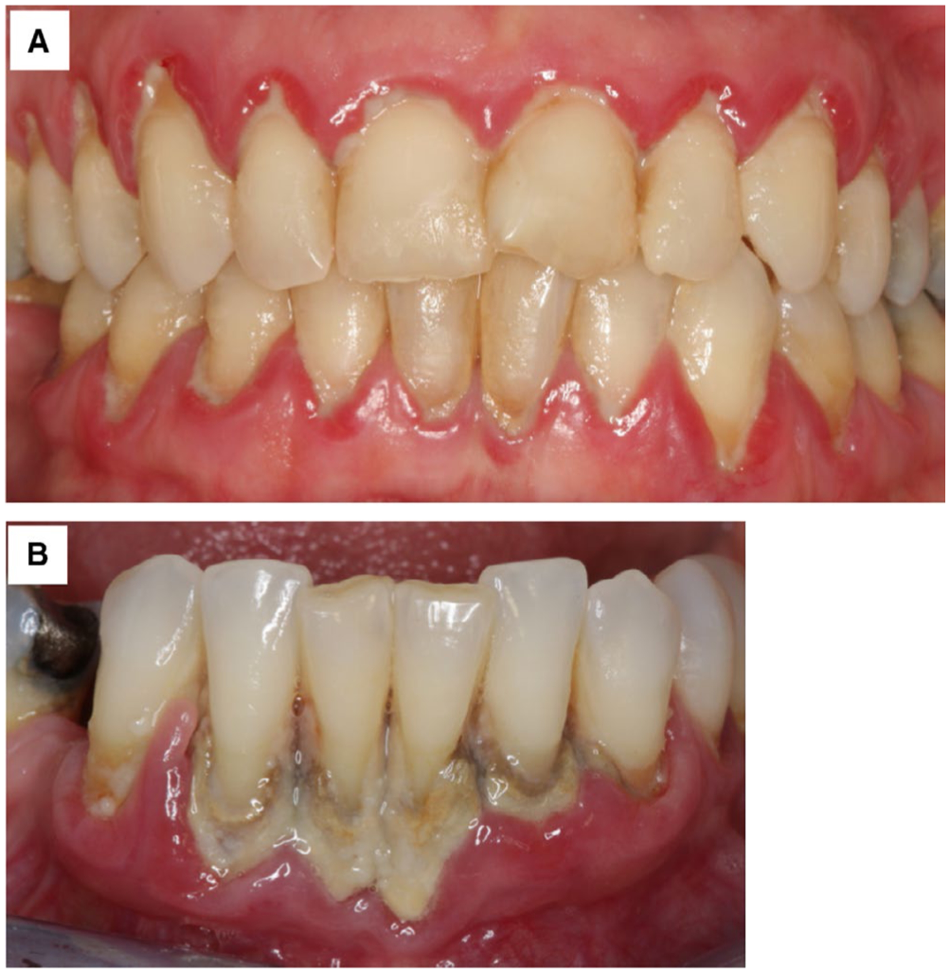
The extracellular matrix is a critical component of microbial biofilms, such as dental plaque, maintaining the spatial arrangement of cells and coordinating cellular functions throughout the structure. The extracellular polymeric substances that comprise the matrix include carbohydrates, nucleic acids, proteins, and lipids, which are frequently organized into macromolecular complexes and/or are associated with the surfaces of microbial cells within the biofilm. Cariogenic dental plaque is rich in glucan and fructan polysaccharides derived from extracellular microbial metabolism of dietary sucrose. By contrast, the matrix of subgingival dental plaque is a complex mixture of macromolecules that is still not well understood. Components of the matrix escape from microbial cells during lysis by active secretion or through the shedding of vesicles and serve to anchor microbial cells to the tooth surface. By maintaining the biofilm in close association with host tissues, the matrix facilitates interactions between microorganisms and the host. The outcome of these interactions may be the maintenance of health or the development of dental disease, such as caries or periodontitis. The matrix affords microbial cells protection against chemical and physical insults and hinders the eradication of pathogenic dental plaque. Therefore, strategies to control the matrix are critical to maintain oral health. This review discusses recent advances in our understanding of the composition, origins, and function of the dental plaque matrix, with a focus on subgingival dental plaque. New strategies to control subgingival dental plaque based on targeting the biofilm matrix are also considered.

Keywords: extracellular DNA; periodontitis; polysaccharides; secretion; subgingival dental plaque; vesicles.


Figures

FIGURE 1
FIGURE 1
Comparison of the structure and function of host vs biofilm extracellular matrix. Host tissue contains a variety of different extracellular matrices. Shown here are the basement membrane and interstitial matrix underlying a layer of keratinized epithelium. Each of these contains proteins, proteoglycans, and glycoproteins that serve a variety of functions in adhesion, sensing, and protection. The basement membrane consists of the basal lamina, which forms the underlying layer for the epithelial cells, and the reticular lamina, composed primarily of collagenous fibers that serve to anchor the basal lamina. The interstitial matrix contains a variety of different cells, including fibroblasts. The biofilm matrix contains macromolecules primarily derived from the resident bacterial cells. These also function in adhesion, sensing, and protection, in addition to acting as a store of nutrient and a potential source of DNA for natural transformation. eDNA, extracellular DNA; LPS, lipopolysaccharide; LTA, lipoteichoic acid; PNAG, poly-N-acetylglutamic acid
FIGURE 2
FIGURE 2
Scanning electron micrograph of subgingival dental plaque on the surface of a tooth extracted due to periodontal disease. Microbial cells are connected by a meshwork of fibrous material (M), apparently the collapsed remains of a hydrated polymeric matrix. Some microbial cells are associated with small particles or vesicles (V) that may provide a source for matrix polymers
FIGURE 3
FIGURE 3
Fluorescent lectin binding analysis of supragingival dental biofilm, grown in situ in the mouth of a volunteer for 48 hours in the absence of dietary sucrose. The biofilm was stained with Morniga-G lectin (MNA-G-fluorescein isothiocyanate, green, recognizes galactose), Helix pomatia lectin (HPA-tetramethylrhodamine, red, recognizes N-acetyl-α-galactosamine), and Syto60 (blue, stains DNA). Maximum projection images are shown; scale bar: 10 μm. The target carbohydrates appear to be primarily associated with microbial cell walls. Image kindly provided by Thomas R. Neu, Pune N. Paqué, and Sebastian Schlafer
FIGURE 4
FIGURE 4
P. gingivalis histone-like protein PG0121 is abundant in dual-species biofilms with Streptococcus gordonii (Sg). Porphyromonas gingivalis (Pg) was seeded into extant S. gordonii biofilms and immunofluorescence was performed to determine the distribution of PG0121 present in dual-species biofilms of a 1:1 ratio of Pg:Sg. All bacterial cells were labeled with the membrane stain FM4-64 and pseudocolored cyan (A), and PG0121 was detected with antibodies directed against PG0121 followed by the addition of secondary antibodies conjugated to Alexa Fluor-488 and pseudocolored magenta (B)
FIGURE 5
FIGURE 5
Interactions between macromolecules in the matrix of dental plaque. Glucans and fructans are associated with the enzymes that produce them (glucosyltransferases, GTFs/fructosyltransferases, FTFs), and insoluble glucans are recognized by glucan-binding proteins (GBPs), including the wall-anchored protein GbpC. Glucans and GTFs associate with extracellular DNA (eDNA) and lipoteichoic acid (LTA), which also binds extracellular DNA. In addition, wall teichoic acids (WTAs) and certain gram-positive cell-surface proteins are covalently linked to peptidoglycan. Therefore, these will remain attached to peptidoglycan fragments in the biofilm matrix. Cell-surface adhesins mediate coaggregation through binding to capsular polysaccharides. Proteins are also associated with extracellular DNA, and extracellular DNA is present on the surface of gram-positive membrane vesicles. Gram-negative outer membrane vesicles contain lipopolysaccharides (LPS), which bind to poly-N-acetyl-d-glucosamine (PNAG) through charge interactions
FIGURE 6
FIGURE 6
Scanning electron micrograph showing erythrocytes and fibrous material on the surface of a tooth extracted due to periodontal disease. Erythrocytes (arrows) are associated with fibrous noncellular material overlying the dental plaque on the tooth surface. Bacterial cells are not visible in this image
FIGURE 7
FIGURE 7
Clinical images of dental plaque. A, A 49-year-old female patient with insufficient oral hygiene, presenting massive amounts of subgingival plaque and clinical signs of plaque-associated gingivitis. B, A 53-year-old female patient with insufficient oral hygiene, presenting massive amounts of subgingival plaque and calculus and clinical signs of advanced periodontitis
FIGURE 8
FIGURE 8
Dental plaque and calculus on the surface of extracted teeth. Teeth 37, 36, and 35 were extracted due to periodontal disease. All teeth present massive amounts of subgingival plaque and calculus

References

    1. Mack WN, Mack JP, Ackerson AO. Microbial film development in a trickling filter. Microb Ecol. 1975;2:215–226. - PubMed
    1. Jones HC, Roth IL, Sanders WM III. Electron microscopic study of a slime layer. J Bacteriol. 1969;99:316–325. - PMC - PubMed
    1. Schroeder HE, de Boever J. The structure of microbial dental plaque. In: McHugh WD, ed. Dental Plaque. Dundee, UK: E. & S. Livingstone; 1969:49–74.
    1. Bennett HS. Morphological aspects of extracellular polysaccharides. J Histochem Cytochem. 1963;11:14–23.
    1. Ito S. Form and function of the glycocalyx on free cell surfaces. Philos Trans R Soc Lond B Biol Sci. 1974;268:55–66. - PubMed

Publication types