Review
. 2021 Jun;86(1):97-112.
doi: 10.1111/prd.12364. Epub 2021 Mar 10.

Spatial scale in analysis of the dental plaque microbiome

Affiliations
Review

Spatial scale in analysis of the dental plaque microbiome

Gary G Borisy et al. Periodontol 2000. 2021 Jun.

Abstract

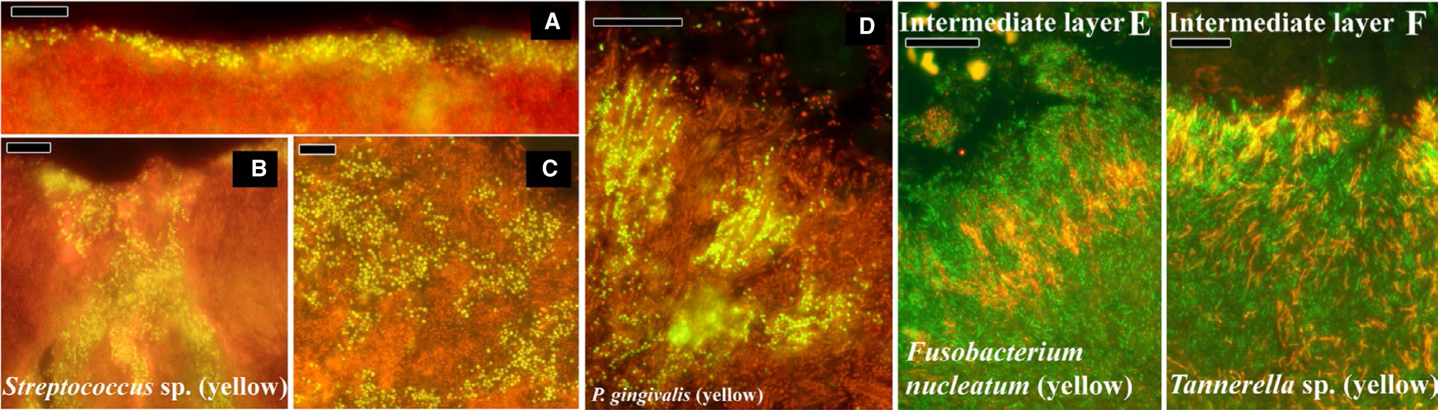
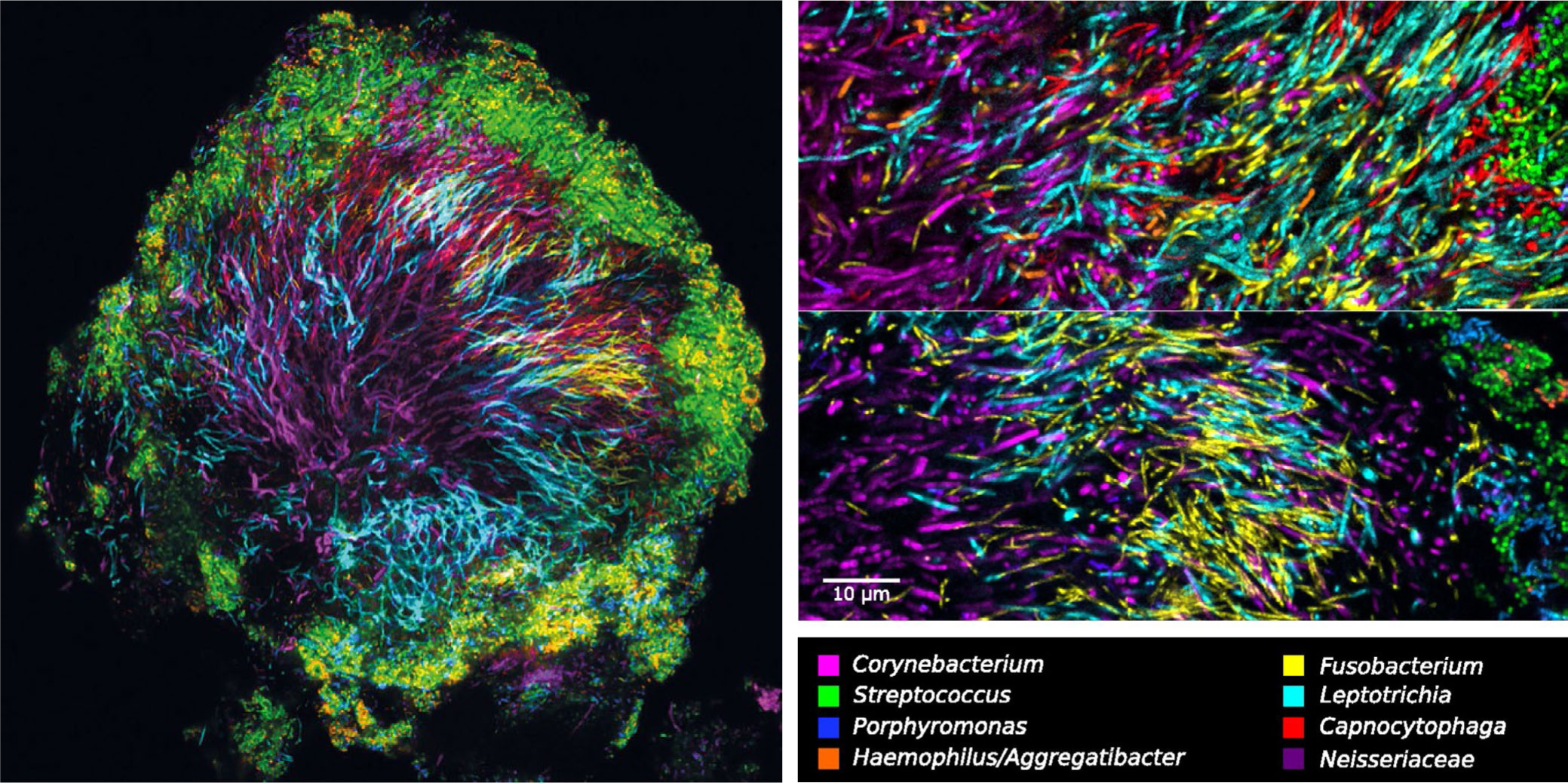
Ecologists have long recognized the importance of spatial scale in understanding structure-function relationships among communities of organisms within their environment. Here, we review historical and contemporary studies of dental plaque community structure in the context of three distinct scales: the micro (1-10 µm), meso (10-100 µm) and macroscale (100 µm to ≥1 cm). Within this framework, we analyze the compositional nature of dental plaque at the macroscale, the molecular interactions of microbes at the microscale, and the emergent properties of dental plaque biofilms at the mesoscale. Throughout our analysis of dental plaque across spatial scales, we draw attention to disease and health-associated structure-function relationships and include a discussion of host immune involvement in the mesoscale structure of periodontal disease-associated biofilms. We end with a discussion of two filamentous organisms, Fusobacterium nucleatum and Corynebacterium matruchotii, and their relevant contributions in structuring dental plaque biofilms.

Keywords: Fusobacterium nucleatum; CLASI-FISH; biofilm; dental plaque; oral microbiome.


Figures

FIGURE 1
FIGURE 1
The significance of scale in dental plaque biology. The structure of dental plaque can be studied at three scales: the micro, meso, and macroscales. Different fundamental forces contribute to the structure of dental plaque at each scale. Micro and macroscale processes that structure dental plaque contribute to the unique emergent properties of dental plaque at the mesoscale. These properties include the creation of ecologic niches, metabolic cooperation, and persistence in its environment
FIGURE 2
FIGURE 2
Early histochemical light micrograph of dental plaque on an epon strip and worn for 2 days by a healthy volunteer. Visible are palisading columns of phenotypically similar organisms, an abundance of cocci with some rods, and the salivary pellicle. Reprinted with permission from Listgarten et al
FIGURE 3
FIGURE 3
Field emission-scanning electron microscopy image of subgingival dental plaque. Mesoscale structure of the subgingival biofilm consists of loosely arranged cells with multiple different morphologies, including rods, filaments, and spirochetes. Also present are extrapolymeric substances and putative outer membrane vesicles arranged in chains, both of which contribute to the spatial structure of plaque biofilms. Image credit: Richard Holliday. Reproduced from Holliday et al
FIGURE 4
FIGURE 4
Fluorescence in-situ hybridization–labeled supra and subgingival dental plaque on teeth from donors with periodontal disease. A-C, Supragingival plaque labeled with a probe for all bacteria; EUB-338 in red, and streptococci in yellow. D, Subgingival plaque labeled with EUB red and a probe for Porphyromonas gingivalis in yellow. E, Subgingival plaque intermediate layer labeled with EUB in green and a probe for Fusobacterium nucleatum in red. F, Intermediate layer of subgingival plaque labeled with EUB in green and a genus-level probe for Tannerella in red. Reprinted from Zijnge et al
FIGURE 5
FIGURE 5
Multispectral fluorescence in-situ hybridization images of a hedgehog structure in supragingival plaque carefully scraped from the tooth of a healthy volunteer. Visible are filamentous organisms in the center of the structure and streptococci and other facultative anaerobes at the saliva-exposed surface of the structure. Zoom images show the abundance of filamentous organisms in the center of the hedgehog structure, which include Corynebacterium, Leptotrichia, and Fusobacterium. Reprinted from Mark Welch et al
FIGURE 6
FIGURE 6
Corncob structures in supragingival plaque. A, B, Scanning electron microscope images of corncob structures showing a central filament encased at the tip with cocci. C, Multispectral fluorescence in-situ hybridization images of corncob structures observed at the periphery of hedgehog structures. Fluorescence in-situ hybridization allows taxonomic identification of the component cells and reveals the central filament to be either unlabeled or labeled with a probe for Corynebacterium. The cocci that decorate the filament show some diversity and include cells of the genera Streptococcus, Haemophilus or Aggregatibacter, and Porphyromonas. A, B, Previously unpublished images courtesy of Professor Charles M. Cobb, see Cobb and Killoy. C, Reprinted from Mark Welch et al

References

    1. Earle KA, Billings G, Sigal M, et al. Quantitative imaging of gut microbiota spatial organization. Cell Host Microbe. 2015;18(4):478–488. - PMC - PubMed
    1. Edwards J, Johnson C, Santos-Medellín C, et al. Structure, variation, and assembly of the root-associated microbiomes of rice. Proc Natl Acad Sci USA. 2015;112(8):E911–E920. - PMC - PubMed
    1. Flintrop CM, Rogge A, Miksch S, Thiele S, Waite AM, Iversen MH. Embedding and slicing of intact in situ collected marine snow. Limnol Oceanogr Methods. 2018;16(6):339–355.
    1. Nunan N, Wu K, Young IM, Crawford JW, Ritz K. Spatial distribution of bacterial communities and their relationships with the micro-architecture of soil. FEMS Microbiol Ecol. 2003;44(2):203–215. - PubMed
    1. Wiens JA. Spatial scaling in ecology. Funct Ecol. 1989;3(4):385–397.

Publication types

Supplementary concepts

LinkOut - more resources