Review
. 2021 Mar 9;54(3):437-453.
doi: 10.1016/j.immuni.2021.01.018.

Autophagy in inflammation, infection, and immunometabolism

Affiliations
Review

Autophagy in inflammation, infection, and immunometabolism

Vojo Deretic. Immunity. .

Abstract

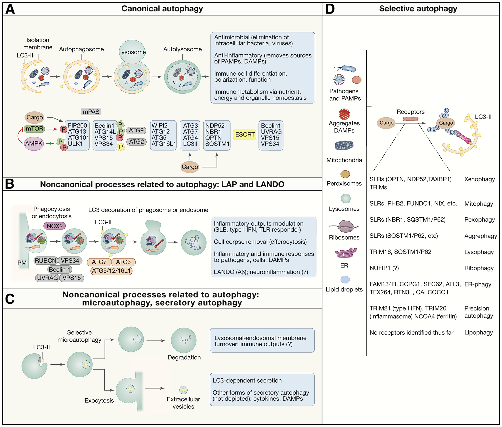
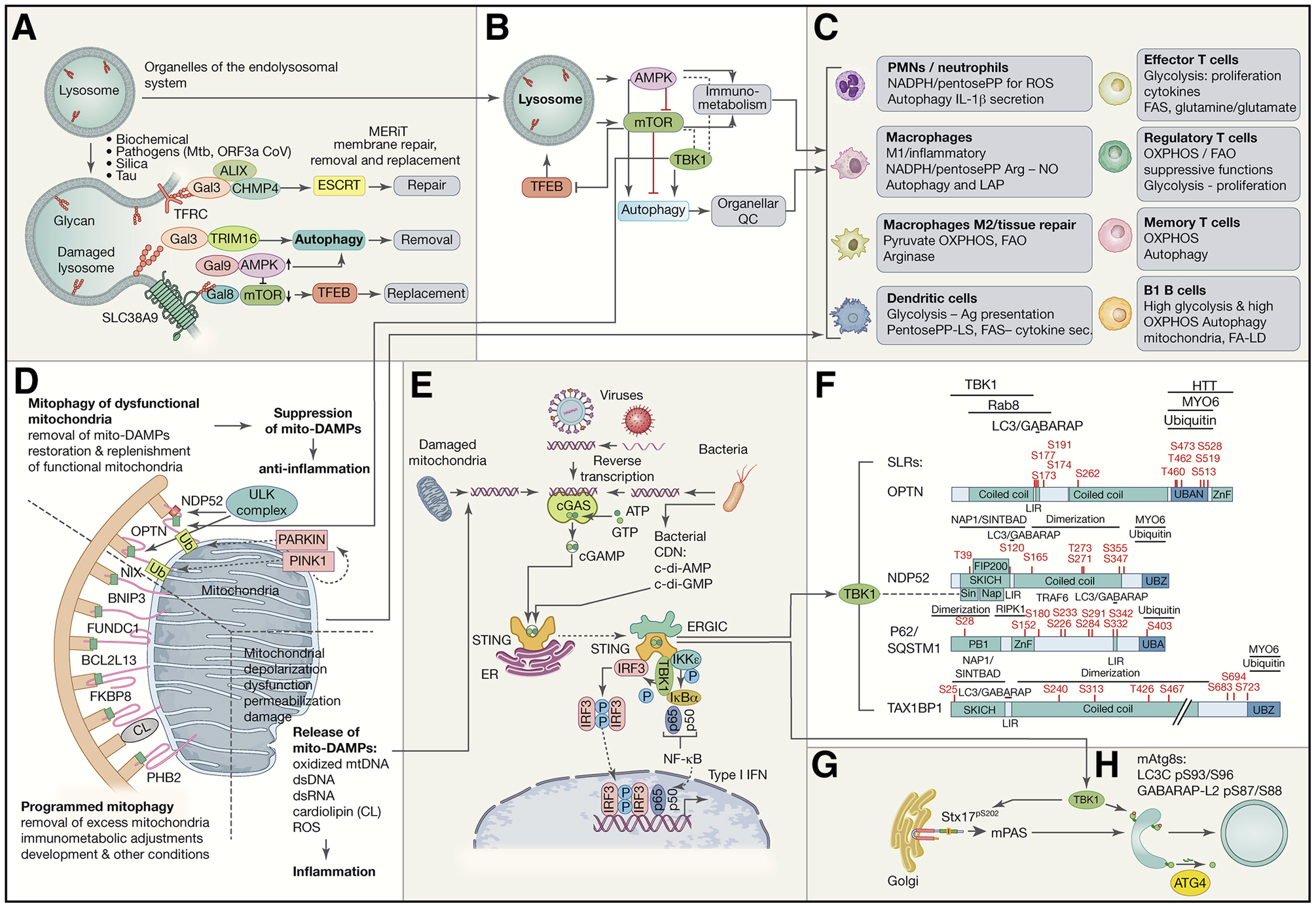
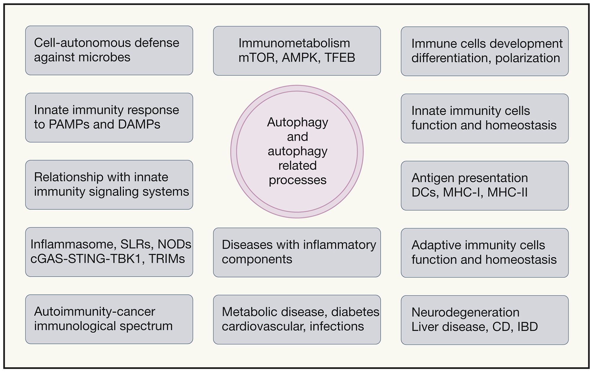
Autophagy is a quality-control, metabolic, and innate immunity process. Normative autophagy affects many cell types, including hematopoietic as well as non-hematopoietic, and promotes health in model organisms and humans. When autophagy is perturbed, this has repercussions on diseases with inflammatory components, including infections, autoimmunity and cancer, metabolic disorders, neurodegeneration, and cardiovascular and liver diseases. As a cytoplasmic degradative pathway, autophagy protects from exogenous hazards, including infection, and from endogenous sources of inflammation, including molecular aggregates and damaged organelles. The focus of this review is on the role of autophagy in inflammation, including type I interferon responses and inflammasome outputs, from molecules to immune cells. A special emphasis is given to the intersections of autophagy with innate immunity, immunometabolism, and functions of organelles such as mitochondria and lysosomes that act as innate immunity and immunometabolic signaling platforms.


Conflict of interest statement

Declaration of interests The author declares no competing interests.

Figures

Figure 1.
Figure 1.. Autophagy and autophagy-related pathways as guardians against excessive inflammation.
A. Simplified morphological stages of canonical autophagy. Autophagosomes have double membrane, sequester diverse cytoplasmic cargo, and typically deliver the cargo to lysosomes for degradation. Principal protein complexes controlling autophagy are shown below in approximate order of activities along the autophagy pathway. Red circles, inhibitory phosphorylation by mTOR. Green squares, activating phosphorylation by AMPK B. LC3-assocate phagocytosis (LAP) and LC-3-associated endocytosis (LANDO): noncanonical autophagy-related processes that originate starting with invaginations at the plasma membrane leading to formation of phagosomes and endosomes decorated with lipidated LC3 (LC3-II). They are single membranes that use only a subset of ATG proteins plus NOX2 (LAP) and Rubicon and are independent of the FIP200 complex. C. Other noncanonical autophagy-related processes. Selective microautophagy: LC3-II on lysosomes and endosomes contributes to formation of intralumenal vesicles that are either digested or exocytosed. Other processes (not depicted) are mentioned in the box. D. List of autophagy cargo, receptors and terminology for various types of autophagy, defined by the targeted cargo. Anti-inflammatory and related immune functions are listed in gray boxes.
Figure 2.
Figure 2.. Lysosomes, mitochondria, and TBK1 in inflammation, immunometabolism and immune responses.
A. Lysosomal integrity is maintained by a system termed MERiT (membrane repair, removal and replacement). Galectins (Gal3, Gal8 and Gal9) recognize β-galactoside glycans exposed on cognate lysosomal membrane proteins: TFRC (transferrin receptor), SLC38A9, and LAMP1 and LAMP2 (not depicted) when lysosomal membrane is breached. They stimulate ESCRTs to repair lysosomes and AMPK to activate autophagy, whereas mTOR is inactivated. TRIM16 is one of lysophagy receptors. TFEB starts transcriptional program to replace lysosomes. B. Lysosome as the hub for AMPK, mTOR and TFEB, the three master regulators of autophagy, lysosomal system, metabolism and immunometabolism. Inhibitory and activating relationships are indicated and include TBK1 (which has positive and negative regulatory connections with AMPK and mTOR). C. Effects of signaling in B in different immune cells. D. Mitophagy, mitophagy receptors, and mitophagic suppression of inflammation caused by mitochondrial DAMPs (‘mito-DAMPs’). Dashed lines separate different types of mitophagy and proinflammatory signalng from damaged mitochiondria. Top, Pink1-Parkin system and receptors that participate in removal of dysfunctional mitochondria. Pink1 kinase and Parkin E3 ligase (phosphorylated by Pink1 to activate latent Parkin activity and recruit it to mitochondria) lead to ubiquitination and generation of phospho-ubiquitin which further amplifies Parkin activity leading to recognition of depolarized or damaged mitochondria by SLRs, NDP52 and OPTN, which bind ubiquitin. Bottom left, other mitophagy receptors act as integral membrane proteins of mitochondria and participate in mitophagy under developmental, differentiation or stress conditions. (E) Molecular machinery involved in STING-TBK1 activation in response to ectopic dsDNA, e.g. mitochondrial DNA that leaks into the cytosol (see panel D), DNA from pathogens (viruses and bacteria), etc. dsDNA binds to and stimulates cGAS that enzymatically generates cGAMP. STING stimulated by cGAMP (and other cyclic dinucleotides that may come from bacteria) activates TBK1. (F) TBK1 phosphorylates SLRs (F) modulating their ability to bind ubiquitin and mAtg8s such as LC3B, and possibly affecting other interactions. (G) TBK1 phosphorylates Syntaxin 17 (Stx17) involved in mPAS (mammalian pre-autophagosomal structure) and subsequent stages such as maturation. (H) TBK1 phosphorylates two members of the mAtg8 family rendering them resistant to delipidation by ATG4.
Figure 3.
Figure 3.. Summary of autophagy relationship with inflammation, innate immunity, adaptive immunity, and diseases.
Autophagy and autophagy related processes (LAP, LANDO, microautophagy, CMA) affect immunometabolism and both innate and adaptive immune cells and immune responses. Left, special functions that autophagy plays: defense against intracellular microbes (xenophagy), response to pathogen associated molecular patterns, relationships with various innate immune response signaling platforms such as inflammasomes, NLRs including NODs, STING-TBK1, and TRIMs. Center top, immunometabolism and connections to AMPK, mTORC1, TFEB and autophagy. Right, contributions of autophagy (through immunometabolism, organelle homeostasis and degradation of specific protein complexes) to differentiation, polarization and function of macrophages, DCs, B1 B cells, plasma cells, T cells, and in antigen presentation. Bottom, examples of diseases with inflammatory components affected by autophagy.

References

    1. Aden K, Tran F, Ito G, Sheibani-Tezerji R, Lipinski S, Kuiper JW, Tschurtschenthaler M, Saveljeva S, Bhattacharyya J, Hasler R, et al. (2018) ATG16L1 orchestrates interleukin-22 signaling in the intestinal epithelium via cGAS-STING. J Exp Med 215, 2868–2886.. - PMC - PubMed
    1. Agudo-Canalejo J, Schultz SW, Chino H, Migliano SM, Saito C, Koyama-Honda I, Stenmark H, Brech A, May AI, Mizushima N, et al. (2020). Wetting regulates autophagy of phase-separated compartments and the cytosol. Nature. - PubMed
    1. Ahmad L, Zhang SY, Casanova JL, and Sancho-Shimizu V (2016). Human TBK1: A Gatekeeper of Neuroinflammation. Trends Mol Med 22, 511–527. - PMC - PubMed
    1. Ahn J, and Barber GN (2019). STING signaling and host defense against microbial infection. Exp Mol Med 51, 1–10. - PMC - PubMed
    1. Ali S, Mann-Nuttel R, Schulze A, Richter L, Alferink J, and Scheu S (2019). Sources of Type I Interferons in Infectious Immunity: Plasmacytoid Dendritic Cells Not Always in the Driver’s Seat. Front Immunol 10, 778. - PMC - PubMed

Publication types