. 2021 Mar:65:103272.
doi: 10.1016/j.ebiom.2021.103272. Epub 2021 Mar 7.

Cell-type-specific imaging of neurotransmission reveals a disrupted excitatory-inhibitory cortical network in isoflurane anaesthesia

Affiliations

Cell-type-specific imaging of neurotransmission reveals a disrupted excitatory-inhibitory cortical network in isoflurane anaesthesia

Juan Guo et al. EBioMedicine. 2021 Mar.

Abstract

Background: Despite the fundamental clinical significance of general anaesthesia, the cortical mechanism underlying anaesthetic-induced loss of consciousness (aLOC) remains elusive.

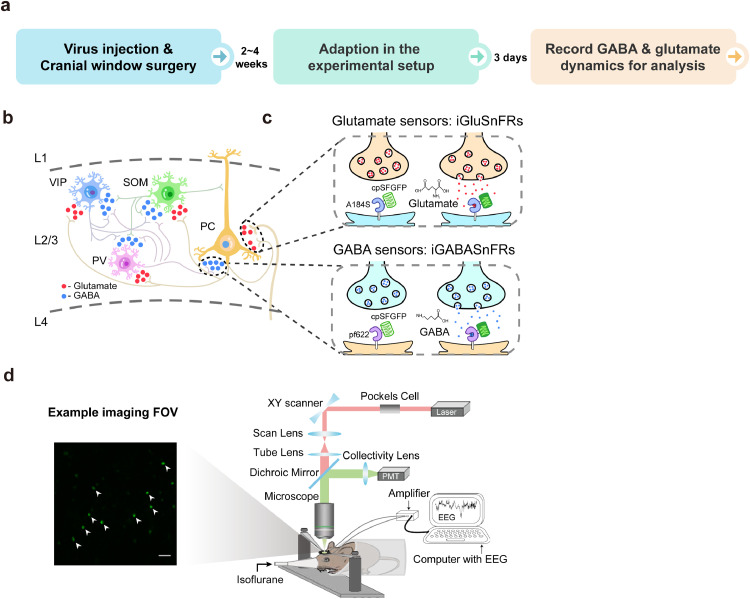
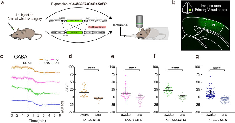
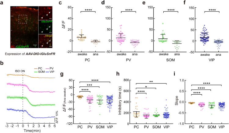
Methods: Here, we measured the dynamics of two major cortical neurotransmitters, gamma-aminobutyric acid (GABA) and glutamate, through in vivo two-photon imaging and genetically encoded neurotransmitter sensors in a cell type-specific manner in the primary visual (V1) cortex.

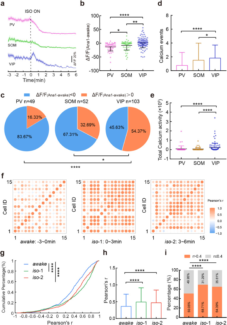
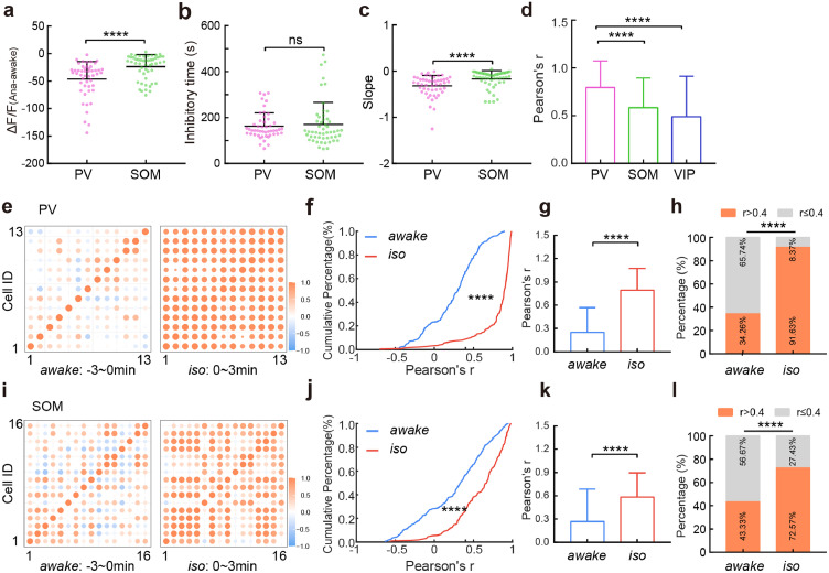
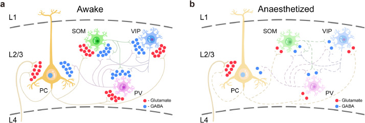
Findings: We found a general decrease in cortical GABA transmission during aLOC. However, the glutamate transmission varies among different cortical cell types, where in it is almost preserved on pyramidal cells and is significantly reduced on inhibitory interneurons. Cortical interneurons expressing vasoactive intestinal peptide (VIP) and parvalbumin (PV) specialize in disinhibitory and inhibitory effects, respectively. During aLOC, VIP neuronal activity was delayed, and PV neuronal activity was dramatically inhibited and highly synchronized.

Interpretation: These data reveal that aLOC resembles a cortical state with a disrupted excitatory-inhibitory network and suggest that a functional inhibitory network is indispensable in the maintenance of consciousness.

Funding: This work was supported by the grants of the National Natural Science Foundation of China (grant nos. 81620108012 and 82030038 to H.D. and grant nos. 31922029, 61890951, and 61890950 to J.H.).

Keywords: GABA glutamate neurotransmission anaesthesia consciousness cortex.


Conflict of interest statement

Declaration of Competing Interests The authors declare no competing interests.

Figures

Fig 1
Fig. 1
In vivo chronic two-photon imaging of cortical GABA and glutamate dynamics with cell-type specificity during aLOC. a Core experimental procedures including virus injection, cranial window surgery, adaption, and recording process. b The GABA and glutamate input to the pyramidal, PV, SOM, and VIP neurons in layer 2/3 of the primary visual (V1) cortex. c Illustration showing the working mechanisms of iGABASnFR and iGluSnFR sensors. d Left image: a representative field of view (FOV) of iGABASnFR from a VIP-ires-cre mouse. Scale bar: 50 μm. Right image: schematic of the experimental setup for two-photon imaging and EEG recording during isoflurane anaesthesia.
Fig 2
Fig. 2
Decayed GABA transmission onto each subtype of cortical neurons under anaesthesia. a Schematic of two-photon imaging after the expression of AAV-DIO-iGABASnFR following virus injection and cranial window surgery. b Virus expression in the imaging area of V1. Scale bar: 200 μm. c Signals of GABA input onto pyramidal (brown), PV (pink), SOM (green), and VIP neurons (blue) in response to isoflurane administration. Colour-shaded areas represent the respective s.e.m. d-g ΔF/F differences in pyramidal (d, n = 26 neurons from 3 mice), PV (e, n = 32 neurons from 5 mice), SOM (f, n = 27 neurons from 4 mice), and VIP (g, n = 82 neurons from 7 mice) neuronal GABA input between the awake state and anaesthetized state. Paired two-tailed Wilcoxon test (d, e, g) and paired two-tailed t test (f). Data with error bars are presented as the mean ± SD, ****p < 0.0001.
Fig 3
Fig. 3
Preserved glutamate transmission to pyramidal cells during aLOC. a Coexpression of iGluSnFR (green) and glutamate (red) within pyramidal cells in layer 2/3 of the V1 cortex. Scale bars: 50 μm (left) and 20 μm (right). Arrowheads indicate coexpressing cells. b Signals of glutamate input to pyramidal (brown), PV (pink), SOM (green), and VIP neurons (blue) in response to isoflurane administration. Colour-shaded areas represent the respective s.e.m. c-f ΔF/F difference in pyramidal (c, n = 24 neurons from 3 mice), PV (d, n = 49 neurons from 6 mice), SOM (e, n = 28 neurons from 4 mice), and VIP (f, n = 84 neurons from 7 mice) neuronal glutamate input between the awake state and anaesthetized state. Paired two-tailed Wilcoxon test. g-i The ΔF/F(Ana-awake) (g), inhibitory time (h), and slope (i) of pyramidal cells, PV, SOM, and VIP neurons. Kruskal-Wallis one-way ANOVA with Dunn's multiple comparisons. Data with error bars are presented as the mean ± SD, *p < 0.05, **p < 0.01, ****p < 0.0001.
Fig 4
Fig. 4
VIP neuronal calcium activity shows delayed inhibition and a synchronized response pattern during aLOC. a Signals of calcium activity of PV (pink), SOM (green), and VIP (blue) neurons in response to isoflurane administration. Colour-shaded areas represent the respective s.e.m. b The ΔF/F(Ana1-awake) of PV(n = 49 neurons from 5 mice), SOM (n = 52 neurons from 7 mice), and VIP (n = 103 neurons from 5 mice) neurons. ΔF/F(Ana1-awake) refers to the difference in ΔF/F value within 1 minute after isoflurane administration minus ΔF/F value within the awake state before isoflurane administration. Kruskal-Wallis one-way ANOVA with Dunn's multiple comparisons. c The percentage of ΔF/F(Ana1-awake) > 0 within the PV, SOM, and VIP interneurons. Chi-square test. (d, e) The calcium events (d) and total calcium activity (e) of PV, SOM and VIP interneurons during the 3 to 6 minutes after isoflurane administration. Kruskal-Wallis one-way ANOVA with Dunn's multiple comparisons. f Matrix of correlation coefficient r of VIP neurons during awake and two isoflurane anaesthesia stages (0-3 minutes and 3-6 minutes after isoflurane administration). (g, h) Cumulative frequency distribution (g) and mean pairwise correlation (h) for Pearson's r of VIP neurons during awake and two isoflurane anaesthesia stages. Kruskal-Wallis one-way ANOVA with Dunn's multiple comparisons. i The percentage of r > 0.4 within VIP neurons during awake and two isoflurane anaesthesia stages. Chi-square test. Data with error bars are presented as the mean ± SD, *p < 0.05, **p < 0.01, ***p < 0.001, ****p < 0.0001.
Fig 5
Fig. 5
Dramatic reduction in and high synchronization of PV neuronal calcium activity during aLOC. a-c The ΔF/F(Ana-awake) (a), inhibitory time (b), and slope (c) of PV and SOM interneurons. Two-tailed Mann-Whitney test. d Mean pairwise correlation for Pearson's r of PV, SOM, and VIP interneurons within 3 minutes after the administration of isoflurane. Kruskal-Wallis one-way ANOVA with Dunn's multiple comparisons. (e, i) Matrix of the correlation coefficient r of PV (e) and SOM (i) interneurons during the awake and isoflurane anaesthesia (0~3 minutes after isoflurane administration) stages. (f, g, j, k) Cumulative frequency distribution (f, j) and mean pairwise correlation (g, k) for Pearson's r of PV (f, g) and SOM (j, k) interneurons during awake and isoflurane anaesthesia stage. Two-tailed Mann-Whitney test. (h, l) The percentage of r > 0.4 within PV (h) and SOM (l) interneurons during awake and isoflurane anaesthesia state. Chi-square test. Data with error bars are presented as the mean ± SD, ****p < 0.0001, ns: not significant.
Fig 6
Fig. 6
Disrupted excitatory-inhibitory network within the V1 cortex during aLOC. a GABA and glutamate transmission of cortical pyramidal, PV, SOM, and VIP neurons during the awake state. b GABA and glutamate transmission within the cortical network during aLOC: general decayed GABA transmission onto each subtype of L2/3 cortical neurons which is mainly from the intralayer inhibitory interneurons; preserved glutamate transmission to L2/3 pyramidal cells which is mainly from the L4 pyramidal cells.

Comment in

Similar articles

Cited by

References

    1. Rose J, Weiser TG, Hider P, Wilson L, Gruen RL, Bickler SW. Estimated need for surgery worldwide based on prevalence of diseases: a modelling strategy for the WHO Global Health Estimate. The Lancet Global Health. 2015;3(Suppl 2):S13–S20. - PMC - PubMed
    1. Wenzel M, Han S, Smith EH, Hoel E, Greger B, House PA. Reduced Repertoire of Cortical Microstates and Neuronal Ensembles in Medically Induced Loss of Consciousness. Cell Syst. 2019;8(5):467–474. e4. - PMC - PubMed
    1. Hudetz AG, Vizuete JA, Pillay S, Mashour GA. Repertoire of mesoscopic cortical activity is not reduced during anesthesia. Neuroscience. 2016;339:402–417. - PMC - PubMed
    1. Lewis LD, Weiner VS, Mukamel EA, Donoghue JA, Eskandar EN, Madsen JR. Proceedings of the national academy of sciences of the United States of America. Vol. 109. 2012. Rapid fragmentation of neuronal networks at the onset of propofol-induced unconsciousness; pp. E3377–E3386. - PMC - PubMed
    1. Meyer K. The role of dendritic signaling in the anesthetic suppression of consciousness. Anesthesiology. 2015;122(6):1415–1431. - PubMed

LinkOut - more resources