

Review
doi: 10.1161/ATVBAHA.120.315697.
Epub 2021 Mar 11.
Calciprotein Particles: Balancing Mineral Homeostasis and Vascular Pathology
Affiliations
- PMID: 33691479
- PMCID: PMC8057528
- DOI: 10.1161/ATVBAHA.120.315697
Item in Clipboard
Review
Calciprotein Particles: Balancing Mineral Homeostasis and Vascular Pathology
Arterioscler Thromb Vasc Biol.
.
Abstract
[Figure: see text].
Keywords: atherosclerosis; calcium; homeostasis; hypercalcemia; hyperphosphatemia; vascular calcification.
Figures
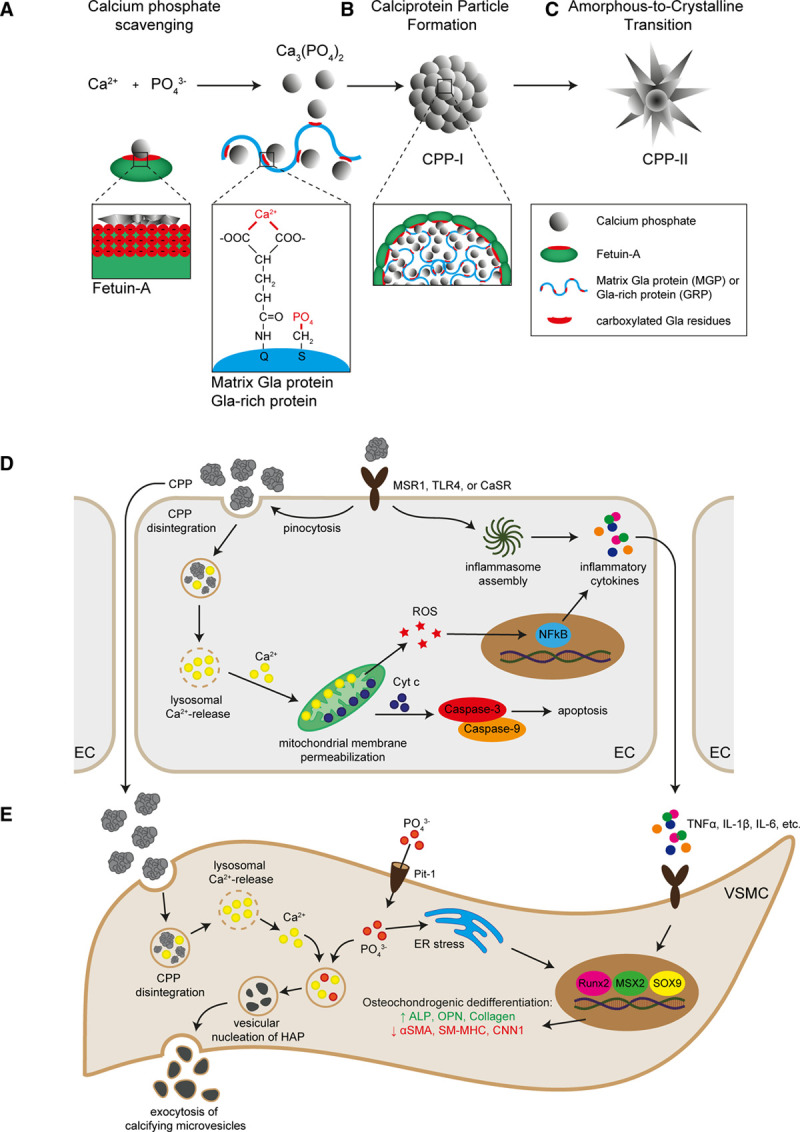
Calciprotein particle (CPP) formation and pathophysiological mechanisms. In the blood, Ca2+ and PO43− form complexes of calcium phosphate that can be scavenged by fetuin-A via the β-sheet of the amino-terminal cystatin-like D1 domain, which contains multiple negatively charged amino acids. MGP (matrix γ-carboxylated glutamate protein) and GRP (γ-carboxylated glutamate–rich protein) scavenge calcium phosphate via their negatively charged amino acids in the γ-carboxylated glutamate residues. Additionally, MGP and GRP scavenge PO43− via the phosphorylation of serine residues (A). The interaction between fetuin-A and MGP integrates calcium phosphate into amorphous spherical particles named primary CPP (B). These primary CPP may ripe into highly crystalline CPP (secondary CPP) under conditions of hypercalcemia and hyperphosphatemia (C). Endothelial cells (ECs) and vascular smooth muscle cells (VSMCs) can internalize CPP via receptor-mediated pinocytosis. In ECs, CPP internalization induces a rise in intracellular Ca2+ level, which results in the inflammatory activation of the ECs, characterized by increased transcellular permeability, oxidative stress, and inflammatory cytokine production (D). In VSMCs, CPP internalization results in a rise in intracellular Ca2+ and PO43− levels that evoke osteochondrogenic dedifferentiation via various mechanisms including inflammatory signaling and oxidative stress. An important molecular consequence of osteochondrogenic dedifferentiation of VSMCs is the production and excretion of calcifying microvesicles, which facilitate vascular calcification (E). α-SMA indicates alpha smooth muscle actin; ALP, alkaline phosphatase; CaSR, calcium-sensing receptor; CNN, calponin; ER, endoplasmatic reticulum; HAP, hydroxyapatite; IL, interleukin; MSR, macrophage scavenge receptor; MSX, homeobox transcription factor muscle segment homeobox; NF, nuclear factor kappa B; OPN, osteopontin; Pit, phosphate transporter; ROS, reactive oxygen species; Runx, runt-related transcription factor; SM-MHC, smooth muscle myosin heavy chain; SOX, sex-determining region Y-box; TLR, toll-like receptor; and TNF, tumor necrosis factor.
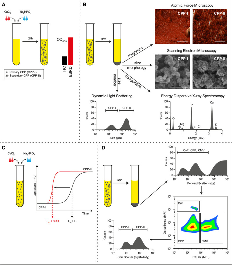
Methods to detect calciprotein particles (CPPs) in clinical samples. Supersaturation of serum with calcium chloride (CaCl2) and sodium diphosphate (Na2HPO4) followed by incubation under culture conditions for 24 h causes the formation of CPPs that can be measured by absorbance at 650 nm. In disease conditions wherein CPP levels are increased, the OD650 readings increase (A). Alternatively, CPPs can be pelleted by centrifugation and investigated by dynamic light scattering to assess particle size, electron and atomic force microscopy to assess morphology, or elemental analysis (EDX) to assess mineral constituent (B). Supersaturation of serum is also used to measure the one-half maximal transition time needed for amorphous-to-crystalline transition (T50). An increased serum propensity for secondary CPP formation is observed as a reduction in T50 (C). A novel flow cytometry-based technique allows for the direct quantification of CPP levels in serum. Here, serum precipitates are labeled with a combination of a fluorescent bisphosphonate (osteoSense) and a fluorescent membrane-intercalating dye (PKH67) and separated based on size, calcium phosphate content, and the presence of membranous lipids. CPPs are observed as OsteoSense+/PKH67− events that fluoresce dim compared to calcium phosphate crystal (CaP) crystals. CPPs are further characterized as primary- or secondary CPPs based on crystallinity (D). CMVs indicates calcifying microvesicles; ESRD, end-stage renal disease sample; HC, healthy control sample; MFI, mean fluorescence intensity; and OD, optical density.
References
-
- Heiss A, DuChesne A, Denecke B, Grötzinger J, Yamamoto K, Renné T, Jahnen-Dechent W. Structural basis of calcification inhibition by alpha 2-HS glycoprotein/fetuin-A. Formation of colloidal calciprotein particles. J Biol Chem. 2003;278:13333–13341. doi: 10.1074/jbc.M210868200 - PubMed
-
- Viegas CSB, Santos L, Macedo AL, Matos AA, Silva AP, Neves PL, Staes A, Gevaert K, Morais R, Vermeer C, et al. Chronic kidney disease circulating calciprotein particles and extracellular vesicles promote vascular calcification: a role for GRP (Gla-Rich Protein). Arterioscler Thromb Vasc Biol. 2018;38:575–587. doi: 10.1161/ATVBAHA.117.310578 - PubMed
-
- Smith ER, Cai MM, McMahon LP, Pedagogos E, Toussaint ND, Brumby C, Holt SG. Serum fetuin-A concentration and fetuin-A-containing calciprotein particles in patients with chronic inflammatory disease and renal failure. Nephrology (Carlton). 2013;18:215–221. doi: 10.1111/nep.12021 - PubMed
-
- Smith ER, Ford ML, Tomlinson LA, Rajkumar C, McMahon LP, Holt SG. Phosphorylated fetuin-A-containing calciprotein particles are associated with aortic stiffness and a procalcific milieu in patients with pre-dialysis CKD. Nephrol Dial Transplant. 2012;27:1957–1966. doi: 10.1093/ndt/gfr609 - PubMed
-
- Goodman WG, Goldin J, Kuizon BD, Yoon C, Gales B, Sider D, Wang Y, Chung J, Emerick A, Greaser L, et al. Coronary-artery calcification in young adults with end-stage renal disease who are undergoing dialysis. N Engl J Med. 2000;342:1478–1483. doi: 10.1056/NEJM200005183422003 - PubMed
Publication types
MeSH terms
Substances
LinkOut - more resources
Full Text Sources
Other Literature Sources
Medical
