

Comparison of volume of the forebrain, subarachnoid space and lateral ventricles between dogs with idiopathic epilepsy and controls using a stereological approach: Cavalieri's principle
- PMID: 33691781
- PMCID: PMC7944915
- DOI: 10.1186/s40575-021-00101-6
Comparison of volume of the forebrain, subarachnoid space and lateral ventricles between dogs with idiopathic epilepsy and controls using a stereological approach: Cavalieri's principle
Abstract
Background: Canine idiopathic epilepsy (IE) is the most common chronic neurological brain disease in dogs, yet it can only be diagnosed by exclusion of all other potential causes. In people, epilepsy has been associated with a reduction in brain volume. The objective was to estimate the volume of the forebrain (FB), subarachnoid space (SAS) and lateral ventricles (LV) in dogs with IE compared to controls using Cavalieri's principle. MRI scans of case and control dogs were identified from two neurology referral hospital databases. Eight breeds with increased odds of having IE were included: Golden Retriever, Labrador Retriever, Cocker Spaniel, Border terrier, German Shepherd dog, Parson Jack Russell terrier, Boxer, and Border Collie. Five dogs of each breed with IE and up to five controls were systematically and uniformly randomly sampled (SURS). The volume of the FB, SAS and LV were estimated from MRI scans by one blinded observer using Cavalieri's principle.
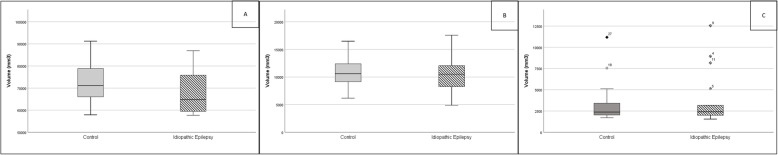
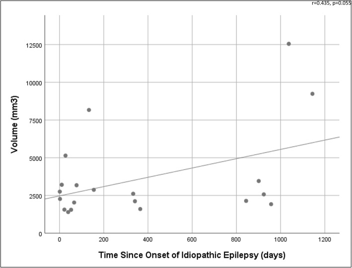
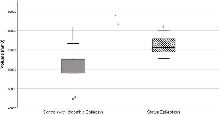
Results: One hundred-two dogs were identified; 56 were diagnosed with IE and 46 were controls. There was no statistically significant difference in FB, SAS and LV volume between dogs with IE and controls. Dogs with a history of status epilepticus had significantly larger FB than those without (p = 0.05). There was a border-line trend for LV volume to increase with increasing length of seizure history in the IE group (p = 0.055).
Conclusion: The volumes of the FB, SAS and LV are not different between dogs with IE and controls, so IE remains a diagnosis of exclusion with no specific neuroanatomical biomarkers identified. This is the first time FB and SAS volume has been compared in dogs with IE. Unfortunately, we have shown that the results reporting significantly larger FBs in dogs with status epilepticus and LV volume increase with length of seizure history were likely confounded by breed and should be interpreted cautiously. Whilst these associations are interesting and clinically relevant, further investigation with breed-specific or larger, breed-diverse populations are required to permit strong conclusions. The Cavalieri principle provided an effective estimation of FB, SAS and LV volumes on MRI, but may be too time-intensive for use in clinical practice.
Keywords: Brain volume; Canine; Cavalieri principle; Design-based stereology; Forebrain; Idiopathic epilepsy; Lateral ventricles; Subarachnoid space.
Conflict of interest statement
HV: Served as contract researcher for: Nestle 2012–2014 and 2017–2019 - dietary modification of epilepsy in dogs; Desitin Pharma, 2012 - the role of levetiracetam in a referral hospital; industrial Funding 2014–2015 - investigating the effects of imepitoin behavioural, physiologic and owner reported indicators of anxiety in dogs treated for idiopathic epilepsy. Received competitive research grants for: RCVS pump primer grant 2010–2013 - pharmacometabonomic profiling of epileptic dogs; Waltham Foundation 2011–2014 - determination of plasma omega-3 fatty acid status in dogs with primary epilepsy and relationship to antiepileptic drug metabolism; CASE BBSRC PhD studentship 2012–2016 - metabolic profiling of epilepsy in dogs; American Kennel Club American Health Foundation, 2016–2018 - Investigating the Effect of a Ketogenic Medium Chain Triglycerides Supplement on the treatment of Canine Idiopathic Epilepsy and its behavioural comorbidities; BBSRC 2017–2020 - Investigating the relationship between epilepsy, drug-resistance and affective disorders in the domestic dog BB/P001874/1.
RMAP: Received industrial funding as a co-applicant from Boehringer Ingelheim (2014–15; Investigating the effects of imepitoin on behavioural, physiologic and owner-reported indicators of anxiety in dogs treated for idiopathic epilepsy) and Nestle (2017–19; Dietary modification of epilepsy in dogs). Received competitive research grants from the American Kennel Club (2016–18; Investigating the effect of a ketogenic medium chain triglycerides supplement on the treatment of canine idiopathic epilepsy and its behavioural comorbidities); BBSRC 2017–20; Investigating the relationship between epilepsy, drug-resistance and affective disorders in the domestic dog; BB/P001874/1 and 2017–2020; Comorbidity and characteristics of canine neurodevelopmental disorders and their impact on animal welfare; BB/P 010881/1.
CR: Served as a paid consultant for Boehringer Ingelheim in 2014.
FW, AC and AT declare that they have no competing interests.
Figures
References
-
- Heske L, Nødtvedt A, Jäderlund KH, Berendt M, Egenvall A. A cohort study of epilepsy among 665,000 insured dogs: incidence, mortality and survival after diagnosis. Vet J. 2014;202(3):471–6. - PubMed
-
- Kearsley-Fleet L, O’Neill D, Volk H, Church D, Brodbelt D. Prevalence and risk factors for canine epilepsy of unknown origin in the UK. Vet Rec. 2012;172(13):338. - PubMed
-
- Zhou SY, Tong L, Song F, Hong XJ, Sun HF, Chang H, et al. Selective medial temporal volume reduction in the hippocampus of patients with idiopathic generalized tonic-clonic seizures. Epilepsy Res. 2015;110:39–48. - PubMed
Grants and funding
LinkOut - more resources
Full Text Sources
Other Literature Sources