Case Reports
. 2021 Mar 10;14(3):e240411.
doi: 10.1136/bcr-2020-240411.

Ectopic sphenoid sinus pituitary adenoma masquerading as metastatic head and neck cancer

Affiliations
Case Reports

Ectopic sphenoid sinus pituitary adenoma masquerading as metastatic head and neck cancer

Augustine Chai et al. BMJ Case Rep. .

Abstract

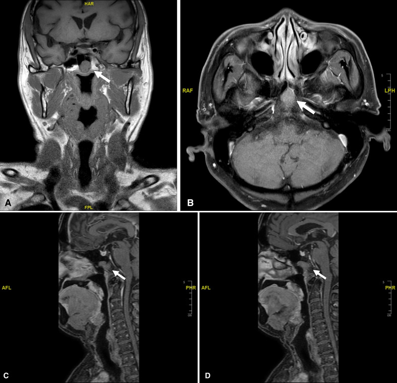
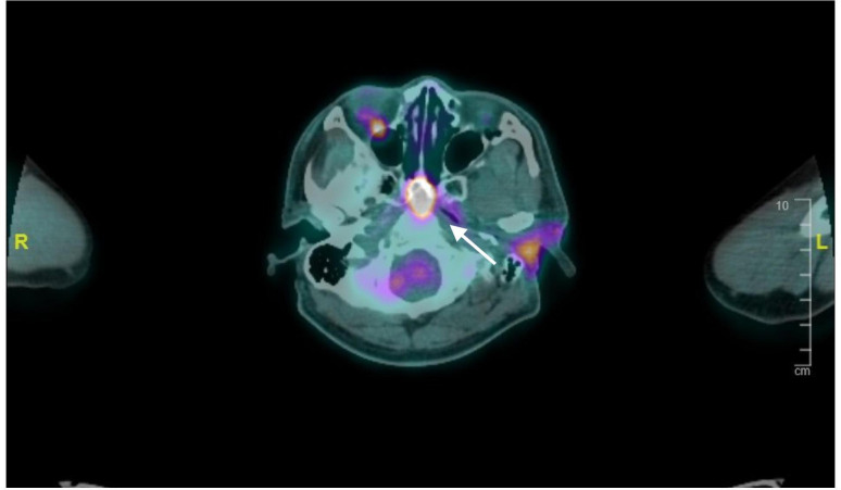
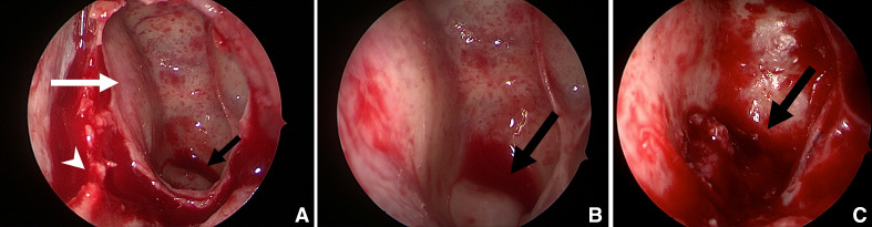
A 68-year-old Chinese man was found to have a lobular mass in the sphenoid sinus which extended to the clivus and the roof of the nasopharynx on a staging MRI scan performed for his high-grade parotid salivary duct carcinoma. Further positron emission tomography scan showed that this lesion was fluorodeoxyglucose (FDG) avid. This proved to be a diagnostic dilemma. The patient underwent a total parotidectomy, left selective neck dissection and a transphenoidal biopsy of his nasal lesion. Final histology revealed that this lesion was a synchronous ectopic sphenoid sinus pituitary adenoma (ESSPA). Initial differential diagnoses that were considered included a chordoma, metastatic carcinoma and nasopharyngeal carcinoma. However, an important differential with a neoplastic appearance and a tendency for positive FDG uptake is an ESSPA. It requires dedicated immunohistochemical staining to diagnose, and its mainstay of treatment is surgical excision.

Keywords: ear; head and neck cancer; neuroendocrinology; nose and throat/otolaryngology; radiology.


Conflict of interest statement

Competing interests: None declared.

Figures

Figure 1
Figure 1
MRI showing sphenoid lesion (white arrow). (A) T1-weighted MRI (coronal). (B) T1-weighted MRI (axial). (C) T1-weighted MRI (sagittal midline) showing a normal sella pituitary and separate sphenoid lesion. (D) T1-weighted MRI (sagittal left side) showing sphenoid lesion extends to the clivus.
Figure 2
Figure 2
Positron emission tomography scan showing flurodeoxyglucose–avid sphenoid sinus lesion (white arrow).
Figure 3
Figure 3
Intraoperative photos of ectopic sphenoid sinus pituitary adenoma (black arrow). (A) Posterior septectomy (white arrowhead), opened left sphenoid sinus (white arrow). (B) Bulge on floor of sphenoid sinus with normal overlying mucosa. (C) Ectopic sphenoid sinus pituitary adenoma with surrounding bony defect seen after removal of left sphenoid sinus mucosa.

References

    1. Rossi ED, Baloch Z, Pusztaszeri M, et al. . The Milan system for reporting salivary gland cytopathology (MSRSGC): an ASC-IAC-sponsored system for reporting salivary gland fine-needle aspiration. J Am Soc Cytopathol 2018;7:111–8. 10.1016/j.jasc.2018.02.002 - DOI - PubMed
    1. Oruçkaptan HH, Senmevsim O, Ozcan OE, et al. . Pituitary adenomas: results of 684 surgically treated patients and review of the literature. Surg Neurol 2000;53:211–9. 10.1016/S0090-3019(00)00171-3 - DOI - PubMed
    1. Moss WJ, Finegersh A, Jafari A, et al. . Isolated sphenoid sinus opacifications: a systematic review and meta-analysis. Int Forum Allergy Rhinol 2017;7:1201–6. 10.1002/alr.22023 - DOI - PubMed
    1. Barrs DM, McDonald TJ, Whisnant JP. Metastatic tumors to the sphenoid sinus. Laryngoscope 1979;89:1239–43. 10.1002/lary.1979.89.8.1239 - DOI - PubMed
    1. Ng YH, Sethi DS. Isolated sphenoid sinus disease: differential diagnosis and management. Curr Opin Otolaryngol Head Neck Surg 2011;19:16–20. 10.1097/MOO.0b013e32834251d6 - DOI - PubMed

Publication types