

Inhibition of CRISPR-Cas12a DNA targeting by nucleosomes and chromatin
- PMID: 33692102
- PMCID: PMC7946368
- DOI: 10.1126/sciadv.abd6030
Inhibition of CRISPR-Cas12a DNA targeting by nucleosomes and chromatin
Abstract
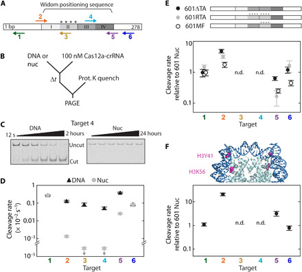
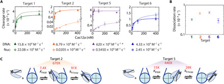
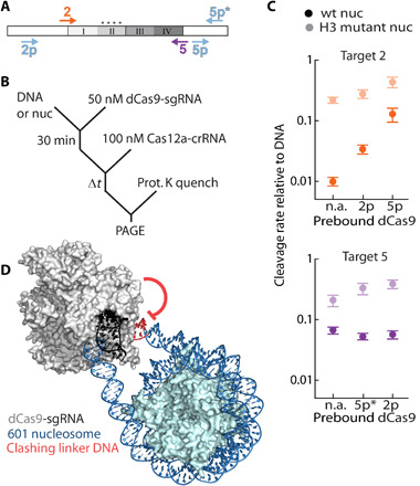
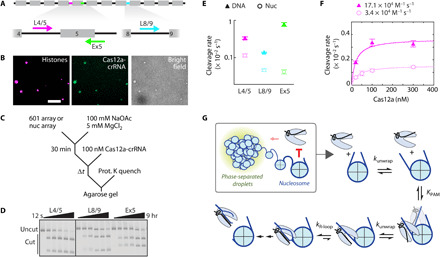
Genome engineering nucleases must access chromatinized DNA. Here, we investigate how AsCas12a cleaves DNA within human nucleosomes and phase-condensed nucleosome arrays. Using quantitative kinetics approaches, we show that dynamic nucleosome unwrapping regulates target accessibility to Cas12a and determines the extent to which both steps of binding-PAM recognition and R-loop formation-are inhibited by the nucleosome. Relaxing DNA wrapping within the nucleosome by reducing DNA bendability, adding histone modifications, or introducing target-proximal dCas9 enhances DNA cleavage rates over 10-fold. Unexpectedly, Cas12a readily cleaves internucleosomal linker DNA within chromatin-like, phase-separated nucleosome arrays. DNA targeting is reduced only ~5-fold due to neighboring nucleosomes and chromatin compaction. This work explains the observation that on-target cleavage within nucleosomes occurs less often than off-target cleavage within nucleosome-depleted genomic regions in cells. We conclude that nucleosome unwrapping regulates accessibility to CRISPR-Cas nucleases and propose that increasing nucleosome breathing dynamics will improve DNA targeting in eukaryotic cells.
Copyright © 2021 The Authors, some rights reserved; exclusive licensee American Association for the Advancement of Science. No claim to original U.S. Government Works. Distributed under a Creative Commons Attribution NonCommercial License 4.0 (CC BY-NC).
Figures
Similar articles
-
Nucleosomes inhibit target cleavage by CRISPR-Cas9 in vivo.Proc Natl Acad Sci U S A. 2018 Sep 18;115(38):9351-9358. doi: 10.1073/pnas.1810062115. Epub 2018 Sep 10. Proc Natl Acad Sci U S A. 2018. PMID: 30201707 Free PMC article.
-
Nucleosomes Inhibit Cas9 Endonuclease Activity in Vitro.Biochemistry. 2015 Dec 8;54(48):7063-6. doi: 10.1021/acs.biochem.5b01108. Epub 2015 Nov 24. Biochemistry. 2015. PMID: 26579937
-
Nucleosome breathing and remodeling constrain CRISPR-Cas9 function.Elife. 2016 Apr 28;5:e13450. doi: 10.7554/eLife.13450. Elife. 2016. PMID: 27130520 Free PMC article.
-
Yeast HMO1: Linker Histone Reinvented.Microbiol Mol Biol Rev. 2016 Nov 30;81(1):e00037-16. doi: 10.1128/MMBR.00037-16. Print 2017 Mar. Microbiol Mol Biol Rev. 2016. PMID: 27903656 Free PMC article. Review.
-
Nucleosome unwrapping and unstacking.Curr Opin Struct Biol. 2020 Oct;64:119-125. doi: 10.1016/j.sbi.2020.06.020. Epub 2020 Jul 29. Curr Opin Struct Biol. 2020. PMID: 32738677 Review.
Cited by
-
Increased Gene Targeting in Hyper-Recombinogenic LymphoBlastoid Cell Lines Leaves Unchanged DSB Processing by Homologous Recombination.Int J Mol Sci. 2022 Aug 16;23(16):9180. doi: 10.3390/ijms23169180. Int J Mol Sci. 2022. PMID: 36012445 Free PMC article.
-
PAM-adjacent DNA flexibility tunes CRISPR-Cas12a off-target binding.Sci Rep. 2025 Feb 10;15(1):4930. doi: 10.1038/s41598-025-87565-9. Sci Rep. 2025. PMID: 39929897 Free PMC article.
-
Decorating chromatin for enhanced genome editing using CRISPR-Cas9.Proc Natl Acad Sci U S A. 2022 Dec 6;119(49):e2204259119. doi: 10.1073/pnas.2204259119. Epub 2022 Dec 2. Proc Natl Acad Sci U S A. 2022. PMID: 36459645 Free PMC article.
-
CRISPR/Cas- and Topical RNAi-Based Technologies for Crop Management and Improvement: Reviewing the Risk Assessment and Challenges Towards a More Sustainable Agriculture.Front Bioeng Biotechnol. 2022 Jun 28;10:913728. doi: 10.3389/fbioe.2022.913728. eCollection 2022. Front Bioeng Biotechnol. 2022. PMID: 35837551 Free PMC article. Review.
-
MARCKSL1-2 reverses docetaxel-resistance of lung adenocarcinoma cells by recruiting SUZ12 to suppress HDAC1 and elevate miR-200b.Mol Cancer. 2022 Jul 21;21(1):150. doi: 10.1186/s12943-022-01605-w. Mol Cancer. 2022. PMID: 35864549 Free PMC article.
References
-
- Swarts D. C., Jinek M., Cas9 versus Cas12a/Cpf1: Structure-function comparisons and implications for genome editing. Wiley Interdisc. Rev. RNA 9, e1481 (2018). - PubMed
-
- Nishimasu H., Nureki O., Structures and mechanisms of CRISPR RNA-guided effector nucleases. Curr. Opin. Struct. Biol. 43, 68–78 (2017). - PubMed
Publication types
MeSH terms
Substances
Grants and funding
LinkOut - more resources
Full Text Sources
Other Literature Sources
Miscellaneous
