. 2021 May 10;95(11):e00083-21.
doi: 10.1128/JVI.00083-21. Epub 2021 Mar 10.

Susceptibility of white-tailed deer (Odocoileus virginianus) to SARS-CoV-2

Affiliations

Susceptibility of white-tailed deer (Odocoileus virginianus) to SARS-CoV-2

Mitchell V Palmer et al. J Virol. .

Abstract

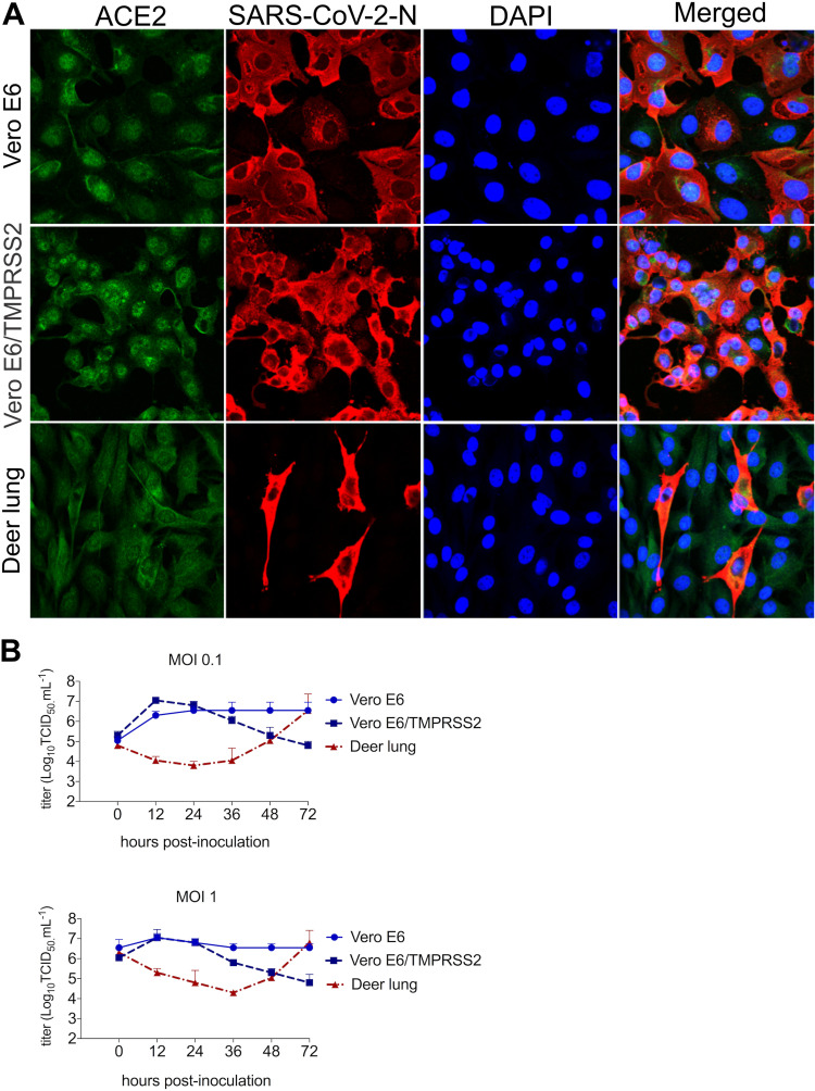
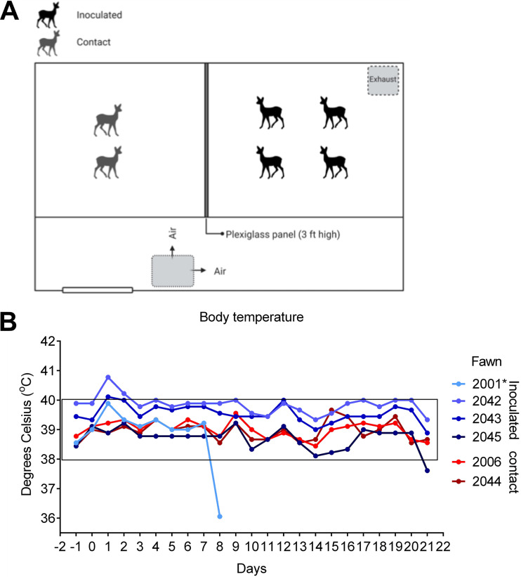
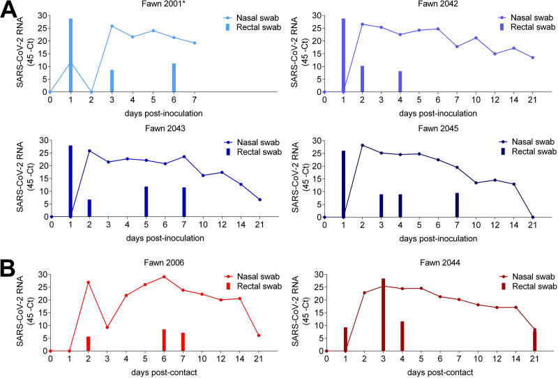
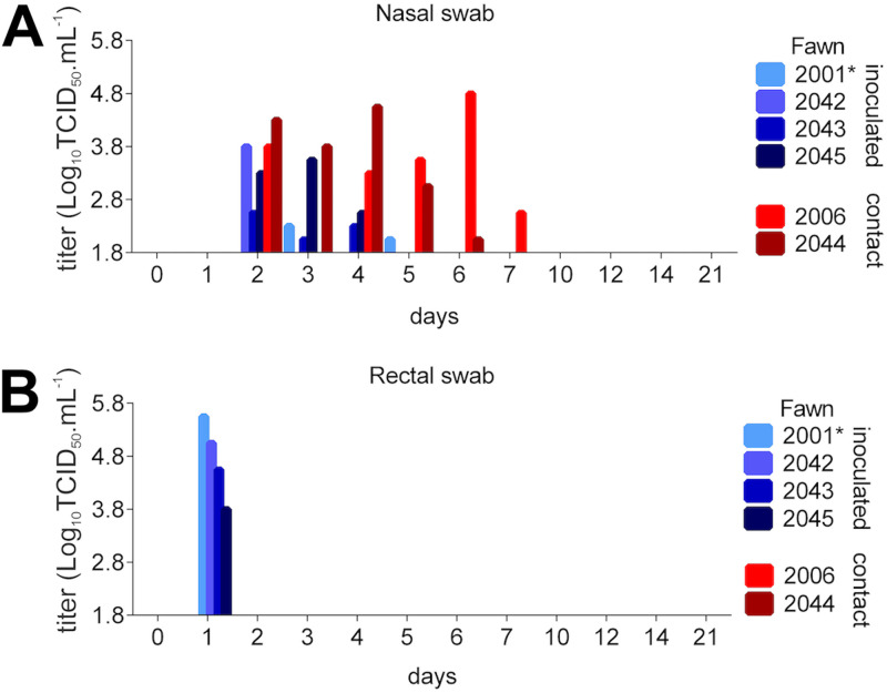
The origin of severe acute respiratory syndrome coronavirus 2 (SARS-CoV-2), the virus causing the global coronavirus disease 19 (COVID-19) pandemic, remains a mystery. Current evidence suggests a likely spillover into humans from an animal reservoir. Understanding the host range and identifying animal species that are susceptible to SARS-CoV-2 infection may help to elucidate the origin of the virus and the mechanisms underlying cross-species transmission to humans. Here we demonstrated that white-tailed deer (Odocoileus virginianus), an animal species in which the angiotensin converting enzyme 2 (ACE2) - the SARS-CoV-2 receptor - shares a high degree of similarity to humans, are highly susceptible to infection. Intranasal inoculation of deer fawns with SARS-CoV-2 resulted in established subclinical viral infection and shedding of infectious virus in nasal secretions. Notably, infected animals transmitted the virus to non-inoculated contact deer. Viral RNA was detected in multiple tissues 21 days post-inoculation (pi). All inoculated and indirect contact animals seroconverted and developed neutralizing antibodies as early as day 7 pi. The work provides important insights into the animal host range of SARS-CoV-2 and identifies white-tailed deer as a susceptible wild animal species to the virus.IMPORTANCEGiven the presumed zoonotic origin of SARS-CoV-2, the human-animal-environment interface of COVID-19 pandemic is an area of great scientific and public- and animal-health interest. Identification of animal species that are susceptible to infection by SARS-CoV-2 may help to elucidate the potential origin of the virus, identify potential reservoirs or intermediate hosts, and define the mechanisms underlying cross-species transmission to humans. Additionally, it may also provide information and help to prevent potential reverse zoonosis that could lead to the establishment of a new wildlife hosts. Our data show that upon intranasal inoculation, white-tailed deer became subclinically infected and shed infectious SARS-CoV-2 in nasal secretions and feces. Importantly, indirect contact animals were infected and shed infectious virus, indicating efficient SARS-CoV-2 transmission from inoculated animals. These findings support the inclusion of wild cervid species in investigations conducted to assess potential reservoirs or sources of SARS-CoV-2 of infection.


Figures

FIG 1
FIG 1
Susceptibility and replication properties of the SARS-CoV-2 in deer cells in vitro. (A) Deer lung (DL), Vero E6, and Vero E6/TMPRSS2 cells were inoculated with SARS-CoV-2 at a multiplicity of infection (MOI) of 1. At 24 h postinoculation (hpi), cells were fixed and subjected to an immunofluorescence assay using a monoclonal antibody (MAb) anti-ACE2 (green) and with a MAb anti-SARS-CoV-2-nucleoprotein (N) (red). Nuclear counterstain was performed with DAPI (blue); ×40 magnification. (B) To assess the kinetics of replication of SARS-CoV-2, DL, Vero E6, and Vero E6/TMPRSS2 cells were inoculated with SARS-CoV-2 isolate TGR/NY/20 (MOI of 0.1 and 1) and harvested at various time points postinoculation (12, 24, 36, 48, and 72 hpi; time zero shown in the graphs represent the back titration of input virus). Virus titers were determined on each time point using endpoint dilutions and the Spearman and Karber's method and expressed as log10 TCID50·ml1. Results represent the average of three independent experiments.
FIG 2
FIG 2
Infection and transmission of SARS-CoV-2 in white-tailed deer. (A) Room setup of animal experiment. Fawns were kept in a room of a biosafety level 3 (agriculture) (BSL-3Ag) facility. Four fawns were inoculated intranasally with a virus suspension containing 5 × 106.3 TCID50 of SARS-CoV-2 isolate TGR/NY/20, and two fawns were maintained as noninoculated room contact animals. All fawns were maintained in a 3.7-m by 3.7-m room and inoculated, and room contact animals were kept in two pens separated by a plexiglass barrier approximately 0.9 m (∼3 ft) in height to prevent direct nose-to-nose contact. Airflow in the room was maintained at 10 to 11 air exchanges per hour and was directional from the contact pen toward the inoculated pen. (B) Fawns were microchipped subcutaneously for identification and monitored daily for clinical signs and body temperature starting on day 1 before inoculation or contact day (day −1). Body temperatures are expressed in degrees Celsius.
FIG 3
FIG 3
Viral RNA in nasal secretion and feces. (A) Dynamics of virus shedding was assessed in nasal secretions (line) and feces (bars) of four inoculated fawns (no. 2001, 2042, 2043, and 2045). Nasal and rectal swabs collected on days 0 to 7, 10, 12, 14, and 21 postinoculation (p.i.) were tested for the presence of SARS-CoV-2 RNA by real-time reverse transcriptase PCR (rRT-PCR). (B) Dynamics of virus shedding of contact fawns (no. 2006 and 2044). Nasal and rectal swabs collected on the same time points as the inoculated animals were subjected to nucleic acid extraction and tested for the presence of SARS-CoV-2 RNA by rRT-PCR.
FIG 4
FIG 4
Shedding of infectious SARS-CoV-2 by inoculated and contact fawns. (A) Infectious virus was assessed by virus isolation in nasal secretions in rRT-PCR-positive samples. (B) Infectious SARS-CoV-2 shedding in feces in rRT-PCR-positive samples. Virus titers were determined using endpoint dilutions and the Spearman and Karber's method and expressed as log10 TCID50·ml1.
FIG 5
FIG 5
Tissue distribution of SARS-CoV-2 RNA. Tissues were collected and processed for rRT-PCR on day 8 postinoculation (p.i.) for animal no. 2001 (which died of an unrelated cause) and 21 p.i. of the remaining animals (animal no. 2042, 2043, 2045, 2006, and 2044).
FIG 6
FIG 6
Tissues from white-tailed deer fawn inoculated intranasally with SARS-CoV-2 and examined 21 days later. (A) Note intense labeling of viral RNA in the centers of lymphoid follicles located subjacent to tonsillar epithelium (upper left). (B and C) Note labeling for SARS-CoV-2 RNA within the medial retropharyngeal lymph node follicle (B) and mediastinal lymph node medulla (C). (D) Nasal turbinate lumen contains aggregate of mucus, cells, and debris with intense labeling for SARS-CoV-2 RNA. (E and F) Adjacent microscopic sections demonstrate intense labeling of lymphoid follicles with probe for SARS-CoV-2 RNA (E) but no labeling using the anti-genomic sense probe (F). ISH-RNAscope.
FIG 7
FIG 7
Antibody responses in white-tailed deer following SARS-CoV-2 infection. (A) Luminex assay to assess IgG anti-SARS-CoV-2 nucleocapsid (N) in serum samples collected on days 0, 7, 14, and 21 postinoculation (p.i.) (fawn no. 2001, 2042, 2043, and 2045), or contact (fawn no. 2006 and 2044). (B) Luminex assay to assess IgG anti-SARS-CoV-2-S-receptor binding domain (RBD) specificity at the same time point and fawns as above. (C) Virus neutralization (VN) assay. Neutralizing antibody (NA) titers were expressed as the reciprocal of the highest dilution of serum that completely inhibited SARS-CoV-2 infection/replication in serum at the same time point and fawns as above. Results represent the geometric mean titers (GMT) of three independent experiments.
FIG 8
FIG 8
Histological examination of lung from white-tailed deer fawns intranasally inoculated with SARS-CoV-2. (A) Note the well-demarcated focus of congestion. Alveolar septal capillaries are engorged with blood surrounded by normal appearing alveolar septa. HE. (B) Multiple alveolar septa are lined by bands of eosinophilic hyalinized proteinaceous material (arrows) consistent with hyaline membranes. Multiple alveoli contain flocculent to fibrillar eosinophilic material consistent with fibrin. HE. (C) Expanded alveolus contains a large collection of fibrin, inflammatory cells, and cell debris (arrow). HE. (D) Alveolar septa are expanded by an inflammatory infiltrate (interstitial pneumonia) composed primarily of lymphocytes (arrows) and macrophages (E). HE. (F) Within a field of congested alveolar septa are irregular regions characterized by hypocellular septa containing few erythrocytes. Septal stroma is fibrillar and lightly eosinophilic. Multiple alveoli within these regions contain flocculent strands of fibrin. HE. (G) There is type II pneumocyte hyperplasia and an increase in alveolar macrophages (arrows). HE. (H) Lumens of cortical tubules in the kidney are filled with necrotic cellular debris. Renal tubules are variably lined by attenuated epithelium, occasionally have hypereosinophilic cytoplasm and pyknotic nuclei (degeneration and necrosis), and overall exhibit increased cytoplasmic basophilia (regeneration). Tubules are separated by interstitial edema and a cellular infiltrate composed of lymphocytes, plasma cells, and fewer macrophages. HE. Tissue sections were examined on day 21 postinoculation.

References

    1. Gorbalenya AE, Baker SC, Baric RS, de Groot RJ, Drosten C, Gulyaeva AA, Haagmans BL, Lauber C, Leontovich AM, Neuman BW, Penzar D, Perlman S, Poon LLM, Samborskiy DV, Sidorov IA, Sola I, Ziebuhr J. 2020. The species severe acute respiratory syndrome-related coronavirus: classifying 2019-nCoV and naming it SARS-CoV-2. Nat Microbiol Nature Res 5:536–544. 10.1038/s41564-020-0695-z. - DOI - PMC - PubMed
    1. Zhu N, Zhang D, Wang W, Li X, Yang B, Song J, Zhao X, Huang B, Shi W, Lu R, Niu P, Zhan F, Ma X, Wang D, Xu W, Wu G, Gao GF, Tan W, China Novel Coronavirus Investigating and Research Team . 2020. A novel coronavirus from patients with pneumonia in China, 2019. N Engl J Med 382:727–733. 10.1056/NEJMoa2001017. - DOI - PMC - PubMed
    1. Zhou P, Yang XL, Wang XG, Hu B, Zhang L, Zhang W, Si HR, Zhu Y, Li B, Huang CL, Chen HD, Chen J, Luo Y, Guo H, Jiang RD, Liu MQ, Chen Y, Shen XR, Wang X, Zheng XS, Zhao K, Chen QJ, Deng F, Liu LL, Yan B, Zhan FX, Wang YY, Xiao GF, Shi ZL. 2020. A pneumonia outbreak associated with a new coronavirus of probable bat origin. Nature 579:270–273. 10.1038/s41586-020-2012-7. - DOI - PMC - PubMed
    1. Wu F, Zhao S, Yu B, Chen YM, Wang W, Song ZG, Hu Y, Tao ZW, Tian JH, Pei YY, Yuan ML, Zhang YL, Dai FH, Liu Y, Wang QM, Zheng JJ, Xu L, Holmes EC, Zhang YZ. 2020. A new coronavirus associated with human respiratory disease in China. Nature 579:265–269. 10.1038/s41586-020-2008-3. - DOI - PMC - PubMed
    1. Andersen KG, Rambaut A, Lipkin WI, Holmes EC, Garry RF. 2020. The proximal origin of SARS-CoV-2. Nat Med 26:450–454. 10.1038/s41591-020-0820-9. - DOI - PMC - PubMed