. 2021 Mar 10;9(1):16.
doi: 10.1038/s41413-020-00124-y.

Piezo1 channel activation in response to mechanobiological acoustic radiation force in osteoblastic cells

Affiliations

Piezo1 channel activation in response to mechanobiological acoustic radiation force in osteoblastic cells

Guangdao Zhang et al. Bone Res. .

Abstract

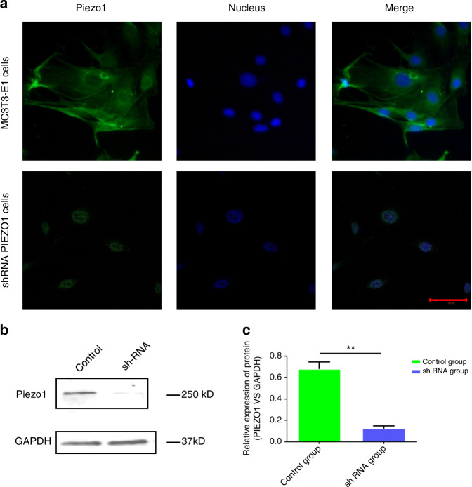
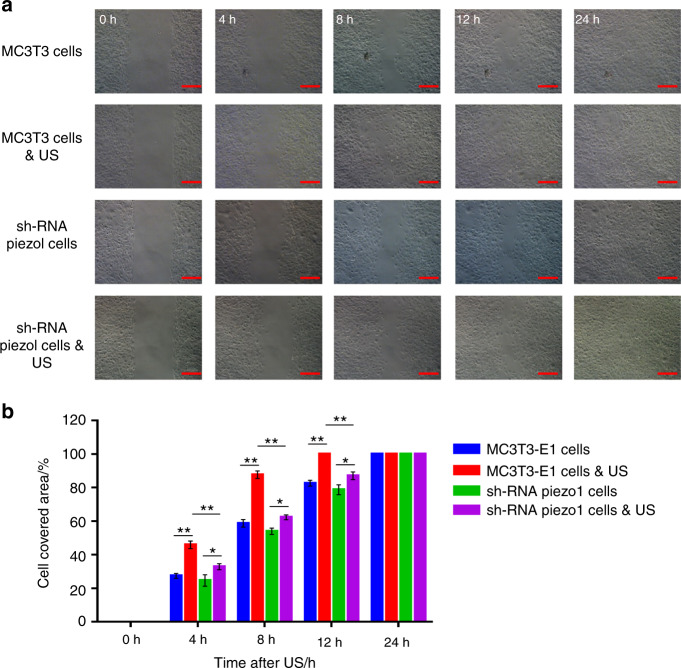
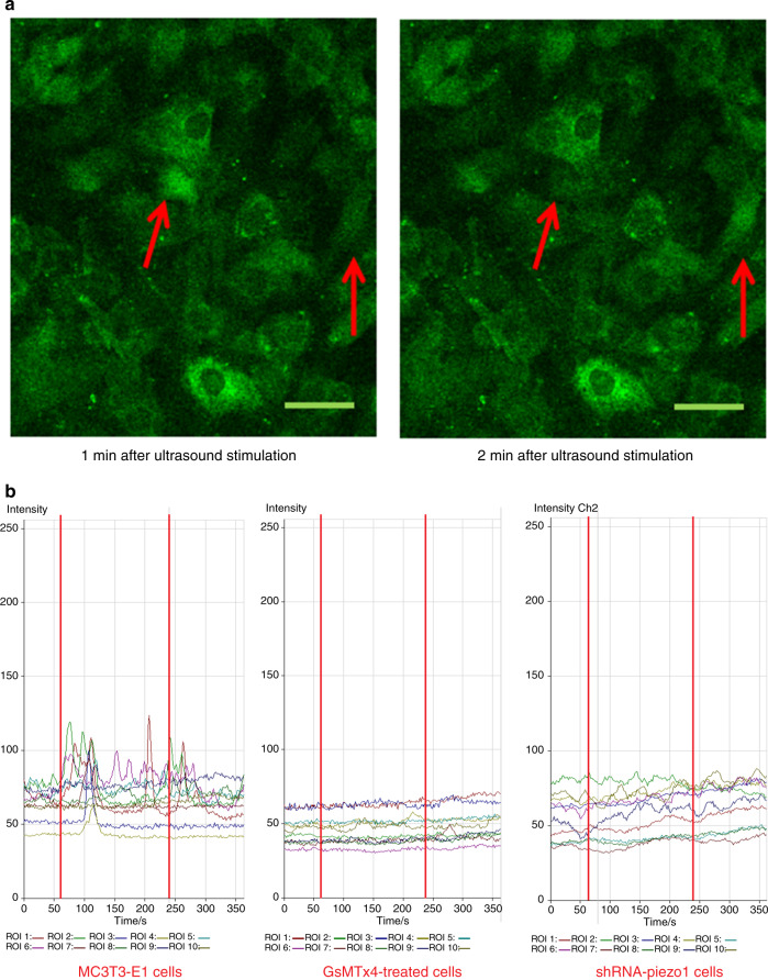
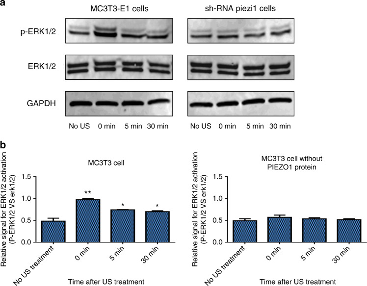
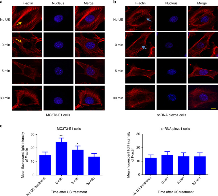
Mechanobiological stimuli, such as low-intensity pulsed ultrasound (LIPUS), have been shown to promote bone regeneration and fresh fracture repair, but the fundamental biophysical mechanisms involved remain elusive. Here, we propose that a mechanosensitive ion channel of Piezo1 plays a pivotal role in the noninvasive ultrasound-induced mechanical transduction pathway to trigger downstream cellular signal processes. This study aims to investigate the expression and role of Piezo1 in MC3T3-E1 cells after LIPUS treatment. Immunofluorescence analysis shows that Piezo1 was present on MC3T3-E1 cells and could be ablated by shRNA transfection. MC3T3-E1 cell migration and proliferation were significantly increased by LIPUS stimulation, and knockdown of Piezo1 restricted the increase in cell migration and proliferation. After labeling with Fluo-8, MC3T3-E1 cells exhibited fluorescence intensity traces with several high peaks compared with the baseline during LIPUS stimulation. No obvious change in the fluorescence intensity tendency was observed after LIPUS stimulation in shRNA-Piezo1 cells, which was similar to the results in the GsMTx4-treated group. The phosphorylation ratio of ERK1/2 in MC3T3-E1 cells was significantly increased (P < 0.01) after LIPUS stimulation. In addition, Phalloidin-iFluor-labeled F-actin filaments immediately accumulated in the perinuclear region after LIPUS stimulation, continued for 5 min, and then returned to their initial levels at 30 min. These results suggest that Piezo1 can transduce LIPUS-induced mechanical signals into intracellular calcium. The influx of Ca2+ serves as a second messenger to activate ERK1/2 phosphorylation and perinuclear F-actin filament polymerization, which regulate the proliferation of MC3T3-E1 cells.


Conflict of interest statement

The authors declare no competing interests.

Figures

Fig. 1
Fig. 1
Expression of Piezo1 in MC3T3-E1 and shRNA-Piezo1 MC3T3-E1 cells. a Piezo1 (green) was expressed on MC3T3-E1 cells and localized at the plasma membrane and nucleus (blue). The protein expression of Piezo1 was decreased in shRNA-Piezo1 cells, especially on the plasma membrane. Piezo1 was expressed only around the nucleus in the shRNA-Piezo1 cells. The scale bar is 50 μm. b, c Western blot analysis showed that the Piezo1 protein expression in the shRNA-Piezo1 MC3T3-E1 group was significantly lower than that in the control group (n = 3, P < 0.01, Student’s t-test)
Fig. 2
Fig. 2
Wound healing and migration assay. a Randomly selected images of the gap (500 μm) at 0, 4, 8, 12, and 24 h after treatment with or without LIPUS stimulation. Scale bars, 200 μm. b Changes in the cell-covered area over time. LIPUS stimulation significantly increased the migration and proliferation of MC3T3-E1 cells (n = 3, **P < 0.01, Student’s t-test). LIPUS stimulation also increased the migration and proliferation of shRNA-Piezo1 cells (n = 3, *P < 0.05, Student’s t-test), but the difference was not as obvious as it was in MC3T3-E1 cells. The cell-covered area of MC3T3-E1 cells was significantly higher than that of shRNA-Piezo1 cells at 4, 8, and 12 h after LIPUS stimulation (n = 3, **P < 0.01, Student’s t-test)
Fig. 3
Fig. 3
Fluorescence imaging of calcium oscillation and the effects of LIPUS stimulation on different groups of cells. a After LIPUS stimulation, the Fluo-8-labeled cells exhibited increased intracellular calcium levels at different time points. The red arrows show the two-cell calcium oscillation phenomenon. Scale bars, 50 μm. b Representative intracellular calcium traces of three groups of cells (MC3T3-E1, shRNA-Piezo1, and GsMTx4-treated cells) are shown as the fold increase in Fluo-8 intensity in response to LIPUS stimulation. The experiment was performed for six total minutes, including 1 min of baseline without LIPUS stimulation, 3 min of active stimulation (between the two red lines), and 2 min of regression. Time-lapse sequences were collected every 1.8 s for 6 min. In each field of interest, the fluorescence intensities of 10 cells were quantified using LSM Image Browser software
Fig. 4
Fig. 4
Western blot analysis of ERK1/2 and p-ERK1/2 in MC3T3-E1 and shRNA-Piezo1 cells after LIPUS stimulation. a Representative western blots of ERK1/2, p-ERK1/2, and GAPDH in MC3T3-E1 and shRNA-Piezo1 cells at the indicated time points (0, 5, and 30 min) after LIPUS stimulation. b Quantitative changes in ERK1/2 activation in MC3T3-E1 and shRNA-Piezo1 cells. The ratio of ERK1/2 phosphorylation to the relative expression of the protein doublet (p-ERK1/2 vs. ERK1/2) is presented as a parameter of ERK1/2 activation (n = 3, *P < 0.05, **P < 0.01, Student’s t-test)
Fig. 5
Fig. 5
Polymerization of perinuclear F-actin after LIPUS stimulation. a, b Representative fluorescence images of Phalloidin-iFluor-labeled F-actin around the nuclei in MC3T3-E1 and shRNA-Piezo1 cells. The orange arrows in a and blue arrows in b indicate perinuclear F-actin. Scale bars: 10 μm. c The mean fluorescence intensity of perinuclear F-actin was measured and analyzed with ImageJ at the indicated time points (0, 5, and 30 min) after LIPUS stimulation (n = 9, *P < 0.05, **P < 0.01, Student’s t-test)
Fig. 6
Fig. 6
Schematic diagram of the experimental setup for the LIPUS stimulation of MC3T3-E1 cells. The setup includes a function generator, a radio-frequency power amplifier, a transducer, and a laser scanning confocal microscope (LSCM). The surface of the transducer was immersed in the medium and located 4 mm away from the cells

References

    1. Calori GM, Mazza E, Colombo M, Ripamonti C, Tagliabue L. Treatment of long bone nonunions with polytherapy: indications and clinical results. Injury. 2011;42:587–590. doi: 10.1016/j.injury.2011.03.046. - DOI - PubMed
    1. Tzioupis C, Giannoudis PV. Prevalence of long-bone nonunions. Injury. 2007;38(Suppl 2):S3–S9. doi: 10.1016/S0020-1383(07)80003-9. - DOI - PubMed
    1. Giannoudis PV, Jones E, Einhorn TA. Fracture healing and bone repair. Injury. 2011;42:549–550. doi: 10.1016/j.injury.2011.03.037. - DOI - PubMed
    1. Lou S, Lv H, Li Z, Zhang L, Tang P. The effects of low-intensity pulsed ultrasound on fresh fracture: a meta-analysis. Medicine. 2017;96:e8181. doi: 10.1097/MD.0000000000008181. - DOI - PMC - PubMed
    1. Hannemann PF, Mommers EH, Schots JP, Brink PR, Poeze M. The effects of low-intensity pulsed ultrasound and pulsed electromagnetic fields bone growth stimulation in acute fractures: a systematic review and meta-analysis of randomized controlled trials. Arch. Orthop. Trauma Surg. 2014;134:1093–1106. doi: 10.1007/s00402-014-2014-8. - DOI - PubMed

LinkOut - more resources