Whole-brain tissue mapping toolkit using large-scale highly multiplexed immunofluorescence imaging and deep neural networks
- PMID: 33692351
- PMCID: PMC7946933
- DOI: 10.1038/s41467-021-21735-x
Whole-brain tissue mapping toolkit using large-scale highly multiplexed immunofluorescence imaging and deep neural networks
Abstract
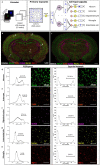
Mapping biological processes in brain tissues requires piecing together numerous histological observations of multiple tissue samples. We present a direct method that generates readouts for a comprehensive panel of biomarkers from serial whole-brain slices, characterizing all major brain cell types, at scales ranging from subcellular compartments, individual cells, local multi-cellular niches, to whole-brain regions from each slice. We use iterative cycles of optimized 10-plex immunostaining with 10-color epifluorescence imaging to accumulate highly enriched image datasets from individual whole-brain slices, from which seamless signal-corrected mosaics are reconstructed. Specific fluorescent signals of interest are isolated computationally, rejecting autofluorescence, imaging noise, cross-channel bleed-through, and cross-labeling. Reliable large-scale cell detection and segmentation are achieved using deep neural networks. Cell phenotyping is performed by analyzing unique biomarker combinations over appropriate subcellular compartments. This approach can accelerate pre-clinical drug evaluation and system-level brain histology studies by simultaneously profiling multiple biological processes in their native anatomical context.
Conflict of interest statement
The authors declare no competing interests.
Figures


References
Publication types
MeSH terms
Associated data
Grants and funding
LinkOut - more resources
Full Text Sources
Other Literature Sources

