. 2021 Mar 10;11(1):5563.
doi: 10.1038/s41598-021-84733-5.

Neutralization of SARS-CoV-2 with IgG from COVID-19-convalescent plasma

Affiliations

Neutralization of SARS-CoV-2 with IgG from COVID-19-convalescent plasma

Kenji Maeda et al. Sci Rep. .

Abstract

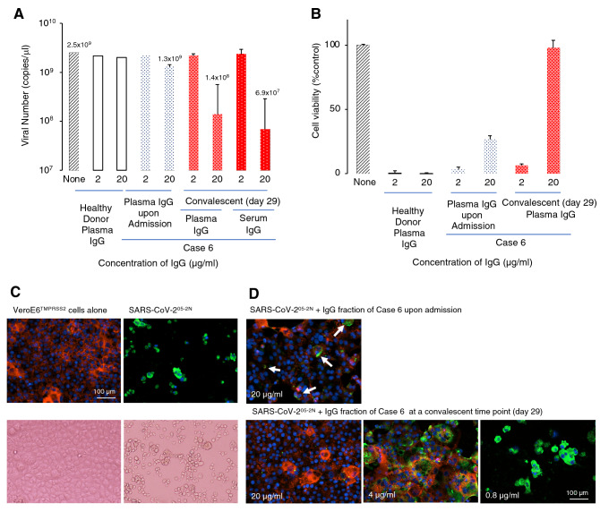
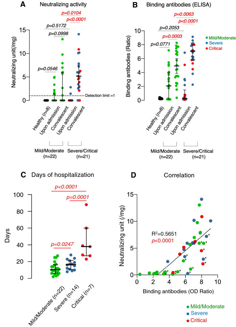
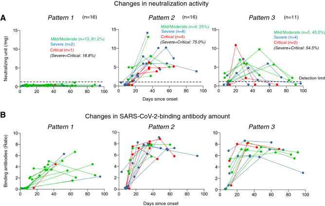
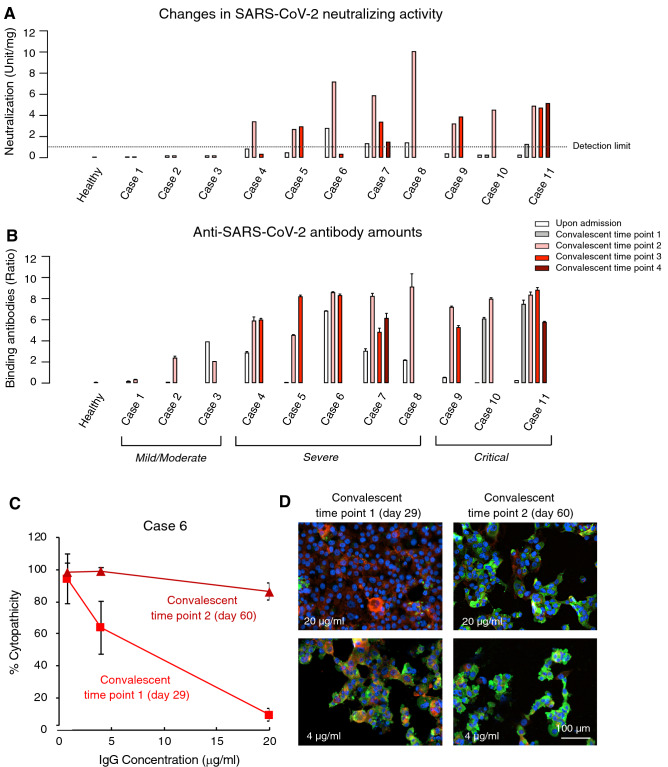
While there are various attempts to administer COVID-19-convalescent plasmas to SARS-CoV-2-infected patients, neither appropriate approach nor clinical utility has been established. We examined the presence and temporal changes of the neutralizing activity of IgG fractions from 43 COVID-19-convalescent plasmas using cell-based assays with multiple endpoints. IgG fractions from 27 cases (62.8%) had significant neutralizing activity and moderately to potently inhibited SARS-CoV-2 infection in cell-based assays; however, no detectable neutralizing activity was found in 16 cases (37.2%). Approximately half of the patients (~ 41%), who had significant neutralizing activity, lost the neutralization activity within ~ 1 month. Despite the rapid decline of neutralizing activity in plasmas, good amounts of SARS-CoV-2-S1-binding antibodies were persistently seen. The longer exposure of COVID-19 patients to greater amounts of SARS-CoV-2 elicits potent immune response to SARS-CoV-2, producing greater neutralization activity and SARS-CoV-2-S1-binding antibody amounts. The dilution of highly-neutralizing plasmas with poorly-neutralizing plasmas relatively readily reduced neutralizing activity. The presence of good amounts of SARS-CoV-2-S1-binding antibodies does not serve as a surrogate ensuring the presence of good neutralizing activity. In selecting good COVID-19-convalescent plasmas, quantification of neutralizing activity in each plasma sample before collection and use is required.


Conflict of interest statement

The authors declare no competing interests.

Figures

Figure 1
Figure 1
Neutralizing activity of IgG fraction from a representative convalescent patient. (A) SARS-CoV-205-2N was mixed with the IgG fraction from a patient (Case 6) or a healthy donor, incubated for 20 min, and then inoculated to VeroE6TMPRSS2 cells. The infectivity of the virus was examined by determining viral copy numbers using RNA-PCR. (B) The inhibition of cytopathic effect of SARS-CoV-205-2N in VeroE6TMPRSS2 cells by the IgG fraction from the plasma of Case 6 or a healthy donor. (C,D) Immunocytochemistry of SARS-CoV-205-2N-infected VeroE6TMPRSS2 cells. The IgG fraction (20 µg/ml) from Case 6 completely blocked the infectivity and cytopathic effect of SARS-CoV-2 and no cells got stained in green. Upper and lower insets in Panel (D) represent merged images, which are composed of images obtained from 3-color fluorescence. Viral antigens, actin filaments, and nuclei are indicated in green, red, and blue. The upper inset in (D) shows that most VeroE6TMPRSS2 cells were protected by the IgG fraction (20 µg/ml), while a few cells were infected (indicated by arrows). Lower right panel shows that the structure of actin filaments was totally destroyed by SARS-CoV-205-2N infection and almost all the cells got stained in green (SARS-CoV-2-positive); however, 20 µg/ml of the IgG fraction from Case 6 completely blocked the infectivity and cytopathicity of the virus. Images of immunocytochemistry were obtained using Cytation5 (BioTek, VT).
Figure 2
Figure 2
Changes in neutralizing activity of IgG fractions and the amounts of SARS-CoV-2-S1-binding antibodies. (A,B) IgG fractions from plasma (or serum) samples of all the 43 patients obtained upon admission and during convalescence were tested for their neutralizing activity and SARS-CoV-2-S1-binding antibody levels. The neutralizing activity of each IgG fraction was normalized by the activity of 100 µg IgG fraction of a patient D008 obtained at a convalescence time point, referred as to be 1 neutralization unit (Supplementary Fig. S2). The dashed line (1 neutralizing unit/mg) denotes the detection limit. The amounts of SARS-CoV-2-S1-binding IgG were measured using ELISA and shown by the ratios between the extinction of the sample and the calibrator (See “Materials and Methods” section for more details on the calculation. For disease severity definitions (Mild/Moderate: n = 22, Severe/Critical: n = 21), see the footnote in Table 1. p-value: Mann–Whitney U test. (C) Correlation of disease severity (Mild/Moderate: 22, Severe: 14, Critical: 7) and days of hospitalization. (D) Correlation of the SARS-CoV-2 binding IgG amounts and the neutralizing activity. Statistical analysis was performed and figures were generated using GraphPad Prism software version 8 (https://www.graphpad.com/scientific-software/prism/) (La Jolla, CA).
Figure 3
Figure 3
Changes in neutralizing activity and SARS-CoV-2-S1-binding antibody levels. (A) Neutralizing activity of each IgG fraction and the amount of SARS-CoV-2-binding antibody in serum/plasma obtained from multiple time points in the 43 patients were determined. The neutralizing activity of each IgG fraction was normalized and presented as a neutralizing unit, as described in the legend to Fig. 2. The definition of Patterns [Pattern 1, in which no neutralizing activity was observed during observation periods; Pattern 2, in which the neutralizing activity was maintained during observation periods, or cases with two time-point data only (shown with dashed line); Pattern 3, in which neutralizing activity was lost after the peak of the activity by < 40% during observation periods]. For the neutralizing activity in Pattern 3 (upper right panel), the average time period from the disease onset to reach the peak of activity was 36 days, and the average time period it took for the same individual to lose the activity by 50% or more from the peak was 24 days. The dashed line in the three upper panels denotes the detection limit value (1 neutralization unit). (B) Changes in SARS-CoV-2-S1-binding antibody amounts in patients in each Pattern group. For the definition of severities of patients, see the footnote to Table 1.
Figure 4
Figure 4
Comparison of neutralizing activity of IgG fractions with SARS-CoV-2-S1-binding IgG levels and changes in neutralizing activity. (A) The neutralizing activity of each IgG fraction was normalized and shown as a neutralizing unit. The broken line denotes the detection limit (1 neutralization unit). Regarding convalescence time points, 1 to 4, see “days of sample collections” in Table S1. (B) The amounts of SARS-CoV-2-S1-binding IgG were measured using ELISA and shown by the ratios between the extinction of the sample and the calibrator. See Materials and Method section for more details. (C) Loss of neutralizing activity of IgG fraction from Case 6 at Convalescence time point 2 (60 days from the onset). Note that the potent neutralization activity of IgG fraction from Case 6 observed at Convalescent time point 1 (29 days from the onset) was lost by Convalescent time point 2. (D) Immunocytochemistry of VeroE6TMPRSS2 cells exposed to SARS-CoV-205-2N and cultured in the presence of IgG fraction from Case 6. Note that while the cells were virtually completely protected by IgG fraction (20 µg/ml) at Convalescence time point 1, such activity was significantly reduced and virtually all the cells were infected and stained in green. Cellular actin filaments, SARS-CoV-205-2N antigens, and cell nuclei stained in red, green, and blue, respectively. Graphs were generated using GraphPad Prism software version 8.

Comment in

References

    1. Zhou P, et al. A pneumonia outbreak associated with a new coronavirus of probable bat origin. Nature. 2020;579:270–273. doi: 10.1038/s41586-020-2012-7. - DOI - PMC - PubMed
    1. Zhu N, et al. A novel coronavirus from patients with pneumonia in China, 2019. N. Engl. J. Med. 2020;382:727–733. doi: 10.1056/NEJMoa2001017. - DOI - PMC - PubMed
    1. World Health Organization. Coronavirus disease (COVID-19) outbreak situation. https://www.who.int/emergencies/diseases/novel-coronavirus-2019. (2020).
    1. Li Q, et al. Early transmission dynamics in Wuhan, China, of novel coronavirus-infected pneumonia. N. Engl. J. Med. 2020;382:1199–1207. doi: 10.1056/NEJMoa2001316. - DOI - PMC - PubMed
    1. Chen D, et al. Recurrence of positive SARS-CoV-2 RNA in COVID-19: A case report. Int. J. Infect. Dis. 2020;93:297–299. doi: 10.1016/j.ijid.2020.03.003. - DOI - PMC - PubMed

Publication types

MeSH terms