. 2021 Feb 22:9:630361.
doi: 10.3389/fcell.2021.630361. eCollection 2021.

Deletion of Kcnj16 in Mice Does Not Alter Auditory Function

Affiliations

Deletion of Kcnj16 in Mice Does Not Alter Auditory Function

Jun Lv et al. Front Cell Dev Biol. .

Abstract

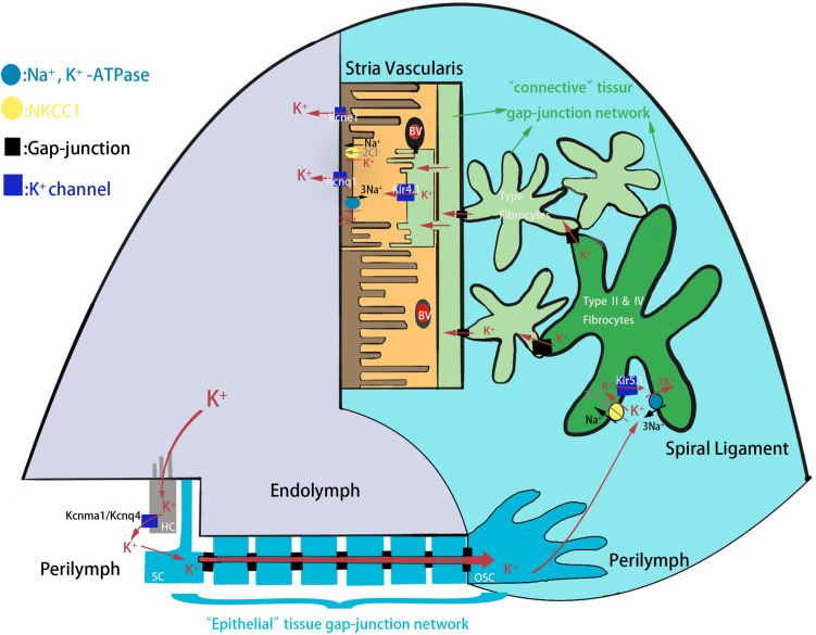
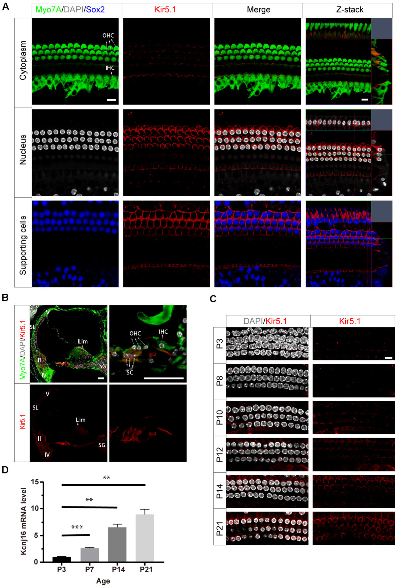
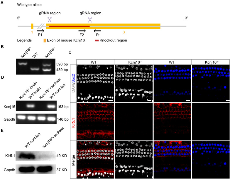
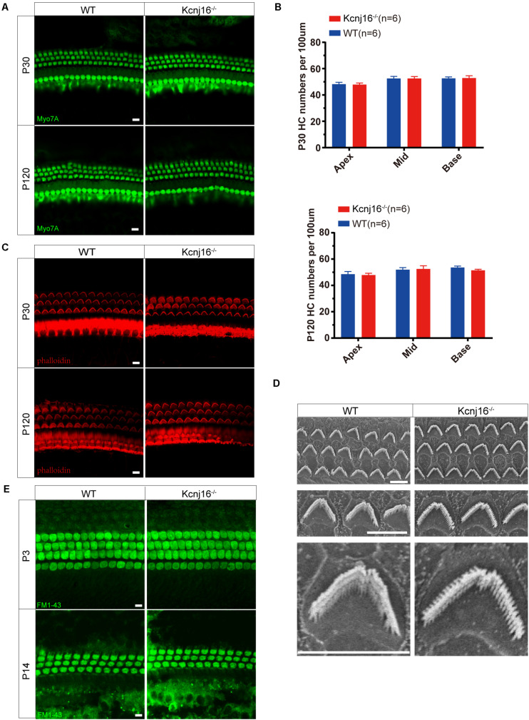
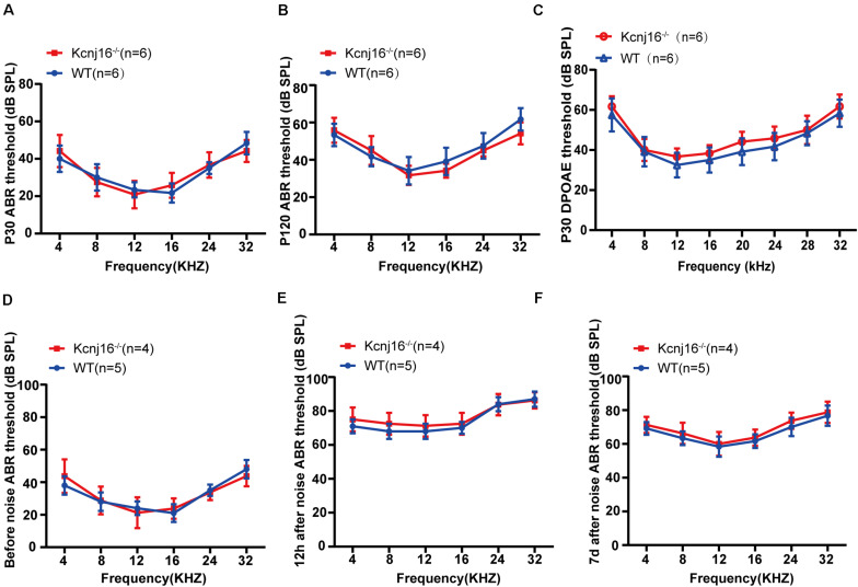
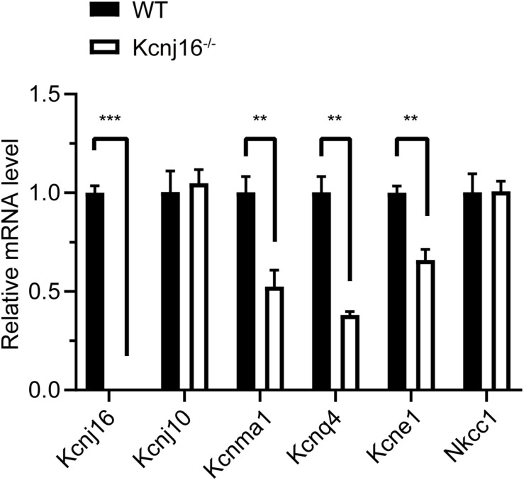
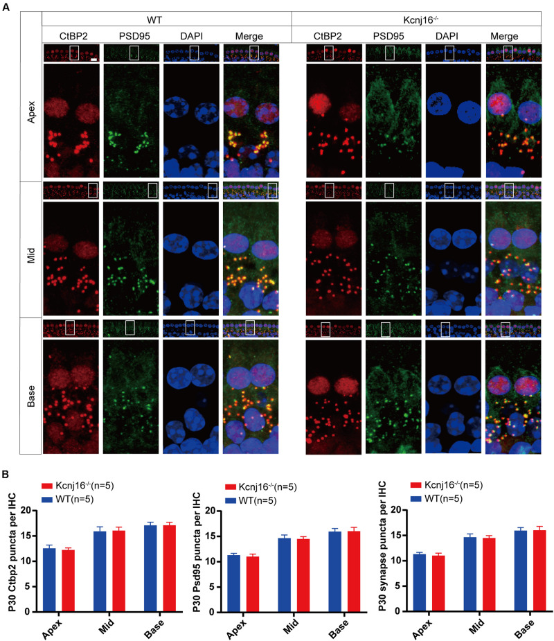
Endolymphatic potential (EP) is the main driving force behind the sensory transduction of hearing, and K+ is the main charge carrier. Kir5.1 is a K+ transporter that plays a significant role in maintaining EP homeostasis, but the expression pattern and role of Kir5.1 (which is encoded by the Kcnj16 gene) in the mouse auditory system has remained unclear. In this study, we found that Kir5.1 was expressed in the mouse cochlea. We checked the inner ear morphology and measured auditory function in Kcnj16 -/- mice and found that loss of Kcnj16 did not appear to affect the development of hair cells. There was no significant difference in auditory function between Kcnj16 -/- mice and wild-type littermates, although the expression of Kcnma1, Kcnq4, and Kcne1 were significantly decreased in the Kcnj16 -/- mice. Additionally, no significant differences were found in the number or distribution of ribbon synapses between the Kcnj16 -/- and wild-type mice. In summary, our results suggest that the Kcnj16 gene is not essential for auditory function in mice.

Keywords: Kir5.1; cochlea; endolymphatic potential; hair cell; hearing loss.


Conflict of interest statement

The authors declare that the research was conducted in the absence of any commercial or financial relationships that could be construed as a potential conflict of interest.

Figures

FIGURE 1
FIGURE 1
The schematic model of K+ circulation in mouse cochlea. K+ exits from HCs into the perilymph and is reabsorbed by the SCs and is further transported to the spiral ligament of the cochlear lateral wall through epithelial tissue gap-junction network. K+ is then reabsorbed by type II and IV fibrocytes, and recycled to the stria vascularis where it is finally secreted into the endolymph. HC, hair cell; SC, supporting cell; OSC, outer sulcus cell; BV, blood vessels.
FIGURE 2
FIGURE 2
Expression of Kir5.1 in the WT mouse cochlea. (A) Immunofluorescence staining with antibodies against Kir5.1 (red), Myo7A (green), and Sox2 (blue) and DAPI staining (white) in the basal turn of the mouse cochlea at P90. There was no difference in the immunolabeling of Kir5.1 from the apical to basal turns. Scale bar = 10 μm. (B) Immunohistochemical staining with antibodies against Kir5.1 (red) and Myo7A (green) and DAPI staining (white). Scale bar = 50 μm. (C) Immunofluorescence staining with antibodies against Kir5.1 (red) and DAPI staining (white) in the cochlea at P3, P8, P10, P12, P14, and P21. Scale bar = 10 μm. (D) Q-PCR results showing the changes in Kcnj16 mRNA in the mouse cochlea from P3 to P21. GAPDH was used as the internal control. Primers are shown in the Supplementary Table 2. Data are presented as the mean ± SD. ∗∗p < 0.01, ∗∗∗p < 0.001, n = 4. OHC, outer hair cell; IHC, inner hair cell; SC, supporting cell; SL, spiral ligament; II, IV, V, type II, IV, and V fibrocytes in the spiral ligament. Lim, spiral limbus; SG, spiral ganglions.
FIGURE 3
FIGURE 3
Analysis of Kir5.1 expression in Kcnj16− /− mice at the protein and mRNA level. (A) Overview of the targeting strategy. (B) PCR results showing genomic DNA of a homozygous (Kcnj16− /−; 598 bp), a wild-type (WT; 489 bp), and a heterozygous (Kcnj16+/−; 489 and 598 bp) mouse. Primers are shown in the Supplementary Table 1. (C) The cochleae of P30 Kcnj16− /− mice were immunolabeled with Kir5.1 antibodies. Scale bar = 10 μm. Images were taken from the middle turns of the sensory epithelium. There was no difference in the immunolabeling of Kir5.1 from the apical to basal turns (data not shown). (D) RT-PCR was performed to identify the presence the Kcnj16 mRNA. RNA was extracted from the cochlea and from total brain tissue of P30 Kcnj16− /− and WT mice, and GAPDH was used as the internal control. Primers are shown in the Supplementary Table 2. (E) Western blot was performed using antibodies against Kir5.1. Proteins from the cochlea were extracted from P30 Kcnj16− /− and WT mice, and GAPDH was used as the internal control.
FIGURE 4
FIGURE 4
Cochlear development and stereocilia structures were normal in the Kcnj16− /− mice. (A) Auditory HCs of P30 and P120 mice were stained with antibodies against Myo7A and imaged using a confocal microscope. Images were taken from the basal turn of the cochlea. There was no difference in the staining from the apical to basal turns. Scale bar = 10 μm. (B) The HCs were counted and compared with age-matched WT mice (p > 0.05, n = 6). Data are presented as the mean ± SD. (C) Auditory HC stereocilia of Kcnj16− /− and WT mice were stained with phalloidin, and images were taken from the basal turn of the cochlea. There was no difference from the apical to basal turns. Scale bar = 10 μm. (D) Low magnification and high magnification scanning electron microscope images of OHC stereocilia bundles in Kcnj16− /− and WT mice. Images were taken from the middle turn of P60 mice. Scale bar = 5 μm. (E) Auditory HCs of P3 and P14 mice were stained with FM1-43, and images were taken from the basal turns. There was no difference in the immunolabeling signals from the apical to basal turns (data not shown). Scale bar = 10 μm.
FIGURE 5
FIGURE 5
Kcnj16− /− mice showed no changes in auditory function. (A,B) ABR thresholds of P30 and P120 Kcnj16– /− and WT mice were measured at 4, 8, 12, 16, 24, 28, and 32 kHz. (C) DPOAE thresholds were measured in response to tone bursts of 4, 8, 16, 20, 24, 28, 32, 36, and 40 kHz in P30 Kcnj16− /− and WT mice. (D–F) The ABR thresholds of P30 Kcnj16− /− and WT mice were measured before, 12 h after, and 7 days after noise exposure. No significant differences were observed between Kcnj16− /− and WT mice (p > 0.05). Data are presented as means ± SD.
FIGURE 6
FIGURE 6
The expression of Kcnma1, Kcnq4, and Kcne1 decreased in P30 Kcnj16− /− mice. Q-PCR was performed with P30 WT and Kcnj16− /− cochleae (p < 0.05, n = 4). Primers are shown in the Supplementary Table 2. Data are presented as the mean ± SD. **p < 0.01, ***p < 0.001, n = 4.
FIGURE 7
FIGURE 7
The morphology of IHC ribbon synapses is preserved in Kcnj16− /− mice. (A) Ribbon synapses of P30 Kcnj16− /− and WT mice were stained with the ribbon synapse-specific markers CtBP2 and PSD95 and imaged under a confocal microscope. Scale bar = 10 μm. (B) The total numbers of CtBP2 and PSD95 puncta and synapses from the apical to basal turns were counted and compared between WT and Kcnj16− /− mice. No significant differences were seen for any measurements (p > 0.05, n = 5). Data are presented as the mean ± SD.

References

    1. Caprara G. A., Mecca A. A., Wang Y., Ricci A. J., Peng A. W. (2019). Hair bundle stimulation mode modifies manifestations of mechanotransduction adaptation. J. Neurosci. 39 9098–9106. 10.1523/jneurosci.1408-19.2019 - DOI - PMC - PubMed
    1. Chen J., Zhao H. B. (2014). The role of an inwardly rectifying K(+) channel (Kir4.1) in the inner ear and hearing loss. Neuroscience 265 137–146. 10.1016/j.neuroscience.2014.01.036 - DOI - PMC - PubMed
    1. Corns L. F., Johnson S. L., Roberts T., Ranatunga K. M., Hendry A., Ceriani F., et al. (2018). Mechanotransduction is required for establishing and maintaining mature inner hair cells and regulating efferent innervation. Nat. Commun. 9:4015. 10.1038/s41467-018-06307-w - DOI - PMC - PubMed
    1. D’Adamo M. C., Shang L., Imbrici P., Brown S. D., Pessia M., Tucker S. J. (2011). Genetic inactivation of Kcnj16 identifies Kir5.1 as an important determinant of neuronal PCO2/pH sensitivity. J. Biol. Chem. 286 192–198. 10.1074/jbc.M110.189290 - DOI - PMC - PubMed
    1. Fang Q., Zhang Y., Da P., Shao B., Pan H., He Z., et al. (2019). Deletion of Limk1 and Limk2 in mice does not alter cochlear development or auditory function. Sci. Rep. 9:3357. 10.1038/s41598-019-39769-z - DOI - PMC - PubMed

LinkOut - more resources