. 2021 Jul 28;144(6):1819-1832.
doi: 10.1093/brain/awab077.

The GGC repeat expansion in NOTCH2NLC is associated with oculopharyngodistal myopathy type 3

Affiliations

The GGC repeat expansion in NOTCH2NLC is associated with oculopharyngodistal myopathy type 3

Jiaxi Yu et al. Brain. .

Erratum in

Abstract

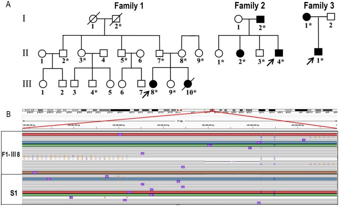
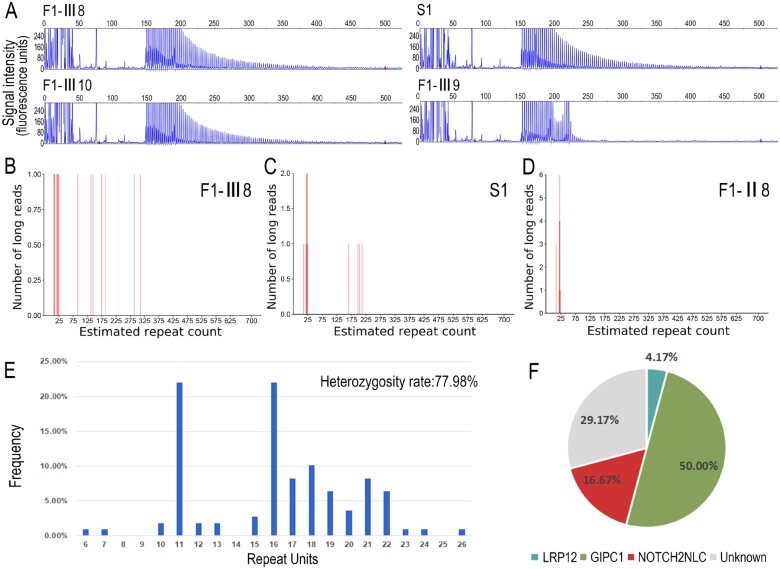
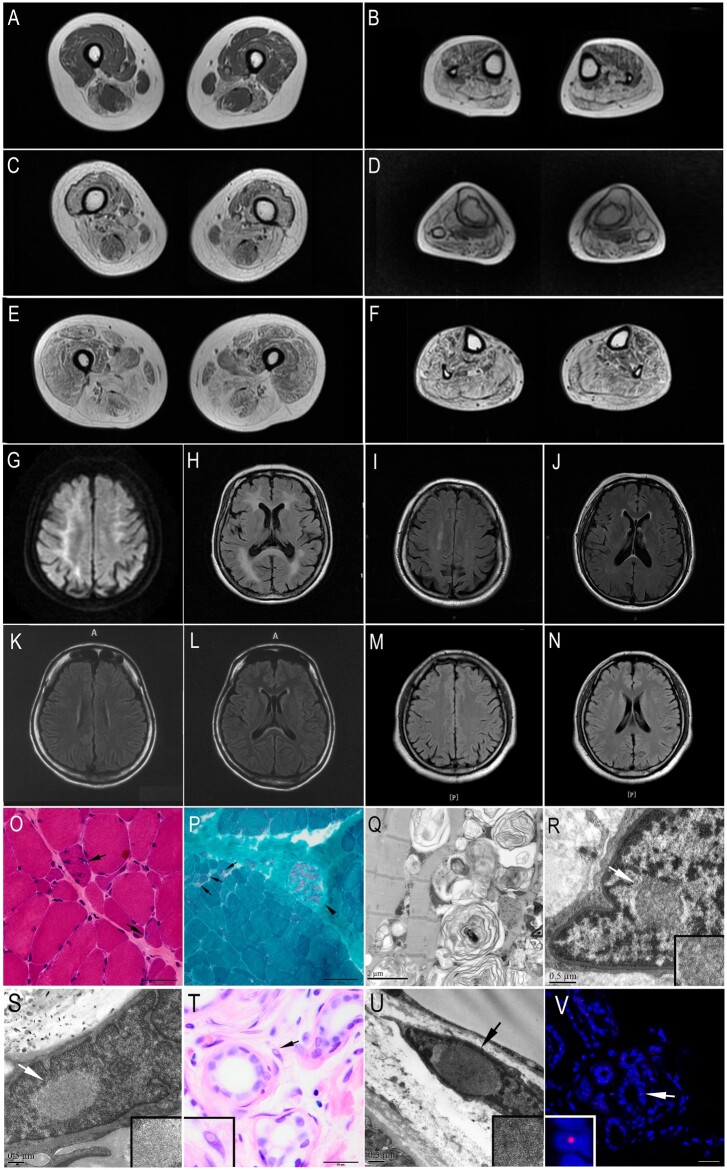
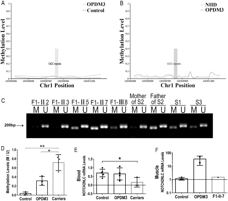
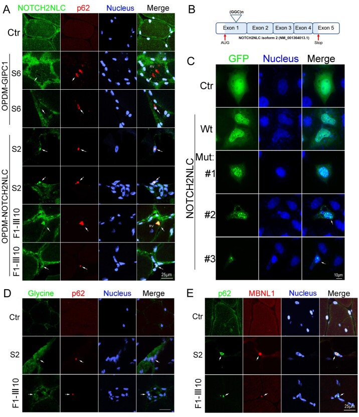
Oculopharyngodistal myopathy (OPDM) is an adult-onset neuromuscular disease characterized by progressive ocular, facial, pharyngeal and distal limb muscle involvement. Trinucleotide repeat expansions in LRP12 or GIPC1 were recently reported to be associated with OPDM. However, a significant portion of OPDM patients have unknown genetic causes. In this study, long-read whole-genome sequencing and repeat-primed PCR were performed and we identified GGC repeat expansions in the NOTCH2NLC gene in 16.7% (4/24) of a cohort of Chinese OPDM patients, designated as OPDM type 3 (OPDM3). Methylation analysis indicated that methylation levels of the NOTCH2NLC gene were unaltered in OPDM3 patients, but increased significantly in asymptomatic carriers. Quantitative real-time PCR analysis indicated that NOTCH2NLC mRNA levels were increased in muscle but not in blood of OPDM3 patients. Immunofluorescence on OPDM muscle samples and expressing mutant NOTCH2NLC with (GGC)69 repeat expansions in HEK293 cells indicated that mutant NOTCH2NLC-polyglycine protein might be a major component of intranuclear inclusions, and contribute to toxicity in cultured cells. In addition, two RNA-binding proteins, hnRNP A/B and MBNL1, were both co-localized with p62 in intranuclear inclusions in OPDM muscle samples. These results indicated that a toxic protein gain-of-function mechanism and RNA gain-of-function mechanism may both play a vital role in the pathogenic processes of OPDM3. This study extended the spectrum of NOTCH2NLC repeat expansion-related diseases to a predominant myopathy phenotype presenting as OPDM, and provided evidence for possible pathogenesis of these diseases.

Keywords: GGC repeat expansion; NOTCH2NLC; RNA gain-of-function mechanism; oculopharyngodistal myopathy; toxic protein gain-of-function mechanism.


Figures

Figure 1
Figure 1
Identification of GGC repeat expansion in the 5′UTR of the NOTCH2NLC gene in OPDM individuals. (A) Pedigree chart of three families with OPDM. The squares indicate males and the circles indicate females. A diagonal line through a symbol indicates a deceased individual. Affected individuals are indicated by filled symbols. All the asterisked individuals had available blood DNA. (B) Visualization on Integrative Genomics Viewer for Patients F1-III8 and S1 revealed DNA reads carrying the mutated repeat unit (chr1:149390780–149390837, hg38 version), corresponding to the GGC repeat expansions in the 5′UTR of the NOTCH2NLC gene.
Figure 2
Figure 2
Validation of GGC repeat expansions and repeat sizes in NOTCH2NLC among OPDM3 patients and normal control subjects. (A) Representative results of RP-PCR analysis showing GGC repeat expansions in Patients F1-III8, F1-III10 and S1. No repeat expansions were shown in one unaffected member Patient F1-III9. Experiments were conducted three times with reproducible results. LRS data showed estimated GGC repeat count of NOTCH2NLC reached more than 100 in Patients F1-III8 (B) and S1 (C) but no more than 25 in the unaffected member Patient F1-II8 (D). (E) Frequency distribution of GGC repeat units in NOTCH2NLC ranging from 6 to 26 among 109 normal control subjects. DNA samples from normal controls were amplified by NOTCH2NLC-specific primers and revealed by fragment analysis. Heterozygosity rate in normal controls was 77.98%. (F) Pie chart for the percentages of disease-causing gene mutations and unknown gene mutations in a cohort of Chinese OPDM patients. Disease-causing genes, trinucleotide repeat expansions in the 5′UTRs of LRP12, GIPC1 and NOTCH2NLC, accounted for 4.17%, 50% and 16.67% of cases in 24 unrelated Chinese OPDM patients, respectively, whereas unknown genetic cause accounted for 29.17% of cases in this cohort.
Figure 3
Figure 3
MRI and pathological changes of the OPDM3 patients. (AF) Muscle MRI of OPDM3 patients showed fatty infiltration of lower limb muscles, with the distal muscles (B, D and F calf level) more severely affected than the proximal muscles (A, C and E thigh level). (A and B) Muscle MRI of Patient F1-III10: moderate weakness with a disease duration of 6 years, ambulatory without support. (C and D) Muscle MRI of Patient F1-III8, moderate weakness with a disease duration of 9 years, ambulatory without support. (E and F) Muscle MRI of Patient S1: severe weakness with a disease duration of 11 years, ambulatory without support. (GN) Brain MRIs of Patients S2 (G and H), S1 (I and J), F1-III8 (K and L), and S3 (M and N). (G) Hyperintense linear lesions in corticomedullary junctions in a diffuse weight image (DWI). (H) Severely diffuse leukoencephalopathy on T2-weighted fluid-attenuated inversion recovery (T2-FLAIR). (IL) Mild white matter signal abnormalities on T2-FLAIR in periventricular white matter and splenium of corpus callosum after 18 years (Patient S1) and 15 years (Patient F1-III8) of the disease, respectively. (M and N) Normal brain MRIs on T2-FLAIR. (O) Haematoxylin and eosin, and (P) modified Gomori trichrome (mGT) stainings of muscle sections from Patient F1-III8, showing dystrophic change with variation in fibre size and endomysial fibrosis, and fibres with rimmed vacuoles (marked by arrow and shown at higher magnification). The intramuscular peripheral nerves were well myelinated (arrowhead) (P). (Q) Electron microscopy of muscle tissue from Subject F1-III10 revealed various myelin figures and autophagic vacuoles containing osmiophilic deposits and amorphous materials. (R and S) Electron microscopy of muscle sample from Patient S2 showed intranuclear inclusions contained filamentous aggregates (marked by arrowhead and shown at higher magnification). (T) Haematoxylin and eosin staining of skin sections from Patient S1 showed intranuclear inclusions in the fibroblasts (marked by arrow and shown at higher magnification). (U) Electron microscopy of skin sample from Patient S1 showed intranuclear inclusions. (V) Immunofluorescence on skin sections from Patient S1 showing p62 positive intranuclear inclusions in the sweat gland (marked by arrow and shown at higher magnification). Scale bars = 50 µm (O and T), 100 µm (P), 2 μm (Q), 0.5 µm (R, S and U) and 25 µm (V).
Figure 4
Figure 4
Methylation and expression at the NOTCH2NLC locus. (A and B) Methylation status across the expanded GGC repeat region was determined using LRS data from three affected individuals (Patients F1-III8, F1-III10 and S1), 10 normal control subjects, and four NIID patients. (A) There were no significant differences between three affected individuals (Patients F1-III8, F1-III10 and S1) and 10 normal controls in methylation. (B) There were no significant differences in methylation between three affected individuals (Patients F1-III8, F1-III10 and S1) and four NIID patients. (C and D) Methylation analysis of the promoter region of expanded GGC repeats in the 5′UTR of the NOTCH2NLC gene in blood. Methylation levels were significantly increased in three asymptomatic carriers (Patients F-II5, F-II7 and father of Patient S2), compared with OPDM3 patients (Patients F-III8, S1 and S3) and controls (F-II2, F-II3 and mother of Patient S2). M = methylated; U = unmethylated. Ratio of methylation level of NOTCH2NLC to that of unmethylation level (M/U) was used to represent the relative methylation status. (E and F) Analysis of transcriptional levels of NOTCH2NLC mRNA in normal control subjects, OPDM3 patients and asymptomatic carriers by RT-qPCR in peripheral blood or muscle samples. No significant difference was detected between control subjects and OPDM3 patients (Patients F1-III8, F1-III10, S1 and S3), while asymptomatic carriers showed significantly lower NOTCH2NLC mRNA levels in peripheral blood (*P < 0.05) (E). The transcriptional levels of NOTCH2NLC mRNA were in an increasing trend in muscle samples (F) from two OPDM3 patients (Patients F1-III10 and S2).
Figure 5
Figure 5
Immunofluorescence on muscle biopsy samples and expression of NOTCH2NLC wild-type or mutant in HEK293 cells. (A) NOTCH2NLC distribution in skeletal muscle of control and OPDM patients. Immunofluorescence showing NOTCH2NLC and p62 co-localized in intranuclear inclusions and rimmed vacuoles of OPDM3 patients (Patients S2 and F1-III10), but not in control or GIPC1-affected OPDM patient (Patient S6), indicated by arrows. (B) Location of GGC repeat expansions in the coding region in the transcript isoform 2 (NM_001364013.1). (C) HEK293 cells were transfected with the control (GFP), NOTCH2NLC wild-type [NOTCH2NLC-(GGC)9] or mutant [NOTCH2NLC-(GGC)69] vectors and subjected to fluorescence observation at 48 h post-transfection. NOTCH2NLC wild-type (Wt) was primarily localized in the nucleus compared to the control, whereas the NOTCH2NLC mutant formed protein aggregates in the nucleus. Different stages of NOTCH2NLC-(GGC)69 aggregation were shown: diffuse small aggregates in Mutant 1, large aggregates in Mutants 2 and 3 (indicated by arrows). The apoptotic cell with condensed nuclei is indicated in Mutant 3. (D) Glycine and p62 co-localized in intranuclear inclusions of OPDM3 patients (Patients S2 and F1-III10), but not in the control (indicated by arrows). (E) RNA binding protein MBNL1 form aggregates in the intranuclear inclusions in OPDM3 patients. Scale bars = 25 μm (A, D and E), and 10 μm (C). Nuclei were counterstained with DAPI.

References

    1. Durmus H, Laval SH, Deymeer F, et al. Oculopharyngodistal myopathy is a distinct entity: Clinical and genetic features of 47 patients. Neurology. 2011;76:227–235. - PubMed
    1. Minami N, Ikezoe K, Kuroda H, Nakabayashi H, Satoyoshi E, Nonaka IJND.. Oculopharyngodistal myopathy is genetically heterogeneous and most cases are distinct from oculopharyngeal muscular dystrophy. Neuromuscul Disord. 2001;11:699–702. - PubMed
    1. Deng J, Yu J, Li P, et al. Expansion of GGC repeat in GIPC1 is associated with oculopharyngodistal myopathy. Am J Hum Genet. 2020;106(6):793–804. - PMC - PubMed
    1. Lu H, Luan X, Yuan Y, Dong M, Sun W, Yan C.. The clinical and myopathological features of oculopharyngodistal myopathy in a Chinese family. Neuropathology. 2008;28:599–603. - PubMed
    1. Satoyoshi E, Kinoshita M.. Oculopharyngodistal myopathy. Arch Neurol. 1977;34:89–92. - PubMed

Publication types

Supplementary concepts