. 2021 Apr 15;217(4):iyab014.
doi: 10.1093/genetics/iyab014.

Split-wrmScarlet and split-sfGFP: tools for faster, easier fluorescent labeling of endogenous proteins in Caenorhabditis elegans

Affiliations

Split-wrmScarlet and split-sfGFP: tools for faster, easier fluorescent labeling of endogenous proteins in Caenorhabditis elegans

Jérôme Goudeau et al. Genetics. .

Abstract

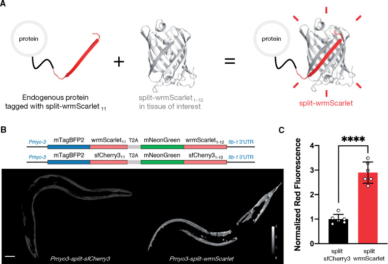
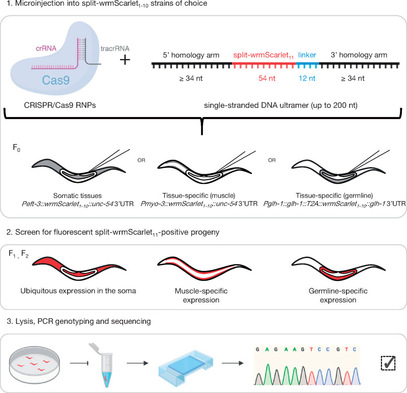
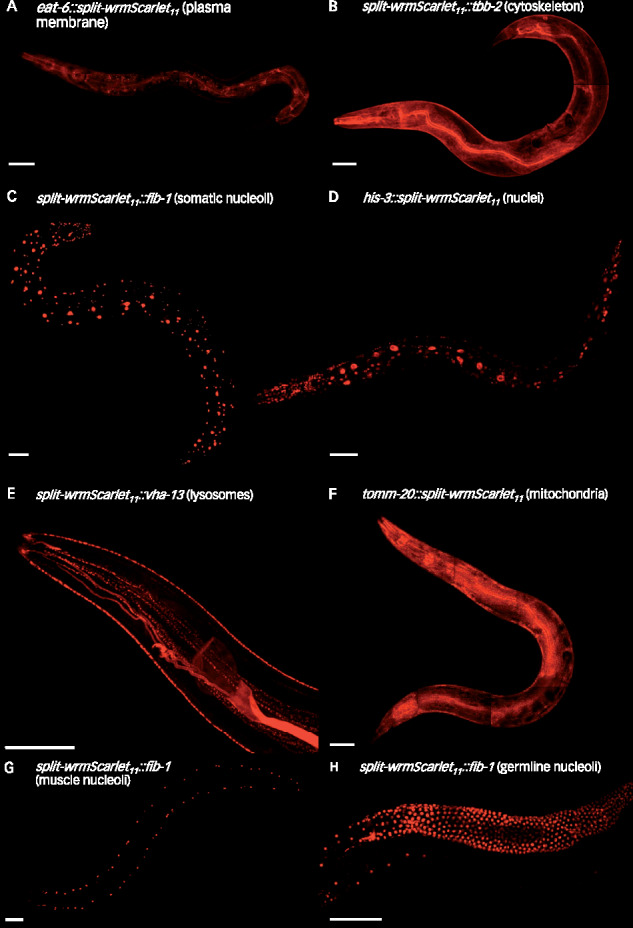
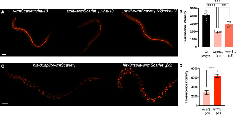
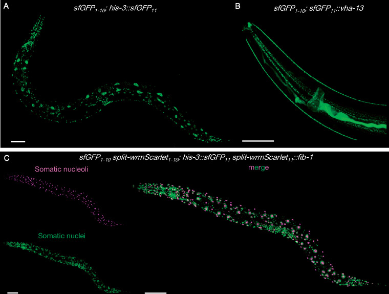
We create and share a new red fluorophore, along with a set of strains, reagents and protocols, to make it faster and easier to label endogenous Caenorhabditis elegans proteins with fluorescent tags. CRISPR-mediated fluorescent labeling of C. elegans proteins is an invaluable tool, but it is much more difficult to insert fluorophore-size DNA segments than it is to make small gene edits. In principle, high-affinity asymmetrically split fluorescent proteins solve this problem in C. elegans: the small fragment can quickly and easily be fused to almost any protein of interest, and can be detected wherever the large fragment is expressed and complemented. However, there is currently only one available strain stably expressing the large fragment of a split fluorescent protein, restricting this solution to a single tissue (the germline) in the highly autofluorescent green channel. No available C. elegans lines express unbound large fragments of split red fluorescent proteins, and even state-of-the-art split red fluorescent proteins are dim compared to the canonical split-sfGFP protein. In this study, we engineer a bright, high-affinity new split red fluorophore, split-wrmScarlet. We generate transgenic C. elegans lines to allow easy single-color labeling in muscle or germline cells and dual-color labeling in somatic cells. We also describe a novel expression strategy for the germline, where traditional expression strategies struggle. We validate these strains by targeting split-wrmScarlet to several genes whose products label distinct organelles, and we provide a protocol for easy, cloning-free CRISPR/Cas9 editing. As the collection of split-FP strains for labeling in different tissues or organelles expands, we will post updates at doi.org/10.5281/zenodo.3993663.

Keywords: C. elegans; CRISPR; Cas9; GFP; genome engineering; germline; mScarlet; protein localization; wrmScarlet.


Figures

Figure 1
Figure 1
Engineering and evaluating split-wrmScarlet. (A) Principle of endogenous protein labeling with split-wrmScarlet. The protein structure from split-wrmScarlet was generated using Phyre2 and PyMOL. (B) Schematic of the plasmids encoding split-wrmScarlet and split-sfCherry3. Each plasmid consists of the large FP1–10 sequence fused to mNeonGreen, and the corresponding small FP11 sequence fused to mTagBFP2. The T2A sequence ensures that mTagBFP2::FP11 and the corresponding mNeonGreen::FP1–10 are separated. The images are representative displays of the ratio of red to green fluorescence intensity from images acquired under identical conditions after background subtraction and masking with the same threshold. Scale bar, 50 µm. (C) Emission intensities from split-sfCherry3 and split-wrmScarlet normalized to mNeonGreen. Mean ± SD. Circles are individuals (n = 6 for each split fluorescent protein). ****P < 0.0001.
Figure 2
Figure 2
Split-wrmScarlet11-mediated tagging. Schematic representation of the split-wrmScarlet tagging workflow to visualize endogenous proteins specifically in muscles, germline, or throughout the soma. Some illustrations were created with BioRender.com.
Figure 3
Figure 3
Tissue-specific split-wrmScarlet labeling of proteins with distinct subcellular locations. Endogenous proteins tagged with split-wrmScarlet11 in animals expressing split-wrmScarlet1–10 in somatic tissues, in muscles or in the germline. (A–F) Confocal images of worms expressing somatic split-wrmScarlet1–10 and (A) EAT-6::split-wrmScarlet11 (plasma membrane), (B) split-wrmScarlet11::TBB-2 (cytoskeleton), (C) split-wrmScarlet11::FIB-1 (nucleoli), (D) HIS-3::split-wrmScarlet11 (nuclei), (E) split-wrmScarlet11::VHA-13 (lysosomes), or (F) TOMM-20::split-wrmScarlet11 (mitochondria). (G) Transgenic worm expressing split-wrmScarlet1–10 in muscle and split-wrmScarlet11::FIB-1. (H) Transgenic worm expressing split-wrmScarlet1–10 in the germline and split-wrmScarlet11::FIB-1. (A–H) Maximum intensity projections of 3D stacks shown. Scale bars, 50 µm.
Figure 4
Figure 4
Split-wrmScarlet11 tandem repeats increase fluorescence. (A) Images of animals carrying either wrmScarlet, split-wrmScarlet11 or two tandem repeats of split-wrmScarlet11 inserted at the endogenous VHA-13 N-terminus. (B) Emission intensities of animals carrying wrmScarlet, split-wrmScarlet11 or dual split-wrmScarlet11 inserted at the VHA-13 N-terminus. Mean ± SD. Circles are individuals. ****P < 0.0001, ***P < 0.001, **P < 0.005. (C) Images of animals carrying either a single split-wrmScarlet11 or three tandem repeats of split-wrmScarlet11 inserted at the HIS-3 C-terminus. (D) split-wrmScarlet emission intensities from animals carrying a single split-wrmScarlet11 or three tandem repeats of split-wrmScarlet11 knock-in at the HIS-3 C-terminus. Mean ± SD. Circles are individuals. ***P < 0.001. Images from each comparison were taken under identical instrument conditions using confocal microscopy and are shown using identical brightness and contrast settings. Images shown are from a single confocal plane. Scale bars, 50 µm.
Figure 5
Figure 5
Split-sfGFP and split-wrmScarlet dual-color protein labeling. Images of animals stably expressing sfGFP1–10 in somatic tissues (A) CF4592 muIs253[Peft-3::sfGFP1–10::unc-54 3'UTR Cbr-unc-119(+)] II; unc-119(ed3) III; his-3(muIs255[his-3::sfGFP11]) V or (B) CF4589 muIs253[Peft-3::sfGFP1–10::unc-54 3'UTR Cbr-unc-119(+)] II; unc-119(ed3) III; vha-13(muIs268[sfGFP11::vha-13]) V. (C) Dual color protein labeling with split-wrmScarlet and split-sfGFP in somatic cells. Composite display of red and green channels of animals expressing split-wrmScarlet1–10 and sfGFP1–10 in somatic tissues, HIS-3::sfGFP11 and split-wrmScarlet11::FIB-1; CF4602 muIs253[Peft-3::sfGFP1–10::unc-54 3'UTR Cbr-unc-119(+)] muIs252[Peft-3::split-wrmScarlet1–10::unc-54 3'UTR Cbr-unc-119(+)] II; unc-119(ed3) III; fib-1(muIs254[split-wrmScarlet11::fib-1]) his-3(muIs255[his-3::sfGFP11]) V. Maximum intensity projections of 3D stacks shown. Scale bars, 50 µm.

References

    1. Bindels DS, Haarbosch L, van Weeren L, Postma M, Wiese KE, et al.2017. mScarlet: a bright monomeric red fluorescent protein for cellular imaging. Nat Methods 14:53–56. - PubMed
    1. Brenner S. 1974. The genetics of Caenorhabditis elegans. Genetics. 77:71–94. - PMC - PubMed
    1. Cabantous S, Terwilliger TC, Waldo GS.. 2005. Protein tagging and detection with engineered self-assembling fragments of green fluorescent protein. Nat Biotechnol. 23:102–107. - PubMed
    1. Dickinson DJ, Goldstein B.. 2016. CRISPR-Based Methods for Caenorhabditis elegans Genome Engineering. Genetics. 202:885–901. - PMC - PubMed
    1. Dokshin GA, Ghanta KS, Piscopo KM, Mello CC.. 2018. Robust genome editing with short single-stranded and long, partially single-stranded DNA donors in Caenorhabditis elegans. Genetics. 210:781–787. - PMC - PubMed

Publication types

MeSH terms

Substances