. 2021 Apr 23;185(4):1617-1637.
doi: 10.1093/plphys/kiab016.

UGT84F9 is the major flavonoid UDP-glucuronosyltransferase in Medicago truncatula

Affiliations

UGT84F9 is the major flavonoid UDP-glucuronosyltransferase in Medicago truncatula

Olubu A Adiji et al. Plant Physiol. .

Abstract

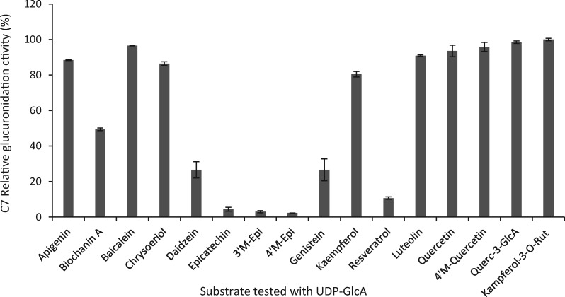
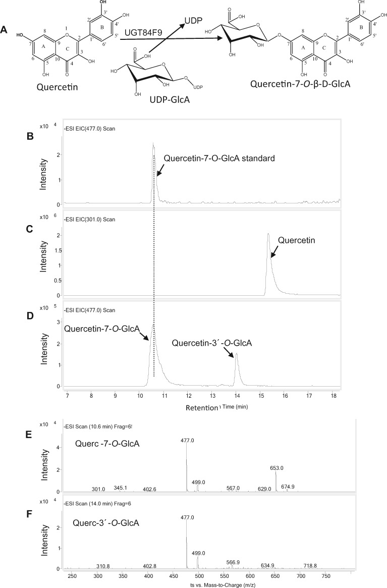
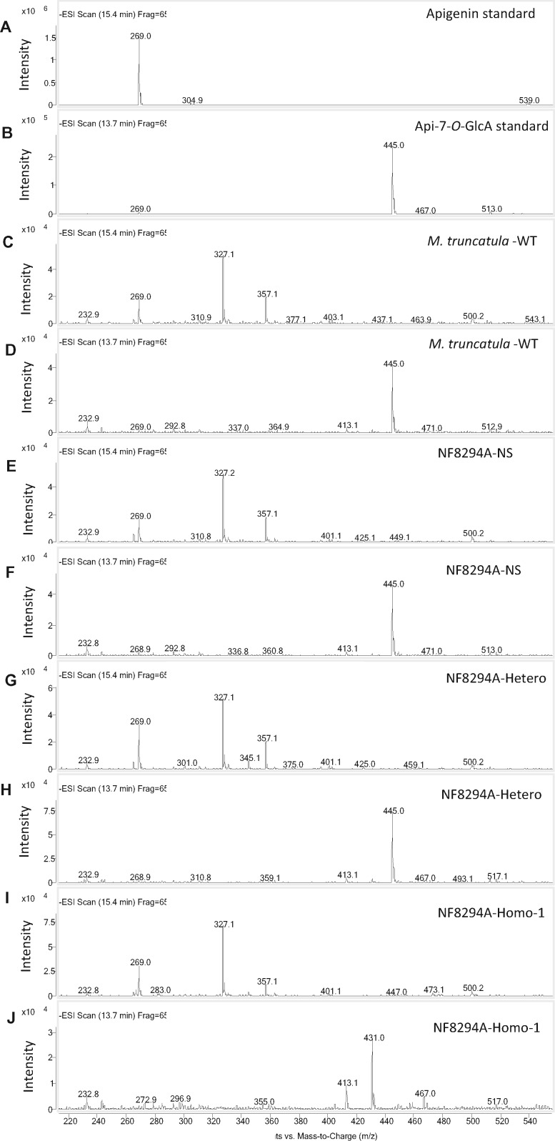
Mammalian phase II metabolism of dietary plant flavonoid compounds generally involves substitution with glucuronic acid. In contrast, flavonoids mainly exist as glucose conjugates in plants, and few plant UDP-glucuronosyltransferase enzymes have been identified to date. In the model legume Medicago truncatula, the major flavonoid compounds in the aerial parts of the plant are glucuronides of the flavones apigenin and luteolin. Here we show that the M. truncatula glycosyltransferase UGT84F9 is a bi-functional glucosyl/glucuronosyl transferase in vitro, with activity against a wide range of flavonoid acceptor molecules including flavones. However, analysis of metabolite profiles in leaves and roots of M. truncatula ugt84f9 loss of function mutants revealed that the enzyme is essential for formation of flavonoid glucuronides, but not most flavonoid glucosides, in planta. We discuss the use of plant UGATs for the semi-synthesis of flavonoid phase II metabolites for clinical studies.


Figures

Figure 1
Figure 1
Amino acid sequence alignment of the N-terminal domains of selected plant UGTs, indicating the presence of a critical catalytic His (indicated by red arrow) that is universally conserved across all plant UGTs. A neighboring residue believed to be important in contributing to sugar donor recognition is indicated with the green arrow. Blue boxes in the left hand column indicated previously identified UDP-glucuronosyltransferases. Orange boxes and binary names (e.g. C3, J5) indicate M. truncatula UG(A)T candidates in this study. Sugar–sugar branching glucuronidating enzymes such as BpUGT94B1 and UGT73P12_canonical have an Arg substitution at this position instead of Ser, Thr, or Pro found in UDP-Glc dependent UGTs. Other amino acid substitutions (His, Gln, or Phe) were found at this location for some of the M. truncatula UGAT candidates we identified. Sequences are: BpUGT94B1, UDP-GlcA dependent UGT from red daisy (Bellis perennis); C7 (UGT84F9), UDP-GlcA and UDP-Glc specific UGT from M. truncatula (this study); VvGT5, UDP-GlcA dependent UGT from V. vinifera, UGT73P12_canonical and UGT73P12_variant are, respectively, UDP-GlcA and UDP-Glc dependent UGTs from licorice (G. uralensis); UGT78G1, UGT71G1, and UGT72L1 are UDP-Glc dependent UGTs from M. truncatula; GuUGAT, UDP-GlcA dependent UGT from licorice; UGT88D-1, 4, 5, 6, 7, and UGT88A7 are UDP-GlcA dependent UGTs from Lamiales species. Blue boxes show previously identified UDP-GlcA-specific enzymes, brown boxes UGAT candidates that utilize UDP-Glc. GenBank accession numbers for these sequences are given in Supplemental Table S2.
Figure 2
Figure 2
Expression analysis of selected candidate UGAT transcripts in different tissues of M. truncatula ecotype R108. A–D, Quantitative real-time PCR analysis of relative transcript levels of UGT84F9 (A), UGT88E28 (B), UGT88E27 (C), and UGT78G3 (D). The latter three were expressed at lower levels in roots than in leaves based on transcriptome analysis in the M. truncatula Gene Expression Atlas (https://mtgea.noble.org/v3/). Transcript levels are expressed relative to those of the housekeeping gene tubulin. Error bars represent standard deviation, n = 3 analytical replicates.
Figure 3
Figure 3
Relative glucuronidation activity of UGT84F9 toward flavonoids and other compounds. Recombinant UGT84F9 was incubated with the individual acceptor substrates (750 µM) and UDP-GlcA (2 mM) for 2 h at 30°C. The activity is expressed as percentage relative to that of the most favored substrate, kaempferol-3-O-rutinoside. 100% activity = 40.055 nmol/min/mg protein. Data are means for three technical replicates, with error bars representing the standard deviation.
Figure 4
Figure 4
UGT84F9 catalyzes the glucuronidation of quercetin. A, The enzymatic reaction catalyzed by UGT84F9. B–D, LC–MS analysis of the enzymatic reaction of UGT84F9 to generate Q-7-O-GlcA. B, Q-7-O-GlcA standard. C, Remaining Q substrate. D, Reaction products of UGT84F9 incubated with quercetin. E, Mass spectrum showing the molecular ions of the glucuronidated product of quercetin at retention time 10. 6 min (m/z = 477). A molecular ion (m/z = 653) matching that of di-glucuronides of Q is also seen. F, Mass spectrum of molecular ions of quercetin glucuronide at retention time 14 min (m/z = 477). The LC–MS was performed in negative ion mode. All reactions were incubated for 2 h.
Figure 5
Figure 5
HPLC-UV analysis of the products of UGT84F9 activity with three flavonoid acceptors using UDP-Glc as sugar donor. A, apigenin. B, luteolin. C, quercetin.
Figure 6
Figure 6
The modeled three-dimensional structure of UGT84F9 with UDP-GlcA and UDP-Glc, showing the possible interactions between the enzyme residues and sugar donor. A, The predicted protein structure of UGT84F9 represented by cartoon (cyan) with UDP-GlcA shown as a stick model (carbon atom in slate blue, oxygen in red, phosphorus in orange). B, The putative active site and substrate binding pocket. Key residues His22 and Trp360, shown as sticks (carbon atom in cyan, nitrogen atom in blue), can potentially interact with the sugar donor, UDP-GlcA, that is also represented in stick model (carbon atom in slate blue, oxygen in red, phosphorus in orange). Catalytic residue His19, shown as a stick model, is in the active pocket and interacts with the acceptor substrate. C and D, The potential three-dimensional structure of UGT84F9 with superimposition of bound UDP-Glc, showing the possible residues interacting with the sugar donor. C, The predicted protein structure of UGT84F9 represented by cartoon (green) with bound UDP-Glc represented in stick model (carbon atoms in slate, oxygen in red, phosphorus in orange, and nitrogen in blue). D, The presumed active site pocket with the bound UDP-Glc showing the possible interaction with residues including His19, Trp360, and His22. SWISS-MODEL software was used to build the 3-dimensional structure of UGT84F9 in comparison to UGT71G1, and the ligands docked into UGT71G1 were superimposed onto UGT84F9 using Wincoot software. PyMOL software was used to visualize the possible catalytic residues interacting with the ligand.
Figure 7
Figure 7
Characterization of Tnt1 transposon insertion mutations in UGT84F9. A, Seven-weeks-old plants with ugt84f9 Tnt1 insertions in NF8294A mutant lines showing smaller size of the homozygote compared with the corresponding heterozygote at the UGT84F9 locus. B, Molecular characterization of the mutation at the UGT84F9 locus in NF8294A and NF5230. Agarose gel electrophoresis analysis of PCR products resulting from genotyping the Tnt1 insertion mutant lines for UGT84F9 gene disruption, compared with the wild-type plant. The first and second columns show the presence of intact UGT84F9 gene, with absence of the Tnt1 insertion (UGT84F9/tnt) in the R108 wild type (the genetic background used for the transposon insertion mutagenesis) and the null-segregant at the UGT84F9 locus. The third column shows the presence of the wild-type and mutant alleles at the UGT84F9 locus indicative of the heterozygote. The fourth and fifth columns show loss of the wild-type allele and presence of the Tnt1 insertion in two independent homozygous lines of NF8294A. In the last two columns, NF5230-WT shows the intact UGT84F9 gene without Tnt1 insertion and NF5230-Homo shows the Tnt1 insertion in the homozygous line of the second allele. UGT88E28 and tubulin served as controls. C, Model showing the Tnt1 insertion in the N-terminal region of the UGT84F9 gene in the NF8294A and NF5230 lines.
Figure 8
Figure 8
Loss of function of UGT84F9 leads to disappearance of apigenin glucuronide in M. truncatula. A–F, LC–MS analysis of flavonoid metabolites in extracts from wild-type and mutant M. truncatula leaf tissue for detection of the flavone apigenin and its mono-glucuronide apigenin-7-O-GlcA. A, Apigenin standard (m/z = 269). D, Apigenin-7-O-GlcA standard (m/z 445). C, E, G, and I, The extracted ion chromatograms from scanning the M. truncatula wild-type R108, the NF8294A-null segregant, the heterozygote and the homozygote extracts, respectively, for apigenin (m/z = 269). D, F, H, and J, The extracted ion chromatograms from scanning the M. truncatula wild-type R108, the NF8294A-null segregant, the heterozygote and the homozygote extracts, respectively, for apigenin-7-O-GlcA (m/z 445). The LC–MS was performed in negative mode.
Figure 9
Figure 9
MS spectra for the metabolites in Figure 8. The peak at retention time 15.4 min is apigenin and the peak at 13.7 min Api-7-O-GlcA. A and B, Apigenin and Api-7-O-GlcA standards. Apigenin, with molecular mass of the parent ion at [M–H]+ = 269, was seen in all the samples (C, E, G, and I); apigenin-7-O-GlcA, [M–H]+ = 455, was present in the extract from M. truncatula wild-type R108, the NF8294A-null segregant, and the heterozygote (D, F, and H) but was not detected in the extract from NF8294A-homozygote (J). The LC–MS was performed in negative mode.

References

    1. Achnine L, Huhman DV, Farag MA, Sumner LW, Blount JW, Dixon RA (2005) Genomics-based selection and functional characterization of triterpene glycosyltransferases from the model legume Medicago truncatula. Plant J 41:875–887 - PubMed
    1. Alonso AP, Piasecki RJ, Wang Y, LaClair RW, Shachar-Hill Y (2010) Quantifying the labeling and the levels of plant cell wall precursors using ion chromatography tandem mass spectrometry Plant Physiol 153:915–924 - PMC - PubMed
    1. Blount JW, Ferruzzi M, Raftery D, Pasinetti GM, Dixon RA (2012) Enzymatic synthesis of substituted epicatechins for bioactivity studies in neurological disorders. Biochem Biophys Res Commun 417:457–461. - PMC - PubMed
    1. Blount JW, Redan BW, Ferruzzi MG, Reuhs BL, Cooper BR, Harwood JS, Shulaev V, Pasinetti G, Dixon RA (2015) Synthesis and quantitative analysis of plasma-targeted metabolites of catechin and epicatechin. J Agric Food Chem 63:2233–2240 - PubMed
    1. Bowles D, Isayenkova J, Lim E-K, Poppenberger B (2005) Glycosyltransferases: managers of small molecules. Curr Opin Plant Biol 8:254–263 - PubMed

Publication types

MeSH terms

LinkOut - more resources