. 2021 Jul 28;144(6):1787-1798.
doi: 10.1093/brain/awab084.

Regional brain iron and gene expression provide insights into neurodegeneration in Parkinson's disease

Affiliations

Regional brain iron and gene expression provide insights into neurodegeneration in Parkinson's disease

George E C Thomas et al. Brain. .

Abstract

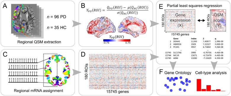
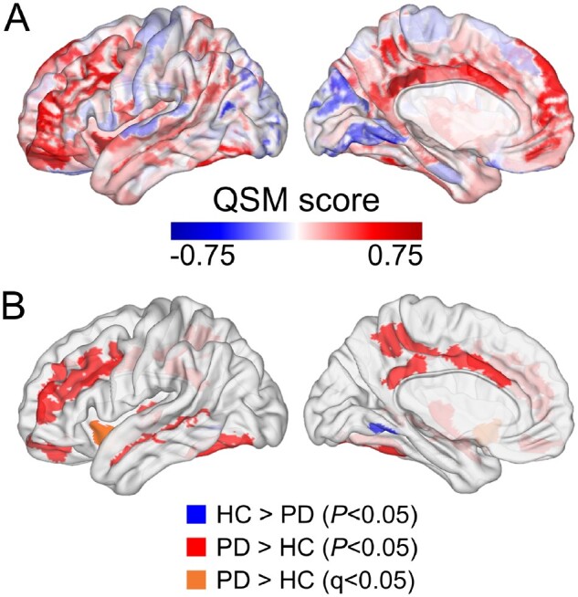
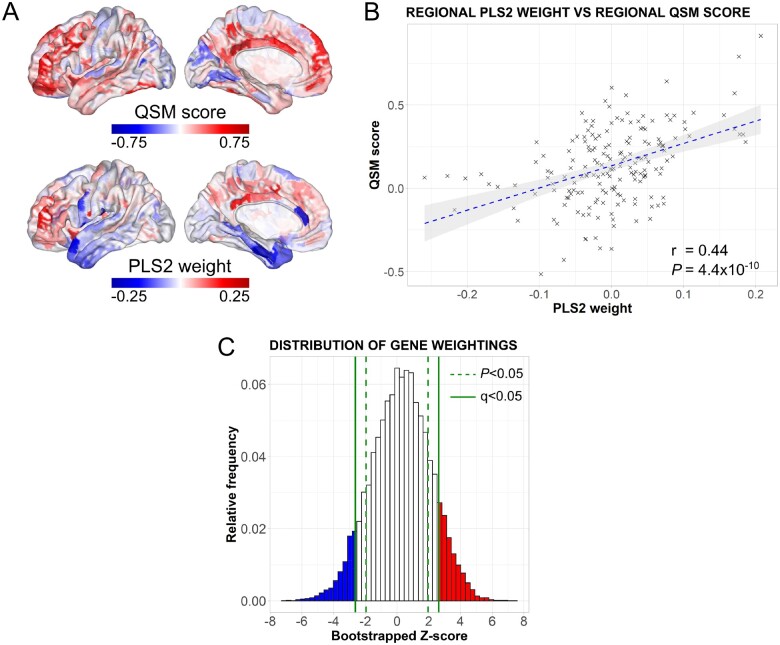
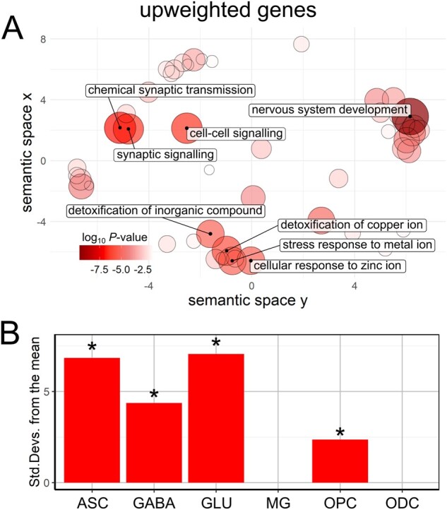
The mechanisms responsible for the selective vulnerability of specific neuronal populations in Parkinson's disease are poorly understood. Oxidative stress secondary to brain iron accumulation is one postulated mechanism. We measured iron deposition in 180 cortical regions of 96 patients with Parkinson's disease and 35 control subjects using quantitative susceptibility mapping. We estimated the expression of 15 745 genes in the same regions using transcriptomic data from the Allen Human Brain Atlas. Using partial least squares regression, we then identified the profile of gene transcription in the healthy brain that underlies increased cortical iron in patients with Parkinson's disease relative to controls. Applying gene ontological tools, we investigated the biological processes and cell types associated with this transcriptomic profile and identified the sets of genes with spatial expression profiles in control brains that correlated significantly with the spatial pattern of cortical iron deposition in Parkinson's disease. Gene ontological analyses revealed that these genes were enriched for biological processes relating to heavy metal detoxification, synaptic function and nervous system development and were predominantly expressed in astrocytes and glutamatergic neurons. Furthermore, we demonstrated that the genes differentially expressed in Parkinson's disease are associated with the pattern of cortical expression identified in this study. Our findings provide mechanistic insights into regional selective vulnerabilities in Parkinson's disease, particularly the processes involving iron accumulation.

Keywords: Parkinson’s disease; genetics; iron; quantitative susceptibility mapping; transcriptomics.


Figures

Figure 1
Figure 1
An overview of the methodology used for regional QSM extraction, estimation of regional gene expression, PLS regression, gene ontological and cell type analyses. (A) Mean, signed QSM values were extracted from 180 left-cortical regions for 96 patients with Parkinson’s disease and 35 controls. (B) A QSM score, YPD, was calculated for each region by a z-score transformation. (C and D) Allen Human Brain Atlas samples of gene expression data were mapped to the 180 left-cortical regions according to the anatomical parcellation and were used to create a matrix containing the average expression of 15 745 genes in those regions. (E) A bootstrapped PLS regression was performed using gene expression (X) as the predictor variable and the QSM score (Y) as the response variable. The second component of X explained maximum variance in Y and bootstrapped z-scores were used to rank each gene’s contribution to this component. (F) Genes that were significant at Q (corrected P) < 0.05 underwent gene ontological analyses for biological processes and expression-weighted cell-type enrichment analyses.
Figure 2
Figure 2
Regional cortical differences in magnetic susceptibility between Parkinson’s disease and controls. (A) The cortical plot of the QSM scores, calculated by z-score transformation of the Parkinson’s disease mean to the control mean for each of the 180 Glasser regions of interest. (B) Glasser regions of interest where a significant difference was observed [blue, controls greater than Parkinson’s disease at P < 0.05; red, Parkinson’s disease greater than controls at P < 0.05; orange, Parkinson’s disease greater than controls at Q (FDR-adjusted P) < 0.05].
Figure 3
Figure 3
Spatial profiles of gene expression and significantly weighted genes associated with cortical iron deposition in Parkinson’s disease. (A) The cortical map of QSM scores had a similar spatial pattern to the regional linearly weighted sum of gene expression scores defined by the PLS2. (B) A scatterplot of regional PLS2 scores versus QSM scores demonstrating a positive correlation; each data point represents one of 180 cortical regions. (C) The distribution of bootstrapped gene weights on PLS2. An FDR inverse quantile transform was used to correct for multiple comparisons, giving a set of 1622 upweighted (red) and 1068 downweighted (blue) genes significant at Q (FDR inverse quantile transform-corrected P) < 0.05 that were used in gene ontological and cell-type analyses.
Figure 4
Figure 4
Enrichment analyses for genes associated with cortical iron deposition in Parkinson’s disease. (A) The gene ontological terms for biological processes that were significantly enriched in significantly upweighted genes defined by PLS2. The terms are plotted in semantic space with more similar terms clustered together. Non-redundant GO terms significant at g:SCS corrected P < 1 × 10⁻⁵ have been labelled in each case. Larger, darker circles indicate greater significance (see colour bar). (B) Expression-weighted cell-type enrichment analyses using the Allen Institute for Brain Science single-cell transcription dataset. Data are presented as standard deviations of the mean expression of upweighted target gene lists from the mean expression of the bootstrap replicates. Cell types in which the target gene lists are significantly enriched are marked with an asterisk (FDR corrected results). ASC = astrocytes; GABA = GABAergic neurons; GLU = glutamatergic neurons; MG = microglia; ODC = oligodendrocytes; OPC = oligodendrocyte precursor cells.

Similar articles

Cited by

References

    1. Hornykiewicz O. Dopamine miracle: From brain homogenate to dopamine replacement. Mov Disord. 2002;17:501-508. - PubMed
    1. Spillantini MG, Schmidt ML, Lee VMY, Trojanowski JQ, Jakes R, Goedert M.. α-synuclein in Lewy bodies. Nature. 1997;388:839-840. - PubMed
    1. Jellinger KA. A critical evaluation of current staging of α-synuclein pathology in Lewy body disorders. Biochim Biophys Acta Mol Basis Dis. 2009;1792:730-740. - PubMed
    1. Surmeier DJ, Obeso JA, Halliday GM.. Selective neuronal vulnerability in Parkinson disease. Nat Rev Neurosci. 2017;18:101-113. - PMC - PubMed
    1. Johnson ME, Stecher B, Labrie V, Brundin L, Brundin P.. Triggers, facilitators, and aggravators: Redefining Parkinson’s disease pathogenesis. Trends Neurosci. 2019;42:4-13. - PMC - PubMed

Publication types