

Evaluation of a variable-aperture full-ring SPECT system using large-area pixelated CZT modules: A simulation study for brain SPECT applications
- PMID: 33704793
- PMCID: PMC8141019
- DOI: 10.1002/mp.14836
Evaluation of a variable-aperture full-ring SPECT system using large-area pixelated CZT modules: A simulation study for brain SPECT applications
Abstract
Purpose: Single photon emission computed tomography (SPECT) scanners using cadmium zinc telluride (CZT) offer compact, lightweight, and improved imaging capability over conventional NaI(Tl)-based SPECT scanners. The main purpose in this study is to propose a full-ring SPECT system design with eight large-area CZT detectors that can be used for a broad spectrum of SPECT radiopharmaceuticals and demonstrate the performance of our system in comparison to the reference conventional NaI(Tl)-based two-head Anger cameras.
Methods: A newly designed full-ring SPECT system is composed of eight large-area CZT cameras (128 mm × 179.2 mm effective area) that can be independently swiveled around their own axes of rotation independently and can have radial motion for varying aperture sizes that can be adapted to different sizes of imaging volume. Extended projection data were generated by conjoining projections of two adjacent detectors to overcome the limited field-of-view (FOV) by each CZT camera. Using Monte Carlo simulations, we evaluated this new system design with digital phantoms including a Derenzo hot rod phantom and a Zubal brain phantom. Comparison of performance metrics such as spatial resolution, sensitivity, contrast-to-noise ratio (CNR), and contrast-recovery ratio was made between our design and conventional SPECT scanners having different pixel sizes and radii of rotation (one clinically well-known type and two arbitrary types matched to our proposed CZT-SPECT geometries).
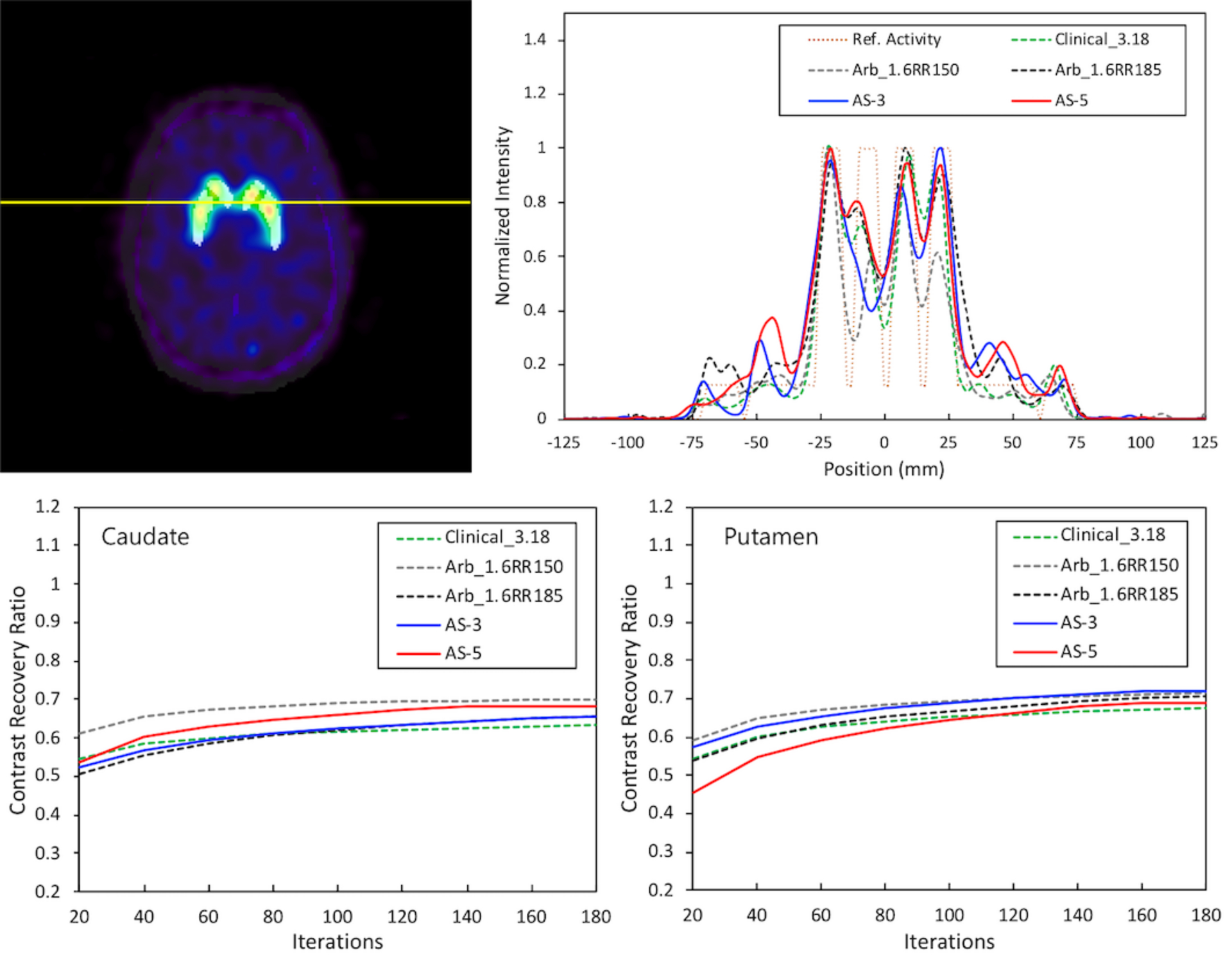
Results: The proposed scanner could result in up to about three times faster in acquisition time over conventional scan time at same acquisition time per step. The spatial resolution improvement, or deterioration, of our proposed scanner compared to the clinical-type scanner was dependent upon the location of the point source. However, there were overall performance improvements over the three different setups of the conventional scanner particularly in volume sensitivity (approximately up to 1.7 times). Overall, we successfully reconstructed the phantom image for both 99m Tc-based perfusion and 123 I-based dopamine transporter (DaT) brain studies simulated for our new design. In particular, the striatal/background contrast-recovery ratio in 3-to-1 reference ratio was over 0.8 for the 123 I-based DaT study.
Conclusions: We proposed a variable-aperture full-ring SPECT system using combined pixelated CZT and energy-optimized parallel-hole collimator modules and evaluated the performance of this scanner using relevant digital phantoms and MC simulations. Our studies demonstrated the potential of our new full-ring CZT-SPECT design, showing reduced acquisition time and improved sensitivity with acceptable CNR and spatial resolution.
Keywords: CZT detector; Monte Carlo simulation; SPECT; brain SPECT; full ring.
© 2021 American Association of Physicists in Medicine.
Conflict of interest statement
CONFLICT OF INTEREST
The authors have no conflict to disclose.
Figures


References
-
- Chowdhury FU, Scarsbrook AF. The role of hybrid SPECT-CT in oncology: current and emerging clinical applications. Clin Radiol. 2008;63(3):241–251. - PubMed
-
- Wu J, Liu C. Recent advances in cardiac SPECT instrumentation and imaging methods. Phys Med Biol. 2019;64(6):06TR01. - PubMed
-
- Zhang L, Villalobos A. Recent advances in the development of PET and SPECT tracers for brain imaging. In: Annual Reports in Medicinal Chemistry. Vol 47. Elsevier; 2012:105–119.
-
- Anger H. A new instrument for mapping gamma-ray emitters. Biology and Medicine Quarterly Report UCRL. 1957;3653:38.
-
- Anger HO. Scintillation camera with multichannel collimators. Journal of Nuclear Medicine. 1964;5(7):515–531. - PubMed
MeSH terms
Substances
Grants and funding
LinkOut - more resources
Full Text Sources
Other Literature Sources
