. 2021 May;45(5):934-947.
doi: 10.1111/acer.14589. Epub 2021 Apr 3.

Alcohol-associated intestinal dysbiosis alters mucosal-associated invariant T-cell phenotype and function

Affiliations

Alcohol-associated intestinal dysbiosis alters mucosal-associated invariant T-cell phenotype and function

Min Gu et al. Alcohol Clin Exp Res. 2021 May.

Abstract

Background: Chronic alcohol consumption is associated with a compromised innate and adaptive immune responses to infectious disease. Mucosa-associated invariant T (MAIT) cells play a critical role in antibacterial host defense. However, whether alcohol-associated deficits in innate and adaptive immune responses are mediated by alterations in MAIT cells remains unclear.

Methods: To investigate the impact of alcohol on MAIT cells, mice were treated with binge-on-chronic alcohol for 10 days and sacrificed at day 11. MAIT cells in the barrier organs (lung, liver, and intestine) were characterized by flow cytometry. Two additional sets of animals were used to examine the involvement of gut microbiota on alcohol-induced MAIT cell changes: (1) Cecal microbiota from alcohol-fed (AF) mice were adoptive transferred into antibiotic-pretreated mice and (2) AF mice were treated with antibiotics during the experiment. MAIT cells in the barrier organs were measured via flow cytometry.

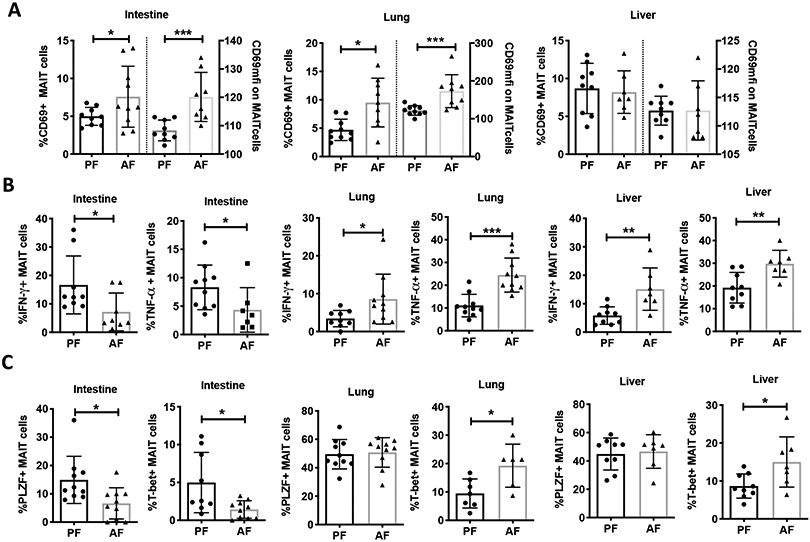
Results: Binge-on-chronic alcohol feeding led to a significant reduction in the abundance of MAIT cells in the barrier tissues. However, CD69 expression on tissue-associated MAIT cells was increased in AF mice compared with pair-fed (PF) mice. The expression of Th1 cytokines and the corresponding transcriptional factor was tissue specific, showing downregulation in the intestine and increases in the lung and liver in AF animals. Transplantation of fecal microbiota from AF mice resulted in a MAIT cell profile aligned to that of AF mouse donor. Antibiotic treatment abolished the MAIT cell differences between AF and PF animals.

Conclusion: MAIT cells in the intestine, liver, and lung are perturbed by alcohol use and these changes are partially attributable to alcohol-associated dysbiosis. MAIT cell dysfunction may contribute to alcohol-induced innate and adaptive immunity and consequently end-organ pathophysiology.

Keywords: Mucosa-associated invariant T cells; alcohol; antibiotics; dysbiosis; fecal transplantation; gut microbiota.


Conflict of interest statement

CONFLICT OF INTEREST

The authors declare no competing financial interests.

Figures

Figure 1:
Figure 1:
Binge-on chronic alcohol reduces MAIT cell number in the intestine, lung and liver. (A) Experimental design of binge-on chronic alcohol exposure on mice. (B) Gating strategy for mouse MAIT cell identification by flow cytometry. (C) MAIT cell count in alcohol drinking mice (counted by flow cytometry and adjusted by counting beads). PF, pair-fed; AF, alcohol-fed. Scatter plots and bar shows the means±SD. * p<0.05, ** p<0.01, *** p<0.001 by Mann-Whitney U test.
Figure 2:
Figure 2:
Binge-on chronic alcohol alters MAIT cell activation, and expression of cytokines and transcriptional factors. (A) MAIT cell expression of activation marker CD69 and CD69 mean fluorescent intensity (MFI) (B) Th1 cytokine expression (IFN-γ and TNF-a), and (C) frequency of transcriptional factor (PLZF and T-bet) expression on MAIT cells isolated from intestine, lung, and liver of pair fed (PF) and alcohol-fed (AF) mice. MFI: mean fluorescence intensity. Scatter plots and bar shows the means±SD. * p<0.05, ** p<0.01, *** p<0.001 by Mann-Whitney U-test.
Figure 3:
Figure 3:
Impact of binge-on chronic alcohol on gut microbiota diversity and function. (A) α-diversity of cecal microbiota from mice with/without alcohol exposure. Observed_OTUs, Chao1 and ACE are estimators of microbiota species richness. (B) β-diversity of cecal microbiota based on sample-wise Bray-Curtis dissimilarity distance. The significance between the PF and AF group was calculated with pairwise ADONIS2 (permutation=10000, p=0.007). Red circles were samples from AF group, and blue triangles were samples from PF group. Principal component (PC) value indicates the percent variation showed by the PC according to the linear correlation between sample microbiota taxonomy and identity. PC1 and PC2 showed 61.4% of the total difference between the two groups. (C) Principle component analysis (PCA) of the predicted functions of cecal microbiota estimated by package PICRUSt2 in R. AF samples clustered separately from the PF, and PC1 and PC2 showed 78% of total difference. Orange squares represent samples from PF mice, and blue circles represent samples from AF mice. (D) Fold change of the bacterial metabolic pathway of purine ribonucleotides degradation. (E) Concentration of riboflavin released in the culture broth by cecal microbiota after 48 h fermentation with sterile Gifu anaerobic medium. Scatter plots and bar shows the means±SD. * p<0.05 by Mann-Whitney U-test.
Figure 4:
Figure 4:
Alcohol dysbiosis incubates with human PBMCs (Bpc=10) for 4 h causing MAIT cell changes in vitro . (A) Representative flow cytometric pseudocolour plots of MAIT cell identification and the distribution of CD4/CD8 receptors on MAIT cells (B) MAIT cell number in human PBMCs after 4 h incubation. (C) The expression of CD38 and CD38 MFI on CD8+ MAIT cells. CD8+ cell is about 80-90% of total MAIT cells in healthy human PBMCs. (D) The expression of granzyme B and IFN-γ on CD8+ MAIT cells after alcohol dysbiosis stimulation. Bpc: bacteria per cell. Scatter plots and bar shows the means±SD. * p<0.05 and ** <0.01 by Mann-Whitney U-test.
Figure 5:
Figure 5:
Alcohol dysbiosis replicates the effect of alcohol feeding on MAIT cells in alcohol naïve mice. (A) Experimental design of mouse to mouse gut microbiota adoptive transfer. After 14 days’ antibiotics treatment, mice received 3 doses of cecal microbiota by gavage. Donor microbiota was collected from binge-on chronic alcohol experiment, and recipients were fed with Lieber-Decarli control liquid diet. (B) MAIT cell number in mice tissues after microbiota recolonization. PF microbiota: mice recolonized with cecal microbiota from pair-fed mice; AF microbiota: mice recolonized with cecal microbiota from alcohol drinking mice. (C) MAIT cell expression of CD69 and CD69 MFI in mice tissues. (D) MAIT cell expression of cytokine IFN-γ and TNF-a in microbiota reconstituted mice. (E) MAIT cell expression of transcriptional factors PLZF and T-bet in tissues. Scatter plots and bar shows the means±SD. * p<0.05 and ** p<0.01 by Mann-Whitney U-test.
Figure 6:
Figure 6:
Binge-on chronic alcohol has limited effects on MAIT cell number, activation, cytokines and transcriptional factor expression in antibiotic-treated mice. (A) Schematic of mice exposure to antibiotics and binge-on chronic alcohol. (B) MAIT cell number, (C) the frequency of CD69 and CD69 MFI on MAIT cells, (D) MAIT cell expression of cytokine IFN-γ and TNF-a, (E) MAIT cell expression of transcriptional factors PLZF and T-bet in the intestine, lung and liver in mice exposed to antibiotics plus binge-on chronic alcohol. PF+Abx, mice exposed to antibiotics and liquid control diet; AF+Abx, mice exposed to and antibiotics and binge-on chronic alcohol. Scatter plots and bar shows the means±SD. Statistical difference was calculated by Mann-Whitney U-test between the two groups.
Figure 7:
Figure 7:
Alcohol increases gut epithelial injury and AF mice serum stimulates blood MAIT cells. Human PBMCs were incubated for 4 h with mice serum (10%, v/v) collected from binge-on chronic alcohol experiment. (A) Serum level of iFABP and circulating bacterial 16S rRNA gene copies in binge-on chronic alcohol exposed mice. (B) MAIT cell number in PBMCs after 4 h incubation. (C) Expression of CD38 and CD38 MFI on CD8+MAIT cells. (D) Production of Granzyme B and IFN-γ by CD8+ MAIT cells. iFABP, intestinal fatty acid binding protein. Scatter plots and bar shows the means±SD. * p<0.05, ** <0.01, and *** p<0.001 by Mann-Whitney U-test.
Figure 8:
Figure 8:
Serum from alcohol-fed mice promotes MAIT cell apoptosis. The frequency of early (Annexin V+Live/dead−), late apoptosis (Annexin V+Live/dead+) and necroptosis (Annexin V-Live/dead+) was measured in MAIT cells, CD4+ and CD8+ T cells by flow cytometry. (A) Representative flow cytometry dot plots of frequency of apoptotic cells (early + late apoptosis and necroptosis) among MAIT cells after co-culture with PF and AF serum for 4 h. (B) Fold change (AF vs. PF) of the frequency of apoptotic cells. (C) Representative dot plots of the frequency of apoptotic cells among MAIT cells, CD4+ and CD8+ T cell. (D) Percentage of live cells among MAIT cells, CD4+ and CD8+ T cells. Scatter plots and bar shows the means±SD. * p<0.05, ** <0.01, and **** p<0.0001 by Mann-Whitney U-test.

References

    1. ADRIAANSE MPM, TACK GJ, PASSOS VL, DAMOISEAUX JGMC, SCHREURS MWJ, VAN WIJCK K, RIEDL RG, MASCLEE AAM, BUURMAN WA, MULDER CJJ & VREUGDENHIL ACE 2013. Serum IFABP as marker for enterocyte damage in coeliac disease and its relation to villous atrophy and circulating autoantibodies. Alimentary Pharmacology & Therapeutics, 37, 482–490. - PubMed
    1. ALFONSO-LOECHES S, PASCUAL M & GUERRI C 2013. Gender differences in alcohol-induced neurotoxicity and brain damage. Toxicology, 311, 27–34. - PubMed
    1. AMOROSO C, PERILLO F, STRATI F, FANTINI MC, CAPRIOLI F & FACCIOTTI F 2020. The Role of Gut Microbiota Biomodulators on Mucosal Immunity and Intestinal Inflammation. Cells, 9. - PMC - PubMed
    1. BACHEM A, MAKHLOUF C, BINGER KJ, DE SOUZA DP, TULL D, HOCHHEISER K, WHITNEY PG, FERNANDEZ-RUIZ D, DAHLING S, KASTEMULLER W, JONSSON J, GRESSIER E, LEW AM, PERDOMO C, KUPZ A, FIGGETT W, MACKAY F, OLESHANSKY M, RUSS BE, PARISH IA, KALLIES A, MCCONVILLE MJ, TUMER SJ, GEBHARDT T & BEDOUI S 2019. Microbiota-Derived Short-Chain Fatty Acids Promote the Memory Potential of Antigen-Activated CD8(+) T Cells. Immunity, 51, 285-+. - PubMed
    1. BELKAID Y & HAND TW 2014. Role of the microbiota in immunity and inflammation. Cell, 157, 121–41. - PMC - PubMed

Publication types

MeSH terms