. 2021 Nov-Dec;18(6):2775-2780.
doi: 10.1109/TCBB.2021.3065361. Epub 2021 Dec 8.

Deep Learning Enables Accurate Diagnosis of Novel Coronavirus (COVID-19) With CT Images

Deep Learning Enables Accurate Diagnosis of Novel Coronavirus (COVID-19) With CT Images

Ying Song et al. IEEE/ACM Trans Comput Biol Bioinform. 2021 Nov-Dec.

Abstract

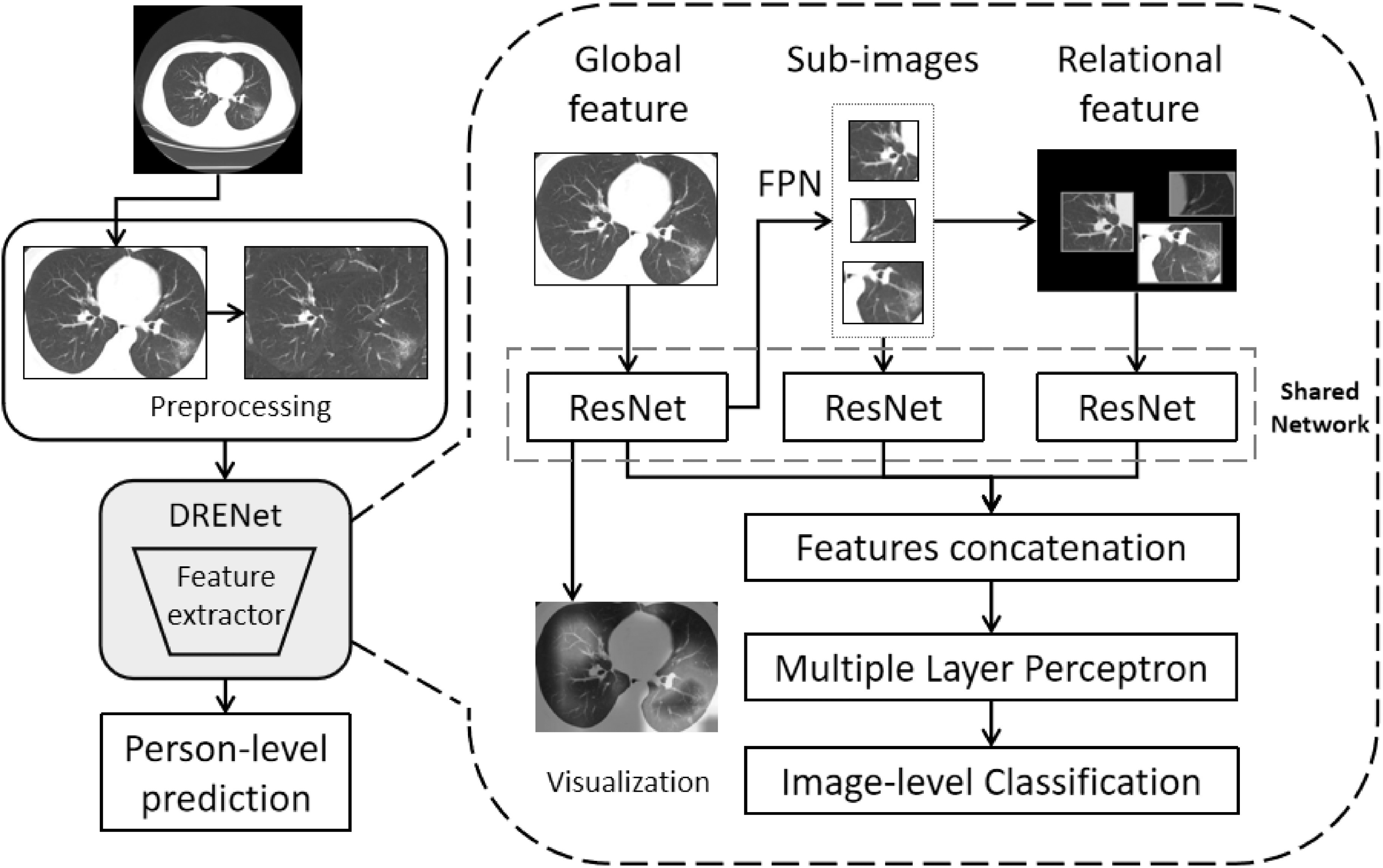
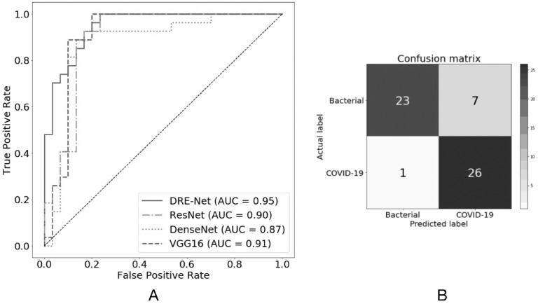
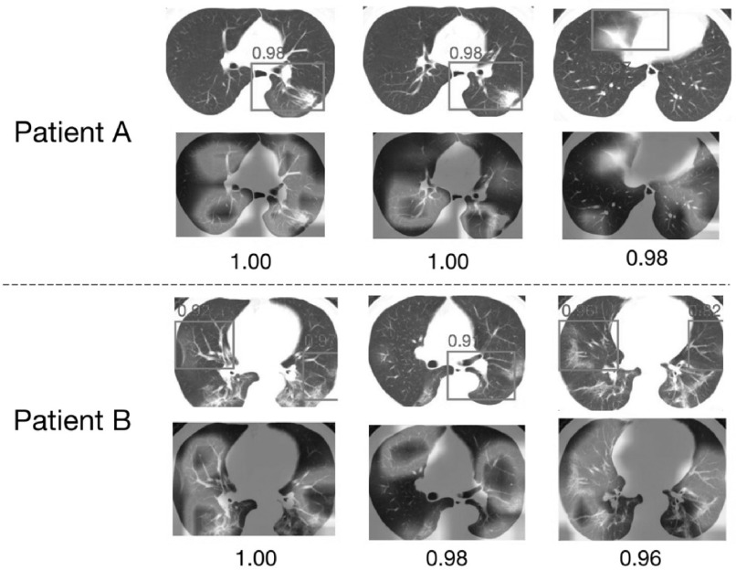
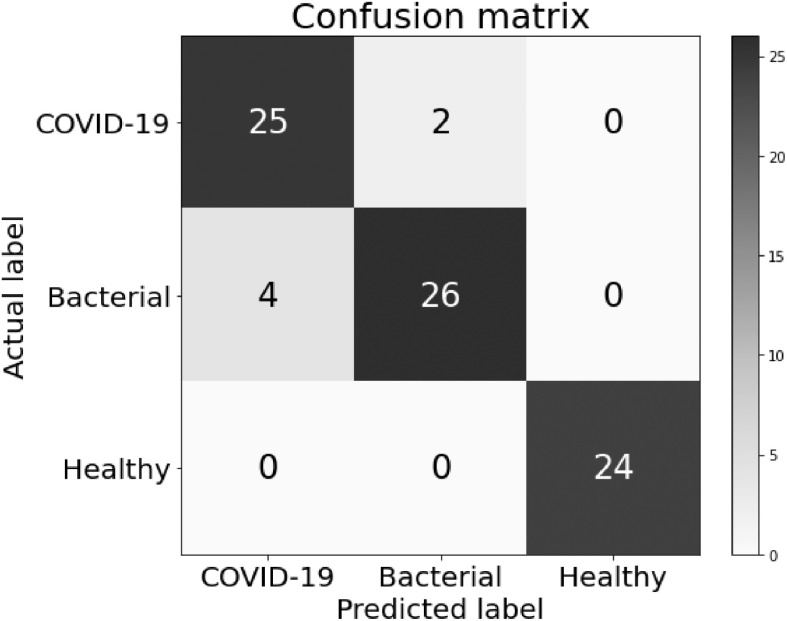
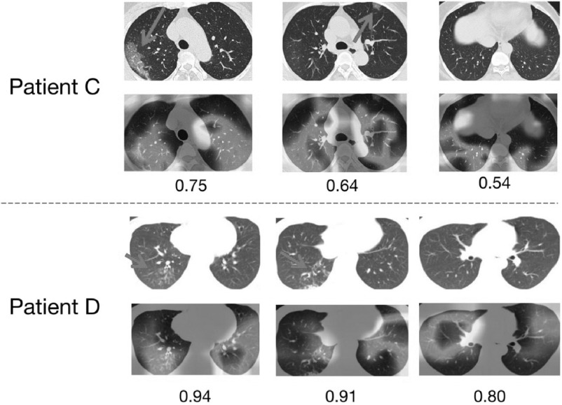
A novel coronavirus (COVID-19) recently emerged as an acute respiratory syndrome, and has caused a pneumonia outbreak world-widely. As the COVID-19 continues to spread rapidly across the world, computed tomography (CT) has become essentially important for fast diagnoses. Thus, it is urgent to develop an accurate computer-aided method to assist clinicians to identify COVID-19-infected patients by CT images. Here, we have collected chest CT scans of 88 patients diagnosed with COVID-19 from hospitals of two provinces in China, 100 patients infected with bacteria pneumonia, and 86 healthy persons for comparison and modeling. Based on the data, a deep learning-based CT diagnosis system was developed to identify patients with COVID-19. The experimental results showed that our model could accurately discriminate the COVID-19 patients from the bacteria pneumonia patients with an AUC of 0.95, recall (sensitivity) of 0.96, and precision of 0.79. When integrating three types of CT images, our model achieved a recall of 0.93 with precision of 0.86 for discriminating COVID-19 patients from others. Moreover, our model could extract main lesion features, especially the ground-glass opacity (GGO), which are visually helpful for assisted diagnoses by doctors. An online server is available for online diagnoses with CT images by our server (http://biomed.nscc-gz.cn/model.php). Source codes and datasets are available at our GitHub (https://github.com/SY575/COVID19-CT).


Figures

Fig. 1.
Fig. 1.
The proposed training architecture including (1) Preprocessing: the CT images are first preprocessed to remove boundary regions around the lungs, and to further fill the blank regions around two lungs with its rotational lung areas (for model training only). (2) Image-level classifications by DRENet: each CT image is input to the pre-trained ResNet50 to extract global features, from which FPN module is employed to identify top-K lesion regions. According to the top-K regions, the shared ResNet is utilized again to extract local features within the sub-images and relational features between the sub-images. These features are concatenated with the learned global features to input into MLP for the image-level prediction. (3) Person-level prediction: the image-level predictions will be aggregated for person-level prediction.
Fig. 2.
Fig. 2.
Performances of different networks on the pneumonia diagnosis with A) Receiver operating characteristic curves for the diagnosis of COVID-19, and B) Confusion matrix of the DRENet on the test set.
Fig. 3.
Fig. 3.
Visualization of two correctly diagnosed nCOV-19 pneumonia patients. For each patient, we showed the top 3 predicted slices and the extracted details (bounding boxes with red color) with normalized predicted scores above 0.8 (ranging from 0 to 1). Moreover, we also used the method proposed by to draw the heat map.
Fig. 4.
Fig. 4.
Confusion matrix of the DRENet on the pneumonia three-class classification.
Fig. 5.
Fig. 5.
Visualization of two wrongly predicted cases on the pneumonia classification for A) one patient of bacterial pneumonia wrongly predicted as the COVID-19 patient, and B) one COVID-19 patient wrongly predicted as bacterial pneumonia. Arrows were added by hand to point to regions containing GGO abnormality.

References

    1. Huang C., et al. , “Clinical features of patients infected with 2019 novel Coronavirus in Wuhan, China,” Lancet, vol. 395, no. 10223, pp. 497–506, 2020. - PMC - PubMed
    1. WHO website WM, Accessed: Feb. 5, 2020. [Online]. Available: https://www.who.int
    1. Lu R., et al. , “Genomic characterisation and epidemiology of 2019 novel coronavirus: Implications for virus origins and receptor binding,” Lancet, vol. 395, 565–574, 2020. - PMC - PubMed
    1. Li Q., et al. , “Early transmission dynamics in Wuhan, China, of novel Coronavirus–infected pneumonia,” New Engl. J. Med., vol. 382, pp. 1199–1207, 2020. - PMC - PubMed
    1. Hui D. S., et al. , “The continuing 2019-nCoV epidemic threat of novel Coronaviruses to global health–The latest 2019 novel Coronavirus outbreak in Wuhan, China,” Int. J. Infect. Dis., vol. 91, pp. 264–266, 2020. - PMC - PubMed

Publication types