. 2021 Jun;191(6):1020-1035.
doi: 10.1016/j.ajpath.2021.02.018. Epub 2021 Mar 8.

The Canonical Wnt Signaling Pathway Inhibits the Glucocorticoid Receptor Signaling Pathway in the Trabecular Meshwork

Affiliations

The Canonical Wnt Signaling Pathway Inhibits the Glucocorticoid Receptor Signaling Pathway in the Trabecular Meshwork

Chenna Kesavulu Sugali et al. Am J Pathol. 2021 Jun.

Abstract

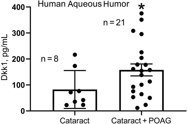
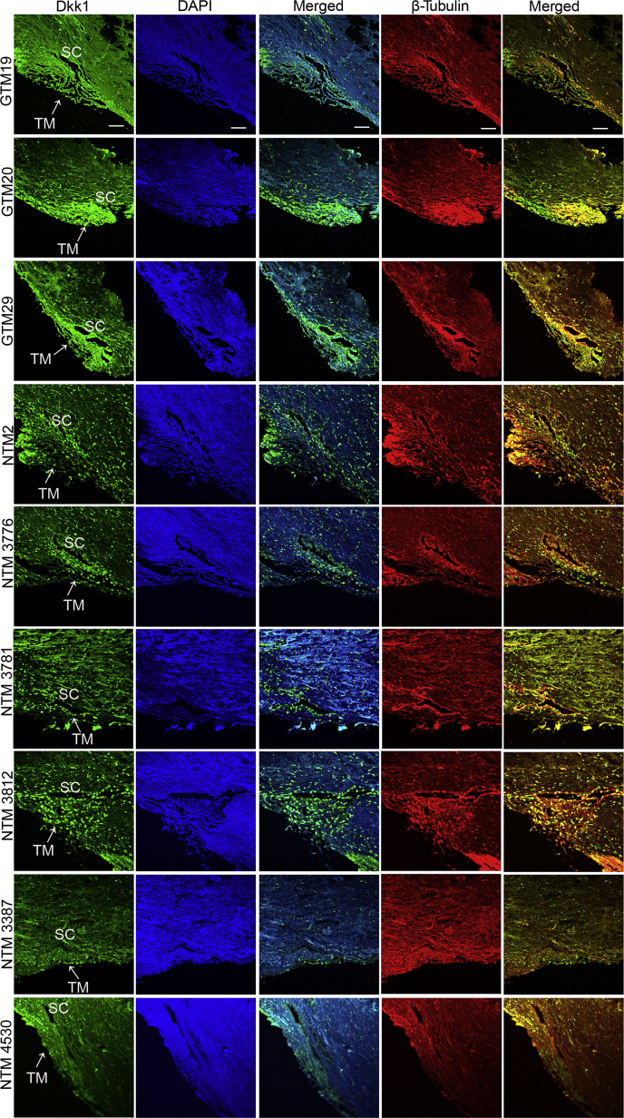
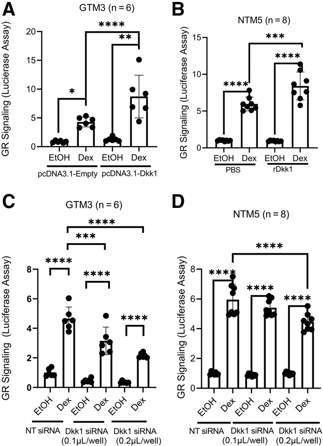
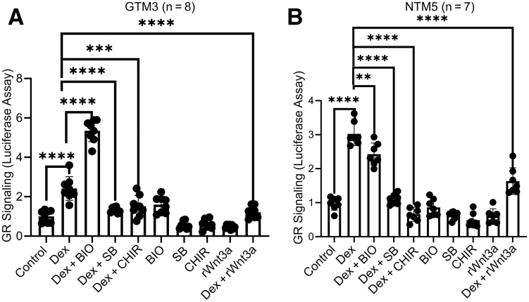
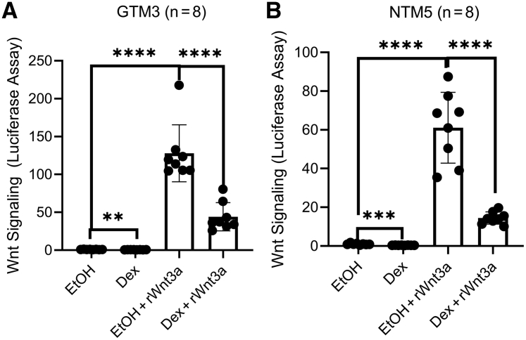
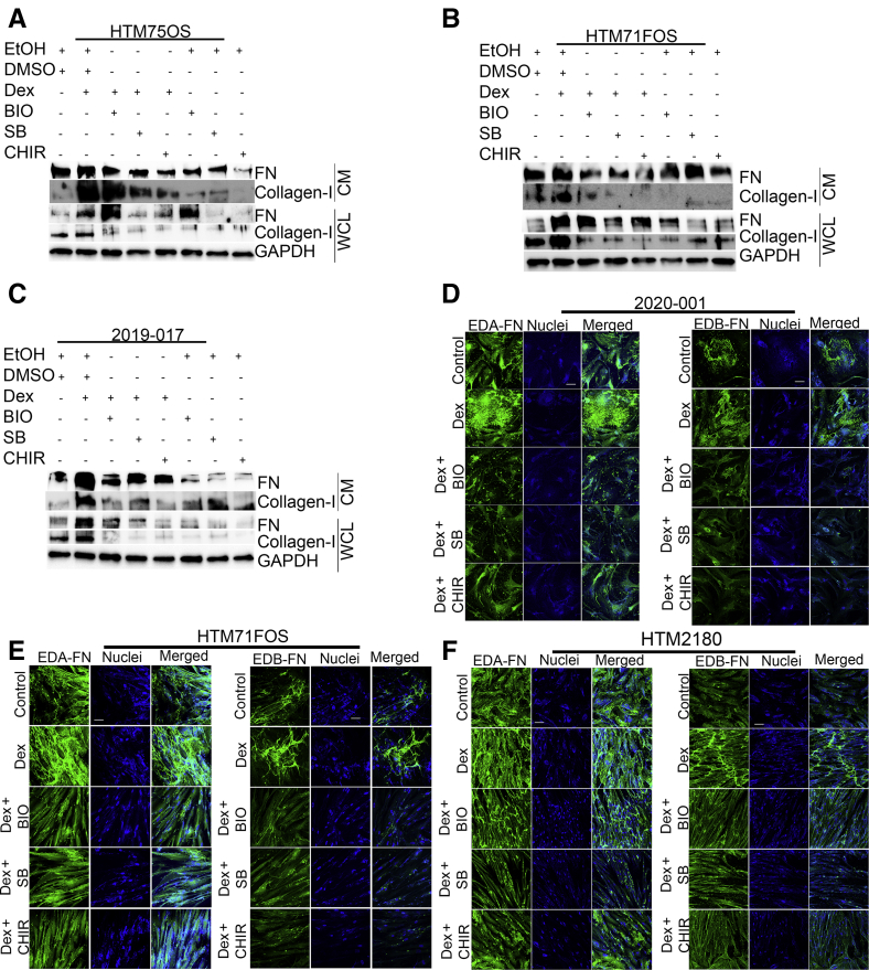
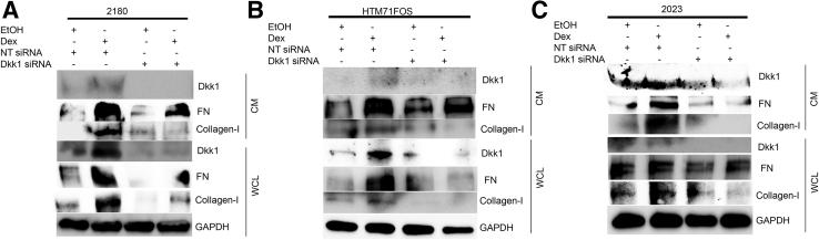
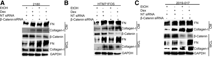
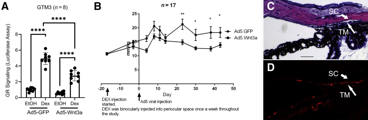
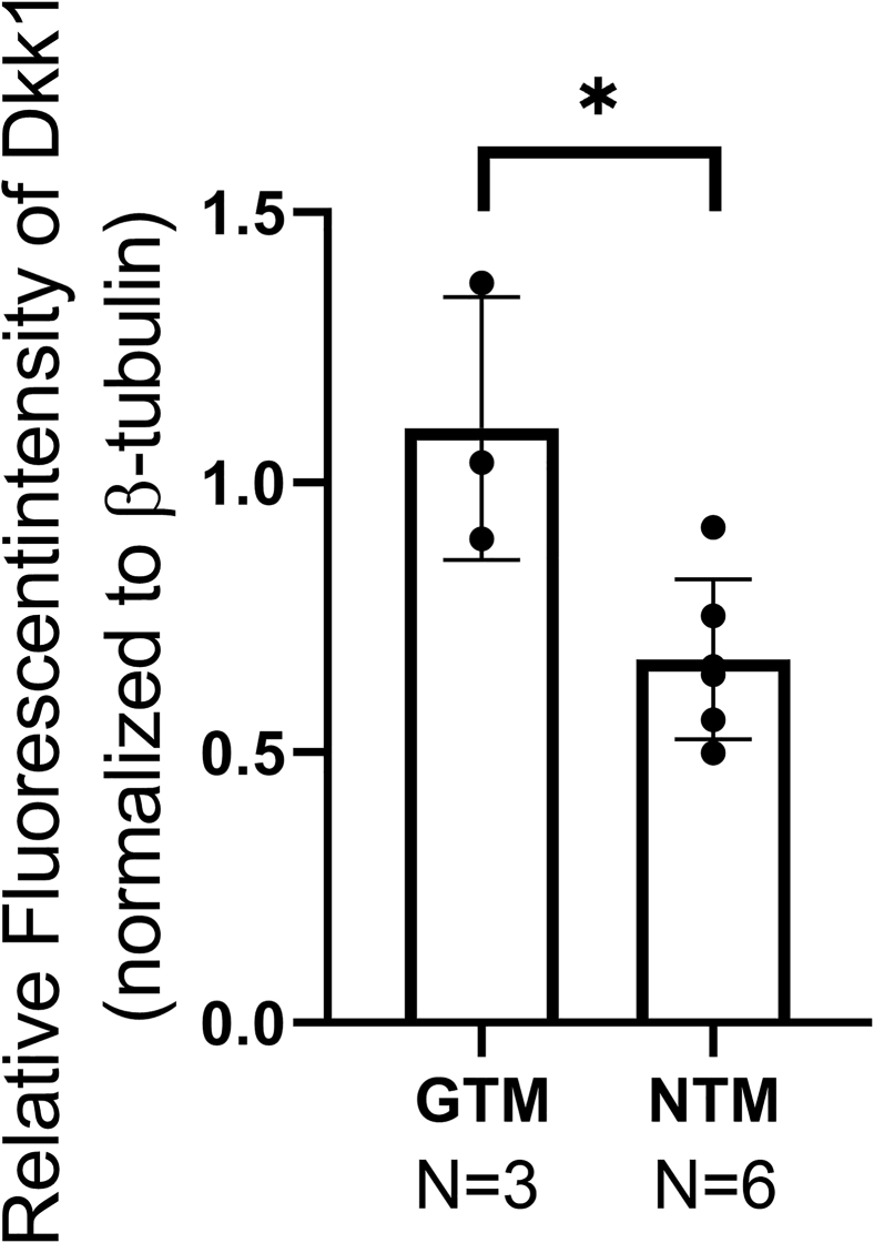
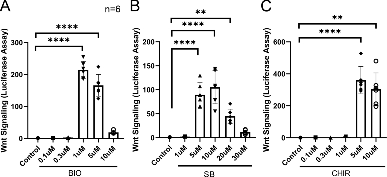
Glucocorticoid-induced glaucoma is a secondary open-angle glaucoma. About 40% of the general population may develop elevated intraocular pressure on prolonged glucocorticoid treatment secondary to damages in the trabecular meshwork (TM), a tissue that regulates intraocular pressure. Therefore, identifying the key molecules responsible for glucocorticoid-induced ocular hypertension is crucial. In this study, Dickkopf-related protein 1 (Dkk1), a canonical Wnt signaling inhibitor, was found to be elevated in the aqueous humor and TM of glaucoma patients. At the signaling level, Dkk1 enhanced glucocorticoid receptor (GR) signaling, whereas Dkk1 knockdown or Wnt signaling activators decreased GR signaling in human TM cells as indicated by luciferase assays. Similarly, activation of the GR signaling inhibited Wnt signaling. At the protein level, glucocorticoid-induced extracellular matrix was inhibited by Wnt activation using Wnt activators or Dkk1 knockdown in primary human TM cells. In contrast, inhibition of canonical Wnt signaling by β-catenin knockdown increased glucocorticoid-induced extracellular matrix proteins. At the physiological level, adenovirus-mediated Wnt3a expression decreased glucocorticoid-induced ocular hypertension in mouse eyes. In summary, Wnt and GR signaling inhibit each other in the TM, and canonical Wnt signaling activators may prevent the adverse effect of glucocorticoids in the eye.


Figures

Figure 1
Figure 1
Dkk1 was elevated in the aqueous humor of primary open-angle glaucoma (POAG) eyes. Aqueous humor samples were collected from cataract and cataract + POAG patients during surgeries. Dkk1 levels were measured using enzyme-linked immunosorbent assay. Data are given as means ± SEM. N = 8 for the cataract group and N= 21 for the cataract+POAG group. ∗P < 0.05 (t-test).
Figure 2
Figure 2
Dkk1 was elevated in the trabecular meshwork (TM) tissue of primary open-angle glaucoma (POAG) eyes. Three glaucomatous TM (GTM) tissues removed during surgeries and six non-glaucomatous TM (NTM) tissues from donor eyes were immunostained with anti-Dkk1 antibody (green) or β-tubulin (red). β-Tubulin was used as a quality control because donor eyes were not as fresh as POAG TM tissues (hours versus days). Nuclei were stained with DAPI (blue). Scale bar = 20 μm. SC, Schlemm canal.
Figure 3
Figure 3
Dkk1 plays a positive role in glucocorticoid receptor (GR) signaling in the human trabecular meshwork (TM). GTM3 cells (A and C) were cotransfected with the glucocorticoid receptor signaling reporter vector together with pcDNA3.1-empty/Dkk1 vector or nontargeting (NT) siRNA/Dkk1 siRNA to overexpress (A) or knock down (C) Dkk1, respectively. Similarly, GRE-NTM5 (B and D), which were stably transduced with glucocorticoid receptor signaling reporter cassettes, were treated/transfected with phosphate-buffered saline (PBS)/recombinant Dkk1 (rDkk1; 200 ng/mL) or NT siRNA/Dkk1 siRNA. All cells were treated with 0.1% ethanol (EtOH) or 100 nmol/L dexamethasone (Dex) before luciferase assays. Data were analyzed using one-way analysis of variance and Sidak post-hoc tests. Data are given as means ± SD of GR signaling activity (AD). N = 6 (A and C); N = 8 (B and D). ∗P < 0.05, ∗∗P < 0.01, ∗∗∗P < 0.001, and ∗∗∗∗P < 0.0001.
Figure 4
Figure 4
Wnt signaling inhibits glucocorticoid receptor (GR) signaling in the human trabecular meshwork (TM). GRE-GTM3 (A) and GRE-NTM5 (B) cells were treated with or without indicated glycogen synthase kinase 3β inhibitors [1 μmol/L 6-bromoindirubin-3′-oxime (BIO), 10 μmol/L SB-216763 (SB), or 5 μmol/L CHIR99021 (CHIR)] together with 0.1% ethanol [vehicle for dexamethasone (Dex)] and/or 100 nmol/L Dex before luciferase assays. Data were analyzed using one-way analysis of variance and Sidak post-hoc tests. Data are given as means ± SD of GR signaling activity (A and B). N = 8 (A); N = 7 (B). ∗∗P < 0.01, ∗∗∗P < 0.001, and ∗∗∗∗P < 0.0001. rWnt3a, recombinant Wnt3a.
Figure 5
Figure 5
Glucocorticoid receptor signaling inhibits Wnt signaling in the human trabecular meshwork (TM). GTM3 (A) and NTM5 (B) cells were transfected with the Wnt signaling reporter vector and treated with 0.1% ethanol (EtOH) or 100 nmol/L dexamethasone (Dex) with or without 100 ng/mL Wnt3a for 72 hours before luciferase assays. Data were analyzed using one-way analysis of variance and Sidak post-hoc tests. Data are given as means ± SD of canonical Wnt signaling activity (A and B). N = 8 (A and B). ∗∗P < 0.01, ∗∗∗P < 0.001, and ∗∗∗∗P < 0.0001. rWnt3a, recombinant Wnt3a.
Figure 6
Figure 6
Activation of Wnt signaling inhibits dexamethasone (Dex)–induced extracellular matrix in primary human trabecular meshwork (HTM) cells. HTM75OS (A), HTM71FOS (B), and 2019-017 (C) primary HTM cells were treated with 0.1% ethanol (EtOH; vehicle), dimethyl sulfoxide (DMSO; vehicle), 100 nmol/L Dex, 1 μmol/L 6-bromoindirubin-3′-oxime (BIO), 10 μmol/L SB-216763 (SB), and/or 5 μmol/L CHIR99021 (CHIR) for 72 hours. Fibronectin (FN) and collagen-I from conditioned medium (CM) and whole cell lysate (WCL) were used for Western immunoblotting. Glyceraldehyde-3-phosphate dehydrogenase (GAPDH) served as a loading control for WCL. The 2020-001 (D), HTM71FOS (E), and HTM2180 (F) primary HTM cells were treated with 0.1% EtOH (vehicle), DMSO (vehicle), 100 nmol/L Dex, and Dex + glycogen synthase kinase 3β inhibitors (1 μmol/L BIO, 10 μmol/L SB, and/or 5 μmol/L CHIR) for 12 to 14 days, and were immunostained with anti–EDA-FN or EDB-FN antibodies (green). Nuclei were stained using Hoechst-33342 and/or DAPI (blue). Scale bar = 20 μm (DF).
Figure 7
Figure 7
Dkk1 knockdown inhibits extracellular matrix in primary human trabecular meshwork (HTM) cells. HTM 2180 (A), HTM71FOS (B), and HTM2023 (C) cells were transfected with nontargeting (NT) siRNA/Dkk1 siRNA. The cells were treated with 0.1% ethanol (EtOH) or 100 nmol/L dexamethasone (Dex) for 3 days, and conditioned medium (CM) and whole cell lysate (WCL) were harvested for Western immunoblotting. Glyceraldehyde-3-phosphate dehydrogenase (GAPDH) served as a loading control for WCL. FN, fibronectin.
Figure 8
Figure 8
Inhibition of canonical Wnt signaling increases extracellular matrix in primary human trabecular meshwork (HTM) cells. HTM 2180 (A), HTM71FOS (B), and 2019-017 (C) HTM cells were transfected with nontargeting (NT) siRNA/β-catenin siRNA. The cells were treated with 0.1% ethanol (EtOH) or 100 nmol/L dexamethasone (Dex) for 2 days, and conditioned medium (CM) as well as whole cell lysate (WCL) were harvested for Western immunoblotting. Glyceraldehyde-3-phosphate dehydrogenase (GAPDH) served as a loading control for WCL. FN, fibronectin.
Figure 9
Figure 9
Wnt3a reduces ocular hypertension in dexamethasone (Dex)–treated mice. A: GTM3 cells were transfected with the glucocorticoid receptor (GR) signaling reporter vector and transduced with the Ad5-CMV-GFP or Ad5-CMV-Wnt3a+mCherry before luciferase assays. Data were analyzed using one-way analysis of variance and Sidak post-hoc tests. B: Three-month–old female C57BL/6J mice received binocular periocular Dex-acetate (Dex-Ac) injection once a week throughout the study (arrow on the left) after baseline intraocular pressure (IOP) establishment. After ocular hypertension development, Ad5-CMV-GFP was injected intravitreally into one eye and Ad5-CMV-Wnt3a+mCherry was injected into the fellow eye (arrow on the right). Mouse IOP was measured under isoflurane anesthesia about once a week throughout the study in a masked manner. Paired t-test was used to compare IOP at each time point. C: Hematoxylin and eosin (H&E) staining of a mouse eye section transduced with Ad5-Wnt3a-mCherry. D: Immunostaining of a section adjacent to C using the anti-mCherry antibody (red; pseudocolor). Data are given as means ± SD of GR signaling activity (A) or means ± SEM of IOP (B). N = 8 (A); N = 17 (B). ∗P < 0.05; ∗∗P < 0.01, and ∗∗∗∗P < 0.0001. Scale bar = 40 μm (C).
Supplemental Figure S1
Fluorescent intensity of Dkk1 protein expression in glaucomatous trabecular meshwork (GTM) and non-glaucomatous trabecular meshwork (NTM). The TM region, as shown in Figure 2, was manually selected, and the fluorescent intensity of Dkk1 and β-tubulin was measured using the ImageJ version 1.8.0_172 software. Dkk1 fluorescent intensity was normalized to β-tubulin fluorescent intensity (relatively fluorescent intensity), and the data were plotted. Data were analyzed using t-test. Data are given as means ± SD of Dkk1 relative fluorescent intensity. N = 3 for GTM; N = 6 for NTM. ∗P < 0.05.
Supplemental Figure S2
Dose-response study of glycogen synthase kinase (GSK) 3β inhibitor concentrations. GTM3 cells were transfected with the Wnt signaling reporter vector and treated without or with indicated GSK3β inhibitors at various concentrations before luciferases assays. A: 6-Bromoindirubin-3′-oxime (BIO). B: SB-216763 (SB). C: CHIR99021 (CHIR). Data were analyzed using one-way analysis of variance and Sidak post-hoc tests. Data are given as means ± SD of canonical Wnt signaling activity (AC). N = 6 (AC). ∗∗P < 0.01, ∗∗∗∗P < 0.0001.

References

    1. Resnikoff S., Pascolini D., Etya'ale D., Kocur I., Pararajasegaram R., Pokharel G.P., Mariotti S.P. Global data on visual impairment in the year 2002. Bull World Health Organ. 2004;82:844–851. - PMC - PubMed
    1. Investigators T.A., The AGIS Investigators The Advanced Glaucoma Intervention Study (AGIS): 7: the relationship between control of intraocular pressure and visual field deterioration. Am J Ophthalmol. 2000;130:429–440. - PubMed
    1. Clark A.F., Wordinger R.J. The role of steroids in outflow resistance. Exp Eye Res. 2009;88:752–759. - PubMed
    1. Armaly M.F. Effect of corticosteroids on intraocular pressure and fluid dynamics, II: the effect of dexamethasone in the glaucomatous eye. Arch Ophthalmol. 1963;70:492–499. - PubMed
    1. Armaly M.F., Becker B. Intraocular pressure response to topical corticosteroids. Fed Proc. 1965;24:1274–1278. - PubMed

Publication types