. 2021 May:126:144-153.
doi: 10.1016/j.actbio.2021.03.007. Epub 2021 Mar 8.

Loss of sarcomeric proteins via upregulation of JAK/STAT signaling underlies interferon-γ-induced contractile deficit in engineered human myocardium

Affiliations

Loss of sarcomeric proteins via upregulation of JAK/STAT signaling underlies interferon-γ-induced contractile deficit in engineered human myocardium

Ren-Zhi Zhan et al. Acta Biomater. 2021 May.

Abstract

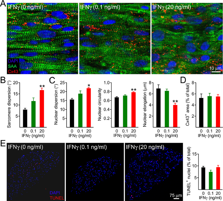
The level of circulating interferon-γ (IFNγ) is elevated in various clinical conditions including autoimmune and inflammatory diseases, sepsis, acute coronary syndrome, and viral infections. As these conditions are associated with high risk of myocardial dysfunction, we investigated the effects of IFNγ on 3D fibrin-based engineered human cardiac tissues ("cardiobundles"). Cardiobundles were fabricated from human pluripotent stem cell-derived cardiomyocytes, exposed to 0-20 ng/ml of IFNγ on culture days 7-14, and assessed for changes in tissue structure, viability, contractile force and calcium transient generation, action potential propagation, cytokine secretion, and expression of select genes and proteins. We found that application of IFNγ induced a dose-dependent reduction in contractile force generation, deterioration of sarcomeric organization, and cardiomyocyte disarray, without significantly altering cell viability, action potential propagation, or calcium transient amplitude. At molecular level, the IFNγ-induced structural and functional deficits could be attributed to altered balance of pro- and anti-inflammatory cytokines, upregulation of JAK/STAT signaling pathway (JAK1, JAK2, and STAT1), and reduced expression of myosin heavy chain, myosin light chain-2v, and sarcomeric α-actinin. Application of clinically used JAK/STAT inhibitors, tofacitinib and baricitinib, fully prevented IFNγ-induced cardiomyopathy, confirming the critical roles of this signaling pathway in inflammatory cardiac disease. Taken together, our in vitro studies in engineered myocardial tissues reveal direct adverse effects of pro-inflammatory cytokine IFNγ on human cardiomyocytes and establish the foundation for a potential use of cardiobundle platform in modeling of inflammatory myocardial disease and therapy. STATEMENT OF SIGNIFICANCE: Various inflammatory and autoimmune diseases including rheumatoid arthritis, sepsis, lupus erythematosus, Chagas disease, and others, as well as viral infections including H1N1 influenza and COVID-19 show increased systemic levels of a pro-inflammatory cytokine interferon-γ (IFNγ) and are associated with high risk of heart disease. Here we explored for the first time if chronically elevated levels of IFNγ can negatively affect structure and function of engineered human heart tissues in vitro. Our studies revealed IFNγ-induced deterioration of myofibrillar organization and contractile force production in human cardiomyocytes, attributed to decreased expression of multiple sarcomeric proteins and upregulation of JAK/STAT signaling pathway. FDA-approved JAK inhibitors fully blocked the adverse effects of IFNγ, suggesting a potentially effective strategy against human inflammatory cardiomyopathy.

Keywords: COVID-19; Fibrin hydrogel; Inflammation; Secretome; hiPSC.


Conflict of interest statement

Declaration of competing interest The authors declare that they have no known competing financial interests or personal relationships that could have appeared to influence the work reported in this paper.

Figures

Figure 1.
Figure 1.
Seven-day IFNγ treatment of human cardiobundles reduces contractile force generation without altering calcium transients. A) Schematics of experimental timeline involving hiPSC-CM differentiation, cardiobundle formation, culture, and assessments. B) Representative force traces during 2 Hz stimulation recoded from 2-week cardiobundles treated with specified doses of IFNγ on culture days 7–14. C-E) Quantified cardiobundle twitch force amplitude (C), contraction time (D), and relaxation time (E) as a function of 7-day application of IFNγ at different doses (n = 5–6 cardiobundles per group). F-G) Representative calcium transients and quantified Ca2+ transient amplitudes (ΔF/F of Fluo-4) of untreated (0 ng/ml) and IFNγ-treated (20 ng/ml) cardiobundles during 1 Hz (F) and 2 Hz (G) stimulation (n = 7–8 cardiobundles per group). *P < 0.05 and **P < 0.01 vs. 0 ng/ml IFNγ.
Figure 2.
Figure 2.
Seven-day IFNγ treatment of cardiobundles induces myofibrillar disarray without loss in cell viability. A) Representative whole-tissue immunostains of cardiobundles treated for 7 days with specified doses of IFNγ. SAA, sarcomeric α-actinin; Cx43, connexin-43; DAPI, nuclei. Note increased cardiomyocyte misalignment and sarcomere disorganization with increased doses of IFNγ. B) Quantitative analysis of sarcomere misalignment (dispersion) as a function of IFNγ dose. C) Quantitative analyses of nuclear misalignment (dispersion), circularity (1 = perfect circle), and elongation (major – minor axis difference in a best fit ellipse). D) Quantification of percent imaged area positive for Cx43 (n = 4–6 cardiobundles per group). E) Representative TUNEL stainings of cardiobundle cross-sections and corresponding quantifications of fractions of TUNEL+ nuclei, shown for specified doses of IFNγ (n = 6–8 cardiobundles per group). *P < 0.05 and **P < 0.001 vs. 0 ng/ml IFNγ.
Figure 3.
Figure 3.
Seven-day IFNγ treatment of cardiobundles decreases expression of sarcomeric proteins and upregulates expression of corresponding genes. A) Quantified expression of sarcomeric genes shown relative to 0 ng/ml group. All mRNA expression levels were first normalized to expression of reference gene HPRT-1 (n = 3 samples per group). MYH6 and MYH7 genes coding for α- and β-myosin heavy chain (MHC), respectively; MYL2 coding for ventricular myosin light chain-2 (MLC-2v); TNN2 coding for cardiac troponin T (cTnT). B) Representative Western blots for MYH, MLC-2v, cTnT, and sarcomeric α-actinin (SAA). C) Corresponding quantification of expressed proteins normalized to GAPDH (n = 6 samples per group). *P < 0.05, **P < 0.01, and ***P < 0.001 vs. 0 ng/ml IFNγ.
Figure 4.
Figure 4.
Four-day IFNγ treatment alters cardiobundle cytokine secretion. Note that concentration of IFNγ is in ng/ml rather than pg/ml (n = 3 samples per group), reflecting the presence of exogenously added IFNγ. *p < 0.05 and **p < 0.01 vs. 0 ng/ml IFNγ.
Figure 5.
Figure 5.
Tofacitinib treatment of cardiobundles prevents contractile force decline and morphological deterioration induced by IFNγ. A) Quantified cardiobundle twitch force amplitude, contraction time, and relaxation time without treatment (Control) or after 7-day treatment with 20 ng/ml IFNγ, 50 nM or 500 nM Tofacitinib (Tofa), or both IFNγ and Tofa (n = 4–11 cardiobundles per group). B) Representative whole-tissue immunostains of cardiobundles treated with IFNγ with and without specified doses of Tofa. F-actin, filamentous actin; Cx43, connexin-43; DAPI, nuclei. Note that cardiomyocyte misalignment due to IFNγ is prevented in the presence of Tofa. **P < 0.01 and ***P < 0.001 vs. control.
Figure 6.
Figure 6.
Tofacitinib treatment of cardiobundles prevents IFNγ-induced upregulation of STAT1 phosphorylation. A-B) Representative Western blots and corresponding quantifications of STAT1 and phosphorylated STAT1 (p-STAT1) expression 48 h (A) and 96 h (B) after treatment with IFNγ with or without Tofa normalized to GAPDH expression and shown relative to untreated (Control) group (n = 3 samples per group). **P < 0.01 and ***P < 0.001 vs. control; $P < 0.01 and $P < 0.001 vs. IFNγ group.
Figure 7.
Figure 7.
Tofacitinib treatment of cardiobundles prevents IFNγ-induced JAK1 and JAK2 phosphorylation. A-C) Representative Western blots (A) and corresponding quantifications of JAK1 (B) and phosphorylated JAK1 (p-JAK1, C) expression normalized to GAPDH expression and shown relative to untreated (Control) group (n = 6–9 samples per group). D-F) Representative Western blots (D) and corresponding quantifications of JAK2 (E) and phosphorylated JAK2 (p-JAK2, F) expression normalized to GAPDH expression and shown relative to untreated (Control) group (n = 6 samples per group). *P < 0.05, **P < 0.01, and ***P < 0.001 vs. control; $P < 0.05 vs. IFNγ group.

Similar articles

Cited by

References

    1. Hotchkiss RS, Moldawer LL, Opal SM, Reinhart K, Turnbull IR, Vincent JL, Sepsis and septic shock, Nat Rev Dis Primers 2 (2016) 16045. - PMC - PubMed
    1. McInnes IB, Schett G, The pathogenesis of rheumatoid arthritis, N Engl J Med 365 (2011) 2205–2219. - PubMed
    1. Kaplan MJ, Cardiovascular complications of rheumatoid arthritis: assessment, prevention, and treatment, Rheum Dis Clin North Am 36 (2010) 405–26. - PMC - PubMed
    1. Liao KP, Cardiovascular disease in patients with rheumatoid arthritis, Trends Cardiovasc Med 27 (2017) 136–140. - PMC - PubMed
    1. Mackey RH, Kuller LH, Moreland LW, Update on cardiovascular disease risk in patients with rheumatic diseases, Rheum Dis Clin North Am 44 (2018) 475–487. - PubMed

Publication types

Substances