. 2021 May:119:102628.
doi: 10.1016/j.jaut.2021.102628. Epub 2021 Mar 9.

HLA class I hyper-expression unmasks beta cells but not alpha cells to the immune system in pre-diabetes

Affiliations

HLA class I hyper-expression unmasks beta cells but not alpha cells to the immune system in pre-diabetes

Mehdi A Benkahla et al. J Autoimmun. 2021 May.

Abstract

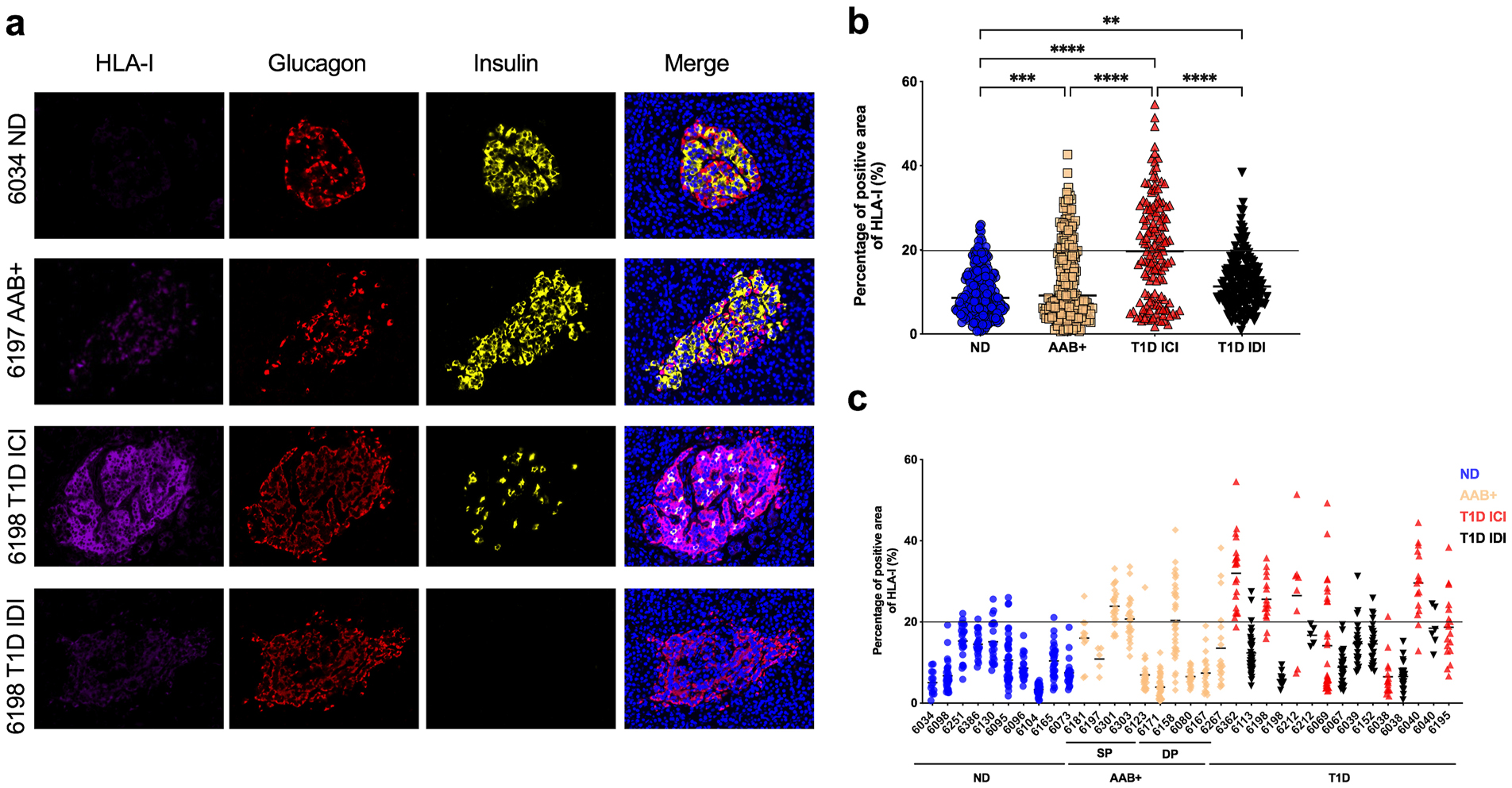
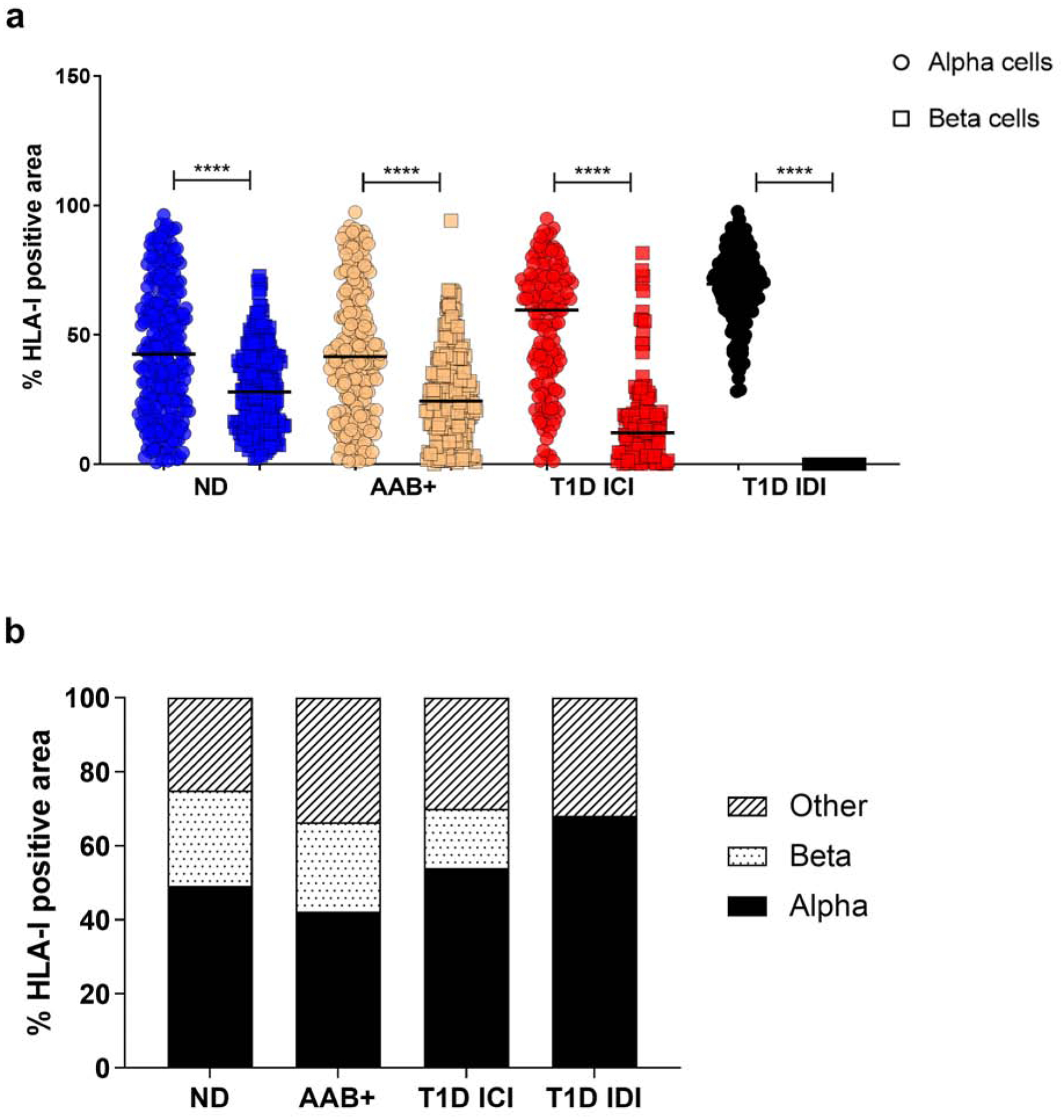
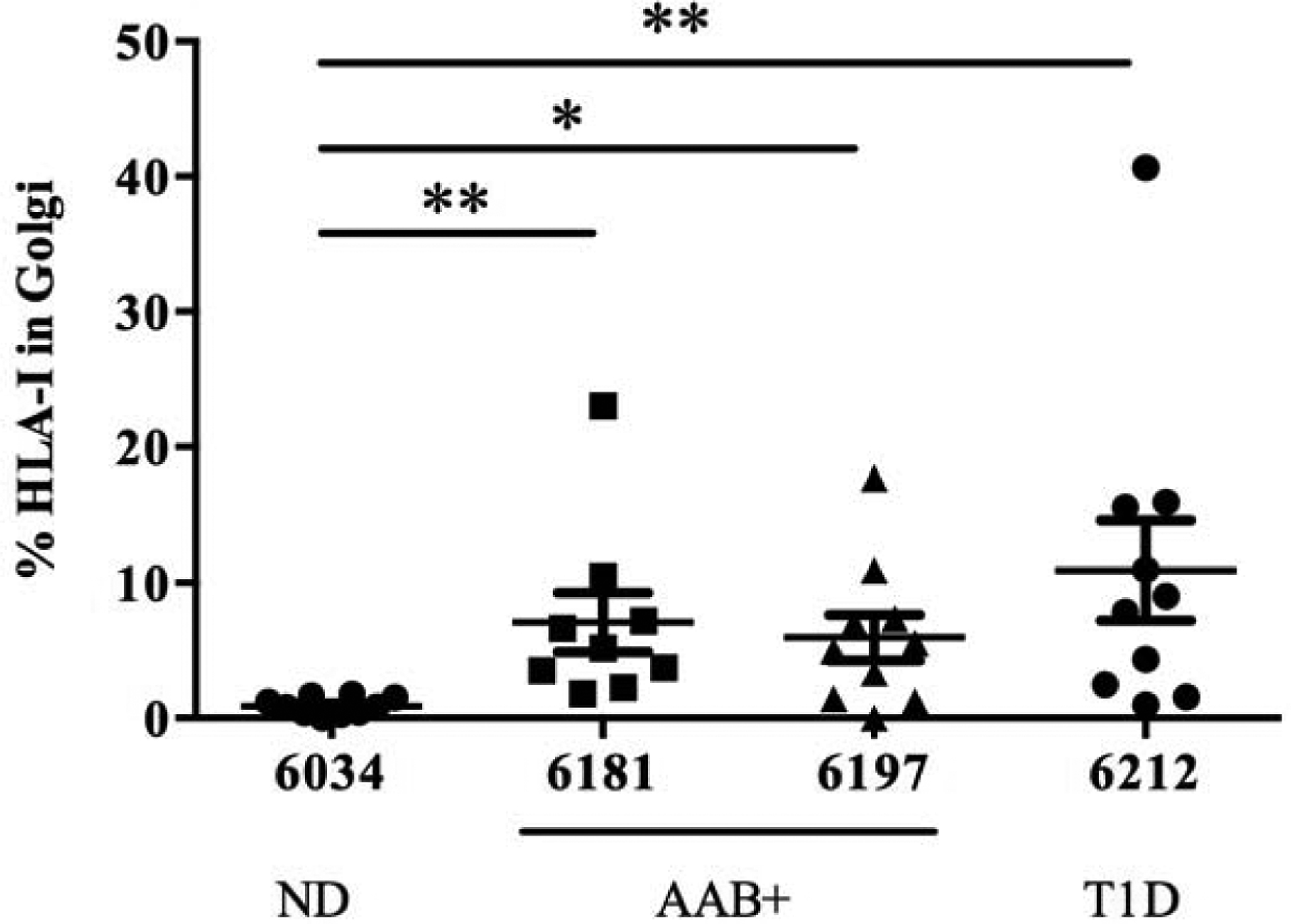
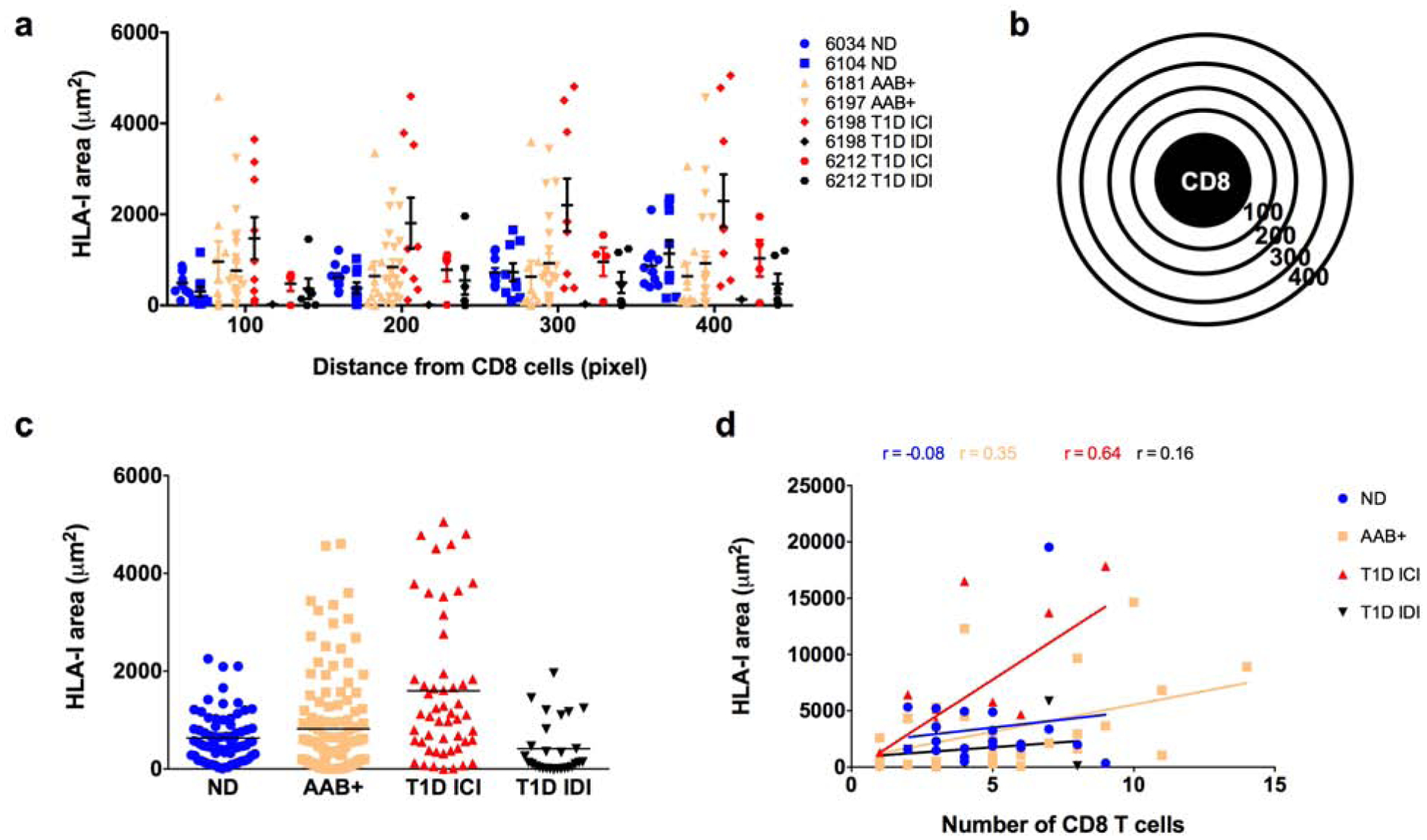
Human leukocyte antigens of class-I (HLA-I) molecules are hyper-expressed in insulin-containing islets (ICI) of type 1 diabetic (T1D) donors. This study investigated the HLA-I expression in autoantibody positive (AAB+) donors and defined its intra-islet and intracellular localization as well as proximity to infiltrating CD8 T cells with high-resolution confocal microscopy. We found HLA-I hyper-expression had already occurred prior to clinical diagnosis of T1D in islets of AAB+ donors. Interestingly, throughout all stages of disease, HLA-I was mostly expressed by alpha cells. Hyper-expression in AAB+ and T1D donors was associated with intra-cellular accumulation in the Golgi. Proximity analysis showed a moderate but significant correlation between HLA-I and infiltrating CD8 T cells only in ICI of T1D donors, but not in AAB+ donors. These observations not only demonstrate a very early, islet-intrinsic immune-independent increase of HLA-I during diabetes pathogenesis, but also point towards a role for alpha cells in T1D.

Keywords: Alpha cells; Beta cells; HLA class I; Type 1 diabetes; nPOD.


Conflict of interest statement

Competing interests: The authors declare no competing interest.

Figures

Fig. 1.
Fig. 1.
HLA class I is hyper-expressed in the islets of T1D as well as in AAB+ donors. FFPE sections of human pancreata were stained with anti-HLA-I (magenta), anti-insulin (green) and anti-glucagon (red) and Hoechst (blue). (a) Representative images of an islet from a non-diabetic (ND) donor (nPOD 6034), an autoantibody positive (AAB+) donor (nPOD 6197), an insulin-containing islet (ICI) of a T1D donor (nPOD 6198) and an insulin-deficient islet (IDI) of a T1D donor (nPOD 6198) are shown. Images were acquired using a confocal microscope LSM 780 with a 63× 1.4na objective. HLA-I was quantified using Zen software. (b) The percentage of total islet area positive for HLA-I was measured in at least 20 islets per section from non-diabetic donors (n=10), single autoantibody positive (n=5), double autoantibody positive (n=5) and type 1 diabetic donors (n=12). (c) Percentage of total islet area positive for HLA-I presented by case. Each data point represents an islet. An arbitrary threshold of 20% was set above which HLA-I was considered hyper-expressed. *** p=0.0004 ****p<0.0001.
Fig. 2.
Fig. 2.
HLA class I is mainly expressed on alpha cells irrespective of disease status. (a) Percentage of colocalization of HLA-I and glucagon (alpha cells- circle) and HLA-I and insulin (beta cells - square) quantified using the Mander’s overlap coefficient using Zen software. Each data point represents an islet. (b) Mean percentage of HLA-I positive area per cell type. ND: non-diabetic; AAB+: autoantibody positive; T1D: type 1 diabetes; ICI: insulin-containing islet; IDI: insulin-deficient islet. **** p<0.0001.
Fig. 3.
Fig. 3.
HLA class I colocalizes with Golgi marker GM130 in the islets of AAB+ and T1D donors. Percentage of colocalization of HLA-I and GM130 quantified using Mander’s overlap coefficient in the islets from non-diabetic donor (nPOD 6034), autoantibody positive donors (nPOD 6181 and 6197) and a type 1 diabetic donor (nPOD 6212). Colocalization analysis was performed in Zen software. ND: Non-diabetic; AAB+: autoantibody positive; T1D: type 1 diabetes. *p<0.05 **p<0.01.
Fig. 4.
Fig. 4.
Proximity analysis of HLA class I in the surroundings of CD8 T cells. Pancreatic sections from organ donors were stained for HLA-I, CD8, insulin and glucagon. CD8 T cells were detected in the pancreatic tissue sections and a proximity analysis was performed to detect the HLA-I expression in the surroundings of CD8 T cells. (a) Quantification of HLA-I using ImagePro premier in the radials surrounding the CD8 T cells, 100 being the closest to the CD8 cell and 400 the furthest presented by case. Each data point represents the level of HLA-I in a radial circle. (b) Schematic representation of CD8 T cell and the radial circles from 100 to 400 used for the proximity analysis. (c) Quantification of HLA-I presented as the sum of the radials from 100 to 400 and presented by group. Each data point represents an islet. Each data point represents the level of HLA-I in a radial circle (d) Correlation between the number of CD8 T cells per field of view and the total expression of HLA-I in the surroundings of those CD8 T cells. Each data point represents a field of view. ND: non-diabetic; AAB+: autoantibody positive; T1D: type 1 diabetes; ICI: insulin-containing islet; IDI: insulin-deficient islet.

References

    1. Katsarou A, Gudbjörnsdottir S, Rawshani A, Dabelea D, Bonifacio E, Anderson BJ, Jacobsen LM, Schatz DA, Lernmark Å, Type 1 diabetes mellitus, Nat Rev Dis Primers 3, 17016 (2017). - PubMed
    1. Leone P, Shin E-C, Perosa F, Vacca A, Dammacco F, Racanelli V, MHC Class I Antigen Processing and Presenting Machinery: Organization, Function, and Defects in Tumor Cells, J Natl Cancer Inst 105, 1172–1187 (2013). - PubMed
    1. Krischer JP, Lynch KF, Schatz DA, Ilonen J, Lernmark Å, Hagopian WA, Rewers MJ, She J-X, Simell OG, Toppari J, Ziegler A-G, Akolkar B, Bonifacio E, TEDDY Study Group, The 6 year incidence of diabetes-associated autoantibodies in genetically at-risk children: the TEDDY study, Diabetologia 58, 980–987 (2015). - PMC - PubMed
    1. Ziegler AG, Rewers M, Simell O, Simell T, Lempainen J, Steck A, Winkler C, Ilonen J, Veijola R, Knip M, Bonifacio E, Eisenbarth GS, Seroconversion to multiple islet autoantibodies and risk of progression to diabetes in children, JAMA 309, 2473–2479 (2013). - PMC - PubMed
    1. Bottazzo GF, Dean BM, McNally JM, MacKay EH, Swift PG, Gamble DR, In situ characterization of autoimmune phenomena and expression of HLA molecules in the pancreas in diabetic insulitis, N. Engl. J. Med 313, 353–360 (1985). - PubMed

Publication types

MeSH terms