. 2021 Mar 11;18(1):69.
doi: 10.1186/s12974-021-02119-w.

Fetal inflammation induces acute immune tolerance in the neonatal rat hippocampus

Affiliations

Fetal inflammation induces acute immune tolerance in the neonatal rat hippocampus

Garima Singh et al. J Neuroinflammation. .

Abstract

Background: Infants born preterm due to chorioamnionitis are frequently affected by a fetal inflammatory response syndrome (FIRS) and then by subsequent postnatal infections. FIRS and postnatal systemic inflammatory events independently contribute to poor neurocognitive outcomes of preterm infants. Developmental integrity of the hippocampus is crucial for intact neurocognitive outcomes in preterms and hippocampally dependent behaviors are particularly vulnerable to preterm systemic inflammation. How FIRS modulates the hippocampal immune response to acute postnatal inflammatory events is not well understood.

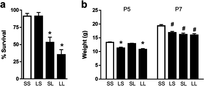
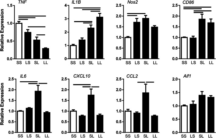
Methods: Prenatal LPS exposed (FIRS) and control neonatal rats received i.p. LPS or saline at postnatal day (P) 5. On P7, immune response was evaluated in the hippocampus of four treatment groups by measuring gene expression of inflammatory mediators and cytosolic and nuclear NFκB pathway proteins. Microglial activation was determined by CD11b+ and Iba1+ immunohistochemistry (IHC) and inflammatory gene expression of isolated microglia. Astrocyte reactivity was measured using Gfap+ IHC.

Results: Postnatal LPS resulted in a robust hippocampal inflammatory response. In contrast, FIRS induced by prenatal LPS attenuated the response to postnatal LPS exposure, evidenced by decreased gene expression of inflammatory mediators, decreased nuclear NFκB p65 protein, and fewer activated CD11b+ and Iba1+ microglia. Isolated microglia demonstrated inflammatory gene upregulation to postnatal LPS without evidence of immune tolerance by prenatal LPS.

Conclusion: Prenatal LPS exposure induced immune tolerance to subsequent postnatal LPS exposure in the hippocampus. Microglia demonstrate a robust inflammatory response to postnatal LPS, but only a partial immune tolerance response.

Keywords: Fetal inflammation; Hippocampus; Immune tolerance; LPS; Microglial activation; Preterm.


Conflict of interest statement

The authors declare that they have no competing interests.

Figures

Fig. 1
Fig. 1
Effect of FIRS and postnatal inflammation on survival and growth. Rat pups were exposed to prenatal i.a. LPS (L) or saline (S) (listed first) and then postnatal i.p. LPS or saline at P5 (listed second) to create 4 treatment groups. a The percentage of pups from each litter surviving at 7 days of life following fostering is depicted (n = 20–22 litters). There was significantly lower survival in both groups that received postnatal LPS (SL and LL); prenatal treatment did not change survival outcomes (*p < 0.05 vs SS and LS). b Weights of pups that received i.a. LPS (LS and LL) were decreased on postnatal (P) day 5 prior to postnatal i.p. LPS injection (*p < 0.05 vs SS and SL; n = 12–30). At P7, all groups that received LPS prenatally or postnatally had lower weights than the controls (#p < 0.05 vs SS; n = 6–17). Data presented as mean ± SEM
Fig. 2
Fig. 2
Effect of FIRS and postnatal inflammation on hippocampal gene expression of pro-inflammatory mediators at P7. Rat pups were exposed to prenatal i.a. LPS (L) or saline (S) (listed first) and then postnatal i.p. LPS or saline at P5 (listed second) to create four treatment groups. Bars above the graphs indicate significant differences between groups (p < 0.05). Data presented as mean ± SEM. Genes are normalized to ribosomal protein S18
Fig. 3
Fig. 3
Effect of FIRS and postnatal inflammation on NFκB pathway gene expression and protein in the hippocampus at P7. Rat pups were exposed to prenatal i.a. LPS (L) or saline (S) (listed first) and then postnatal i.p. LPS or saline at P5 (listed second) to create four treatment groups. a Relative gene expression of indicated NFκB pathway genes in the hippocampus at P7 normalized to ribosomal protein S18 (n = 5–7/group). Protein product of each gene is in parentheses. b Western blot analyses of nuclear and cytosolic protein fractions of NFκB p65 in the hippocampus at P7 (n = 3–6/group). c Western blot analyses of nuclear and cytosolic protein fractions of NFκB p50 in the hippocampus at P7 (n = 4–6/group). d Western blot analyses of cytosolic fractions of IκBα and IκBβ proteins in the hippocampus at P7 (n = 3–5/group). In all graphs, bars above the graphs indicate significant differences between groups (p < 0.05). Data presented as mean ± SEM
Fig. 4
Fig. 4
Postnatal LPS increases activated microglia in the hippocampus, but the effect is attenuated by FIRS. Rat pups were exposed to prenatal i.a. LPS (L) or saline (S) (listed first) and then postnatal i.p. LPS or saline at P5 (listed second) to create four treatment groups. On P7, microglial activation was quantified in each treatment group by counting activated microglia and total microglia. a Activated microglia count (top bar graph) and total microglia count (bottom bar graph) of CD11b+ cells. Activated microglia were identified by thickened cell bodies and blunted processes. Examples of surveillance (non-activated) and activated microglia are shown below pictures. b Activated microglia count of Iba1+ cells identified by thickened cell bodies and blunted process. Bars above the graphs indicate significant differences between groups (p < 0.05; n = 5–7/group). Data presented as mean ± SEM. Pictures representative of each group are shown at × 20. Scale bar represents 100 μm
Fig. 5
Fig. 5
Effect of FIRS and postnatal inflammation on astrocytes in the hippocampus at P7. Rat pups were exposed to prenatal i.a. LPS (L) or saline (S) (listed first) and then postnatal i.p. LPS or saline at P5 (listed second) to create four treatment groups. a On P7, reactive astrocytosis was quantified in each treatment group by calculating areal coverage ratio of Gfap+ cells in the hippocampus. Scale bar represent 100 μm. Pictures representative of each group are shown at × 20. b Expression of reactive astrocyte genes in the hippocampus at P7. Bars above the graphs indicate significant differences between groups (p < 0.05). Data presented as mean ± SEM
Fig. 6
Fig. 6
Effect of FIRS and postnatal inflammation on gene expression of M1 activation markers in isolated microglia at P7. Rat pups were exposed to prenatal i.a. LPS (L) or saline (S) (listed first) and then postnatal i.p. LPS or saline at P5 (listed second) to create four treatment groups. Bars above the graphs indicate significant differences between groups (p < 0.05). Data presented as mean ± SEM. Genes are normalized to the ribonuclease RPP30
Fig. 7
Fig. 7
Co-localization of M1 marker MHCII with Iba1+ microglia in the hippocampus at P7. Rat pups were exposed to prenatal i.a. LPS (L) or saline (S) (listed first) and then postnatal i.p. LPS or saline at P5 (listed second) to create four treatment groups. Total double positive Iba1 (green) and MHCII (red) cells were quantified in each treatment group (blue = DAPI). Bars above the graph indicate significant differences between groups (p < 0.05; n = 4–6/group). Data presented as mean ± SEM. Pictures representative of each group are shown at × 20. Scale bar represents 100 μm

References

    1. Gravett MG, Rubens CE, Nunes TM, GAPPS Review Group Global report on preterm birth and stillbirth (2 of 7): discovery science. BMC Pregnancy Childbirth. 2010;10 Suppl 1:S2. doi: 10.1186/1471-2393-10-S1-S2. - DOI - PMC - PubMed
    1. Hamilton BE, Martin JA, Osterman MJ, Curtin SC, Matthews TJ. Births: Final Data for 2014. Natl Vital Stat Rep. 2015;64:1–64. - PubMed
    1. Stoll BJ, Hansen N. Infections in VLBW infants: studies from the NICHD Neonatal Research Network. Semin Perinatol. 2003;27:293–301. doi: 10.1016/S0146-0005(03)00046-6. - DOI - PubMed
    1. Gotsch F, Romero R, Kusanovic JP, Mazaki-Tovi S, Pineles BL, Erez O, Espinoza J, Hassan SS. The fetal inflammatory response syndrome. Clin Obstet Gynecol. 2007;50:652–683. doi: 10.1097/GRF.0b013e31811ebef6. - DOI - PubMed
    1. Gomez R, Romero R, Ghezzi F, Yoon BH, Mazor M, Berry SM. The fetal inflammatory response syndrome. Am J Obstet Gynecol. 1998;179:194–202. doi: 10.1016/S0002-9378(98)70272-8. - DOI - PubMed