Review
. 2021:142:197-231.
doi: 10.1016/bs.ctdb.2020.10.009. Epub 2020 Nov 19.

Dorsal commissural axon guidance in the developing spinal cord

Affiliations
Review

Dorsal commissural axon guidance in the developing spinal cord

Sandy Alvarez et al. Curr Top Dev Biol. 2021.

Abstract

Commissural axons have been a key model system for identifying axon guidance signals in vertebrates. This review summarizes the current thinking about the molecular and cellular mechanisms that establish a specific commissural neural circuit: the dI1 neurons in the developing spinal cord. We assess the contribution of long- and short-range signaling while sequentially following the developmental timeline from the birth of dI1 neurons, to the extension of commissural axons first circumferentially and then contralaterally into the ventral funiculus.

Keywords: Axon guidance; BMP; Chemotaxis; Commissural axons; Ephrin; Floor plate; Haptotaxis; Netrin1; Neural development; Robo; Roof plate; Slit.


Figures

Fig. 1
Fig. 1
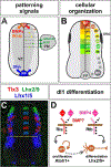
Organization and specification of dI1s in the spinal cord. (A, B) Six domains of dorsal progenitor neurons (dP1–dP6) arise in the ventricular zone (VZ) in response to BMP/Wnt signals from the roof plate (RP) and retinoic acid (RA) from paraxial mesoderm (PM). This process results in six distinct classes of post-mitotic dorsal neurons (dI1–dI6). The ventral spinal cord is patterned by a gradient of Shh from the floor plate FP. (C) Dorsal interneurons can be distinguished by their expression of distinct complements of transcription factors (Andrews et al., 2017). (D) Model for the specification of dI1s. Both BMP4 and BMP7 can promote Atoh1+ dP1 patterning through BmprIa or BmprIb (chicken), but only BMP4 directs progenitors to differentiate as Lhx2+ dI1s through BmprIb (mouse and chicken) (Andrews et al., 2017).
Fig. 2
Fig. 2
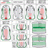
Models for the long- and short-range activities of netrin1. (A) In the long-range gradient model, the RP-derived Bmps act as a chemorepellent “pushing” commissural axons away from the dorsal midline, while FP-derived netrin1 and Shh function as chemoattractants “pulling” commissural axons to the ventral midline. (B) In the short-range model, pial-netrin1 first orients early born commissural axons to extend ventrally. NPCs transcribe netrin1 (red) and then deposit netrin1 protein (green) on the pial surface, where it may act as a growth substrate to promote axon extension (Varadarajan et al., 2017). (C) As development progresses, pre-crossing commissural axons extend into the ventral spinal cord, and no longer grow adjacent to pial-netrin1 (dotted green line). Rather, they project precisely around a “hederal” boundary of netrin1 expressing NPCs (orange line). We have proposed that the netrin1 hederal boundary promotes directed axon fasciculation while preventing innervation of netrin1 expressing cells. This activity permits commissural axons to grow around the VZ. (D) Commissural axons extend across the FP in a highly fasciculated bundle within a narrow corridor bounded by hederal-netrin1 and pial-netrin1. Post-crossing commissural axons then turn rostrally to extend in the ventral funiculus, again growing adjacent to a pial-netrin1 substrate (solid green line). Concomitantly, a domain of netrin1 expressing cells (red) emerges adjacent to the DREZ, which continue to sculpt axonal trajectories within the spinal cord. (E–G) We propose that the netrin1 produced by neural progenitors is transported to the pial surface in their progenitor endfeet, and then transfers from this pial-substrate to Dcc+ commissural axons (E) (Varadarajan et al., 2017; Varadarajan & Butler, 2017). Dcc and netrin1 then interact in cis to promote the selective fasciculation and growth commissural axons around netrin1 expressing NPCs (F). In absence of Dcc, netrin1 does accumulate on the pial surface, but does not transfer to commissural axons. These axons fail to fasciculate and grow randomly, including into the VZ (G).
Fig. 3
Fig. 3
Short- and long-range phenotypes observed in the absence of NPC- or FP-derived netrin1. (A) Summary of the distribution of netrin1 transcript (red) and protein (green). NPCs and FP cells express netrin1, while netrin1 protein is present on the pial surface and on commissural axons. (B) In netrin1 loss of function mutants, spinal axons are highly defasciculated, extending into the VZ, and wandering across the motor column. (C, D) Short range phenotypes for NPC-derived netrin1: When netrin1 expression is removed from either a large (C) or small (D) number of NPCs using conditional genetic approaches (C=Pax3::cre driver; D=Dbx1::cre driver) (Varadarajan et al., 2017), axons defasciculate specifically and locally in the region where netrin1 activity is absent. In particular, the introduction of ectopic netrin1 on::off boundaries (D) locally reshapes axon trajectories in a manner consistent with the hederal boundary model. Long-range phenotypes for NPC-derived netrin1: none observed. (E–I) Short-range phenotypes for FP-derived netrin1: In netrin1ΔFP mice (Shh::cre driver), commissural axons project ventrally toward the FP (E) in a manner largely indistinguishable from controls. However, errors occur ~10μm from the FP (yellow box, G), when axons reach the “off” edge of the ventral domain pial-netrin1, where they locally defasciculate (arrowheads, I). Axons start to cross the FP from this “off” edge, resulting in a laterally displaced “U” shaped trajectory (G), distinct from the “V” shape observed in controls (F). Control commissural axons then turn rostrally to project longitudinally beside the FP in the ventral funiculus (H). Most axons correctly make the rostral turn in netrin1ΔFP mice, however, we (S.A. and S.J.B., personal communication) and others (Moreno-Bravo et al., 2019) have observed that a subset of commissural axons fail to cross the FP, and turn ipsilaterally (I), resulting in a thinning of the FP commissure (Wu et al., 2019). It remains unresolved whether this ipsilateral turn is the result of axons following an ectopic hederal boundary created by the specific loss of netrin1 in the FP (I), or a direct requirement for FP-netrin1 in axon crossing. Long-range phenotypes for FP-derived netrin1: While we have not observed this phenotype, other reports (Wu et al., 2019) have shown that commissural axons are modestly defasciculated in the ventral spinal cord in the absence of FP-derived netrin1 (yellow arrowheads, G) i.e. ~100μm from the FP. This phenotype is significantly enhanced when netrin1ΔFP is combined with a Boc mutation (G), the non-canonical receptor that mediates the axon guidance activities of Shh (Wu et al., 2019).

Similar articles

Cited by

References

    1. Akin O, & Zipursky SL (2016). Frazzled promotes growth cone attachment at the source of a Netrin gradient in the Drosophila visual system. eLife, 5. - PMC - PubMed
    1. Alaynick WA, Jessell TM, & Pfaff SL (2011). SnapShot: Spinal cord development. Cell, 146. 178–178 e171. - PMC - PubMed
    1. Alther TA, Domanitskaya E, & Stoeckli ET (2016). Calsyntenin 1-mediated trafficking of axon guidance receptors regulates the switch in axonal responsiveness at a choice point. Development (Cambridge, England), 143, 994–1004. - PubMed
    1. Altman J, & Bayer SA (1984). The development of the rat spinal cord. - PubMed
    1. Andermatt I, Wilson NH, Bergmann T, Mauti O, Gesemann M, Sockanathan S, et al. (2014). Semaphorin 6B acts as a receptor in post-crossing commissural axon guidance. Development (Cambridge, England), 141, 3709–3720. - PMC - PubMed

Publication types