. 2021 Apr;18(4):711-722.
doi: 10.1016/j.jsxm.2021.01.175. Epub 2021 Mar 9.

Caspase Signaling in ED Patients and Animal Models

Affiliations

Caspase Signaling in ED Patients and Animal Models

Sarah Martin et al. J Sex Med. 2021 Apr.

Abstract

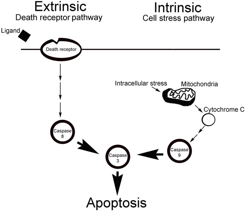
Background: Current treatments for erectile dysfunction (ED) are ineffective in prostatectomy and diabetic patients due to cavernous nerve (CN) injury, which causes smooth muscle apoptosis, penile remodeling, and ED. Apoptosis can occur via the intrinsic (caspase 9) or extrinsic (caspase 8) pathway.

Aim: We examined the mechanism of how apoptosis occurs in ED patients and CN injury rat models to determine points of intervention for therapy development.

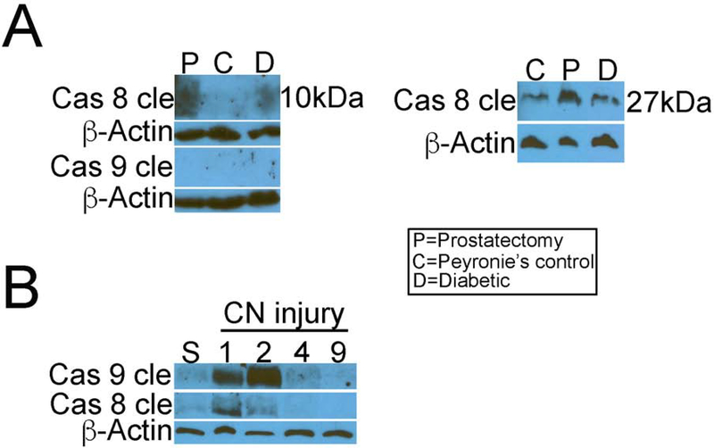
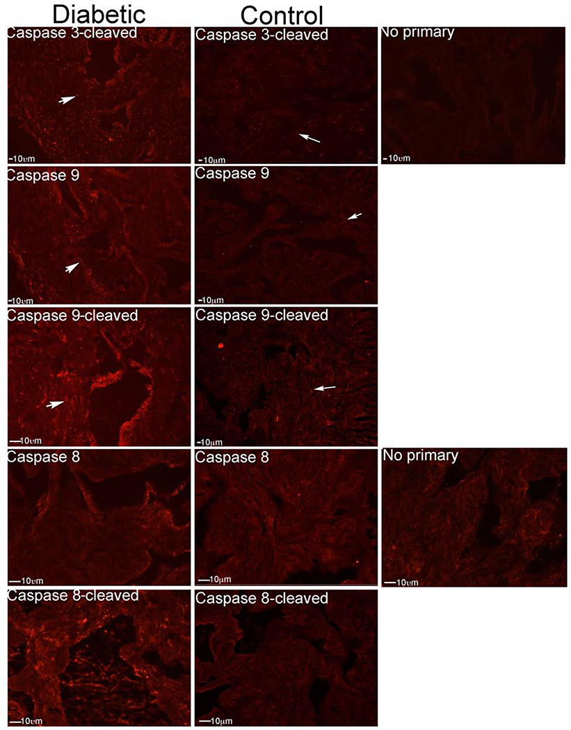
Methods and outcomes: Immunohistochemical and western analyses for caspase 3-cleaved, caspase-8 and caspase-9 (pro and active forms) were performed in corpora cavernosal tissue from Peyronie's, prostatectomy and diabetic ED patients (n = 33), penis from adult Sprague Dawley rats that underwent CN crush (n = 24), BB/WOR diabetic and control rats (n = 8), and aged rats (n = 9).

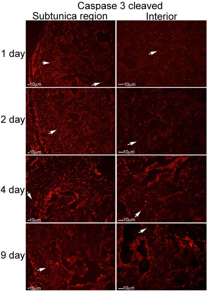
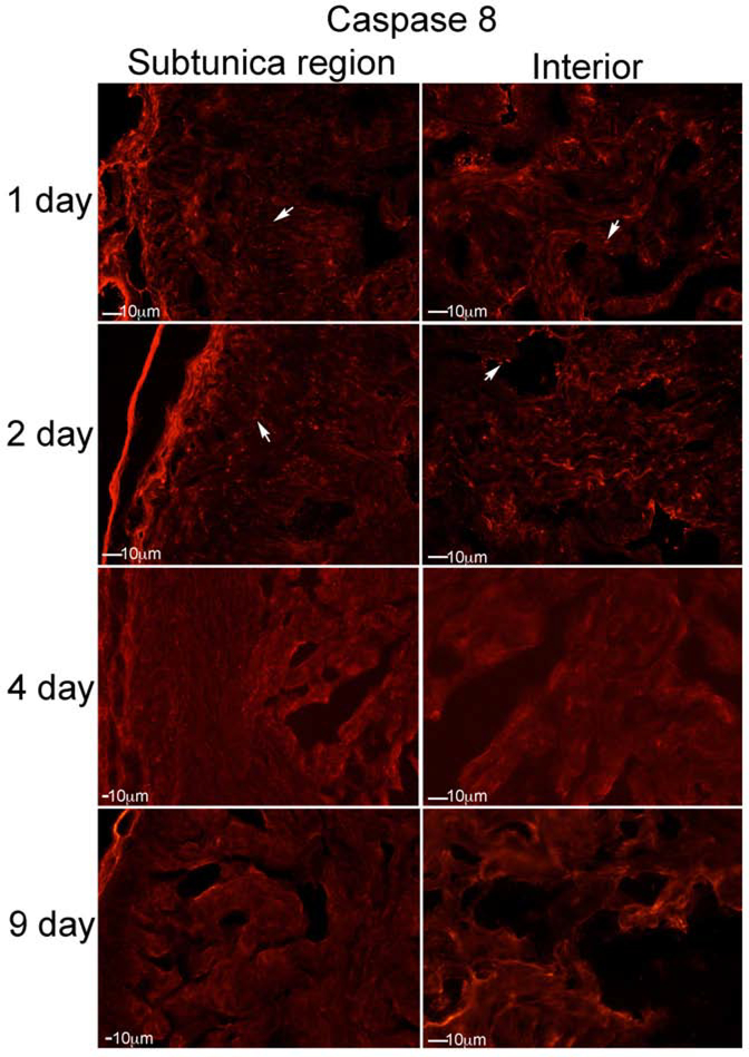
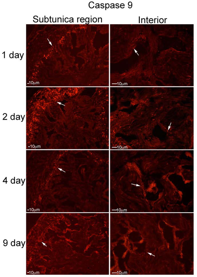
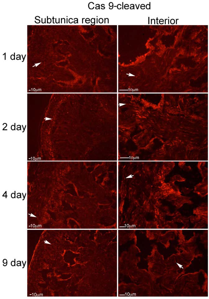
Results: Caspase 3-cleaved was observed in corpora cavernosa from Peyronie's patients and at higher abundance in prostatectomy and diabetic tissues. Apoptosis takes place primarily through the extrinsic (caspase 8) pathway in penis tissue of ED patients. In the CN crushed rat, caspase 3-cleaved was abundant from 1-9 days after injury, and apoptosis takes place primarily via the intrinsic (caspase 9) pathway. Caspase 9 was first observed and most abundant in a layer under the tunica, and after several days was observed in the lining of and between the sinuses of the corpora cavernosa. Caspase 8 was initially observed at low abundance in the rat corpora cavernosa and was not observed at later time points after CN injury. Aged and diabetic rat penis primarily exhibited intrinsic mechanisms, with diabetic rats also exhibiting mild extrinsic activation.

Clinical translation: Knowing how and when to intervene to prevent the apoptotic response most effectively is critical for the development of drugs to prevent ED, morphological remodeling of the corpora cavernosa, and thus, disease management.

Strengths and limitations: Animal models may diverge from the signaling mechanisms observed in ED patients. While the rat utilizes primarily caspase 9, there is a significant flux through caspase 8 early on, making it a reasonable model, as long as the timing of apoptosis is considered after CN injury.

Conclusions: Apoptosis takes place primarily through the extrinsic caspase 8 dependent pathway in ED patients and via the intrinsic caspase 9 dependent pathway in commonly used CN crush ED models. This is an important consideration for study design and interpretation that must be taken into account for therapy development and testing of drugs, and our therapeutic targets should ideally inhibit both apoptotic mechanisms. Martin S, Harrington DA, Ohlander S, et al. Caspase Signaling in ED Patients and Animal Models. J Sex Med 2021;18:711-722.

Keywords: Apoptosis; Caspase; Cavernous Nerve Injury; Erectile Dysfunction; Extrinsic; Intrinsic; Penis; Peripheral Nerve Regeneration.


Figures

Figure 1:
Figure 1:
Diagram of caspase dependent apoptotic mechanism. Caspases are activated through two distinct signaling mechanisms; (1) these are the cell-intrinsic pathway (mitochondrial pathway), initiated by internal sensors for severe distress (caspase 9 dependent), and (2) the cell-extrinsic pathway (caspase 8 dependent), triggered by extracellular ligands through cognate death receptors at the surface of target cells
Figure 2:
Figure 2:
IHC analysis was performed on corpora cavernosal tissue from ED patients with Peyronie’s (control, n=8), diabetes (n=7), or prostatectomy (n=7), assaying for caspase 3-cleaved (active form), caspase 8, caspase 9, caspase 8-cleaved (active form), and caspase 9-cleaved (active form). Caspase 3-cleaved was identified in corpora cavernosa of all three patient groups, but was more abundant in corpora cavernosa from the diabetic and prostatectomy patients, indicating increased apoptosis. Caspase 8 and caspase 8-cleaved were identified in penis of Peyronie’s patients, and were increased in penis of diabetic and prostatectomy patients, indicating extrinsic apoptotic signaling. Caspase 9 and caspase 9-cleaved were not present in Peyronie’s and prostatectomy patients, indicating that the intrinsic apoptotic pathway was not active. Only a few cells were identified that stained positively for caspase 9 in the diabetic group. The absence of artifact staining was confirmed by omitting the rabbit (bottom left) and mouse (bottom right) primary antibodies. Arrows indicate staining. 200X magnification.
Figure 3:
Figure 3:
IHC analysis was performed assaying for caspase 3-cleaved, on penis tissue from rats that underwent CN crush and were euthanized 1 (n=5), 2 (n=5), 4 (n=4) and 9 (n=4) days after CN injury (Left: subtunica region of corpora cavernosa, Right: interior of corpora cavernosa). Caspase 3-cleaved increased from 1–9 days after CN injury, indicating apoptosis was taking place. Staining was first observed in a discreet layer under the tunica, at one day after CN injury. At 2 days after CN injury, caspase 3-cleaved became abundant in the interior of the corpora cavernosa, in smooth muscle between the sinuses. By 4 days after CN injury, staining was abundant both under the tunica, in between the sinuses, and within the sinusoidal smooth muscle. By 9 days after injury, staining was decreased, indicating tapering of the apoptotic response. Arrows indicate staining. 200X magnification.
Figure 4:
Figure 4:
IHC analysis was performed assaying for caspase 8, on penis tissue from rats that underwent CN crush and were euthanized 1 (n=5), 2 (n=5), 4 (n=4) and 9 (n=4) days after CN injury (Left: subtunica region of corpora cavernosa, Right: interior of corpora cavernosa). Caspase 8 was identified at low abundance after CN injury. It was observed one day after CN injury, primarily under the tunica. Caspase 8 was most abundant at two days after CN injury, and was present both under the tunica and within the smooth muscle lining of the sinuses. Caspase 8 was not identified at 4 and 9 days after CN injury. Arrows indicate staining. 200X magnification.
Figure 5:
Figure 5:
IHC analysis was performed assaying for caspase 8 cleaved (active form), on penis tissue from rats that underwent CN crush and were euthanized 1 (n=5), 2 (n=5), 4 (n=4) and 9 (n=4) days after CN injury (Left: subtunica region of corpora cavernosa, Right: interior of corpora cavernosa). Caspase 8 cleaved was identified at low abundance at 1 and 2 days after CN injury, was barely detectable by 4 days, and was not identified at 9 days after CN injury. Arrows indicate staining. 200X magnification.
Figure 6:
Figure 6:
IHC analysis was performed assaying for caspase 9, on penis tissue from rats that underwent CN crush and were euthanized 1 (n=5), 2 (n=5), 4 (n=4) and 9 (n=4) days after CN injury (Left: subtunica region of corpora cavernosa, Right: interior of corpora cavernosa). Caspase 9 was abundant in the rat penis after CN injury. It was identified primarily in a region under the tunica of the corpora cavernosa at one day after CN injury, with limited staining within sinusoidal smooth muscle. Caspase 9 was most abundant at 2 days after CN injury, started decreasing by 4 days after CN injury, and was present at lower abundance by 9 days after CN injury. Arrows indicate staining. 200X magnification.
Figure 7:
Figure 7:
IHC analysis was performed assaying for caspase 9-cleaved (active form), on penis tissue from rats that underwent CN crush and were euthanized 1 (n=5), 2 (n=5), 4 (n=4) and 9 (n=4) days after CN injury (Left: subtunica region of corpora cavernosa, Right: interior of corpora cavernosa). Caspase 9-cleaved was identified in the region of the corpora cavernosa under the tunica at 1–9 days after CN injury, with highest abundance at 2 and 4 days after injury. However caspase 9-cleaved was also abundant in corpora cavernosal tissue between the sinuses, and within the sinusoidal lining. Arrows indicate staining. 200X magnification.
Figure 8:
Figure 8:
(A) Western analysis for caspase 8-cleaved and caspase 9-cleaved (active form) were performed in corpora cavernosa from ED patients with Peyronie’s (control, n=4), prostatectomy (n=4) and diabetes (n=4). Caspase 8 cleaved was increased with prostatectomy and diabetes in comparison to Peyronie’s controls. β-ACTIN was used as a housekeeper protein for comparison to ensure equal loading of proteins. Caspase 9-cleaved was not identified in human corpora cavernosal tissue. (B) Western analysis was performed for caspase 8-cleaved and caspase 9-cleaved in corpora cavernosal tissue from rats that underwent sham or CN crush surgery and were euthanized 1 (n=5), 2 (n=5), 4 (n=4) and 9 (n=4) days after CN injury. Caspase 9-cleaved was detectable at 1 day after CN injury, was most abundant at 2 days after CN injury, and remained identifiable at 4 days after injury. Caspase 8-cleaved was transiently expressed at low abundance at 1 day after CN injury, and was barely detectable by two days after CN injury. Legend: P=Prostatectomy, C=Peyronie’s, D=Diabetic.
Figure 9:
Figure 9:
IHC analysis was performed for caspase 3-cleaved, caspase 9, caspase 9-cleaved, caspase 8, and caspase 8-cleaved in aged Sprague Dawley rats (P200–329, n=9) and adult (P115–120, n=9) that had not undergone surgical CN injury. Caspase 3-cleaved was elevated without CN injury in aged rat penis, indicating apoptosis was taking place. Caspase 9 and caspase 9-cleaved were increased in aged rat corpora cavernosa. Only a few cells stained for caspase 8 and caspase 8-cleaved. Secondary artifact staining was not observed when the primary antibodies were omitted. Arrows indicate staining. 200–400X magnification.
Figure 10:
Figure 10:
IHC analysis was performed for caspase 3-cleaved, caspase 9, caspase 9-cleaved, caspase 8, and caspase 8-cleaved in BB/WOR diabetic (n=) and control diabetes resistant BB/WOR (n=4) rat penis. Caspase 3-cleaved was elevated, indicating apoptosis was taking place. Caspase 9 and caspase 9-cleaved were increased in the sinusoidal lining, and around the sinusoids of the corpora cavernosa in the diabetic penis. Caspase 8 and caspase 8-cleaved were also upregulated in a limited manner in the lining of the sinusoids and the tissue in between the sinuses. No primary controls did not exhibit artifact staining. Arrows indicate staining. 200X magnification.

Similar articles

Cited by

References

    1. Feldman HA, Goldstein I, Hatzichristou DG, Krane RJ, McKinlay JB. Impotence and its medical and psychosocial correlates: results of the Massachusetts Male Aging Study. J Urol 1994; 151: 54–61. - PubMed
    1. Heruti R, Shochat T, Tekes-Manova D, Ashkenazi I, Justo D (2004) Prevalence of erectile dysfunction among young adults: results of a large-scale 525 survey. J Sex Med 2004; 1: 284–291. - PubMed
    1. Nguyen HMT, Gabrielson AT, Hellstrom WJG. Erectile dysfunction in young men-A review of the prevalence and risk factors. Sex Med Rev 2017; 5: 508–520. - PubMed
    1. Selvin E, Burnett AL, Platz EA. Prevalence and risk factors for erectile dysfunction in the US. American J Medicine 2007; 120: 151–157. - PubMed
    1. Hakim LS, Goldstein I. Diabetic sexual dysfunction. Endocrinol Metab Clin North Am 1996; 25:379–400. - PubMed

Publication types