. 2021 Apr 2;372(6537):52-56.
doi: 10.1126/science.abg3074. Epub 2021 Mar 11.

Structure of the human Mediator-bound transcription preinitiation complex

Affiliations

Structure of the human Mediator-bound transcription preinitiation complex

R Abdella et al. Science. .

Abstract

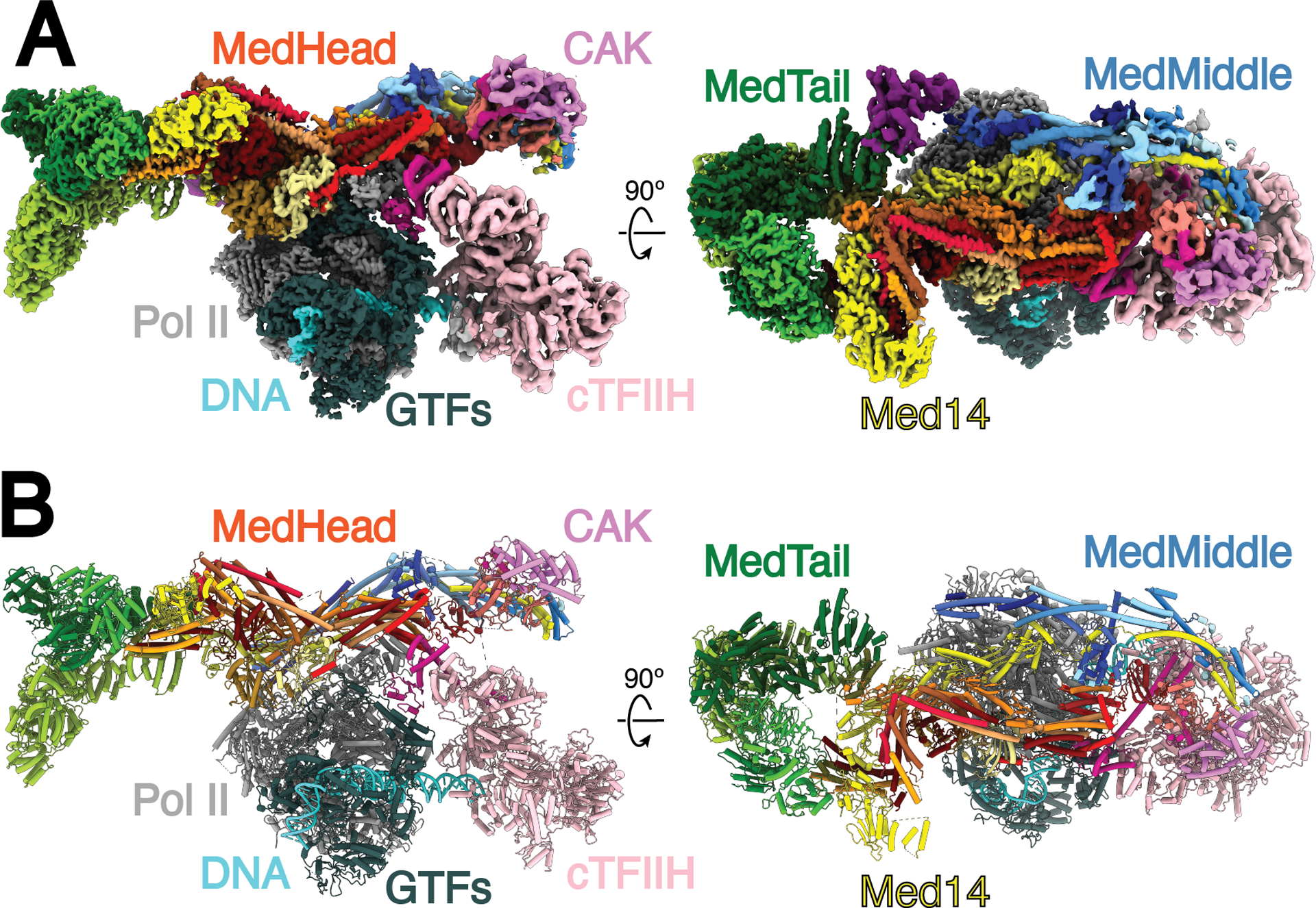
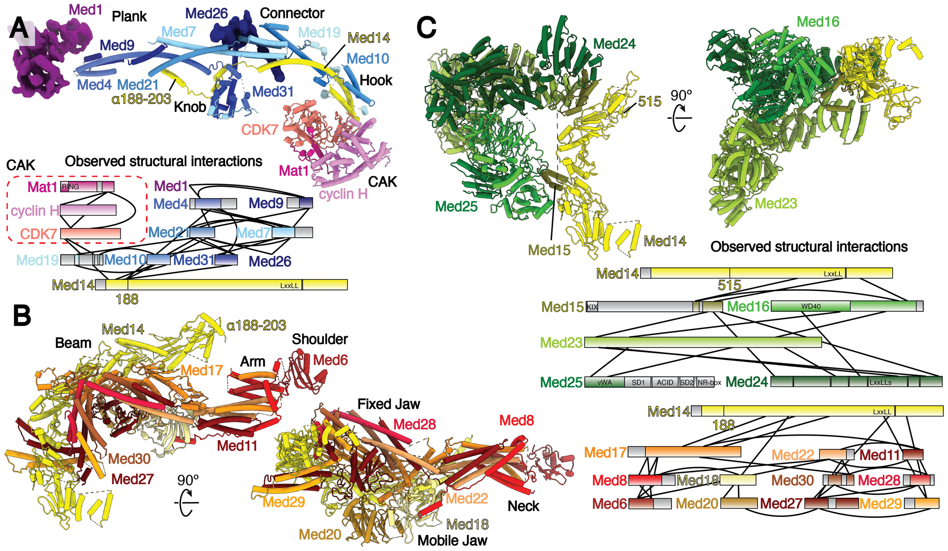
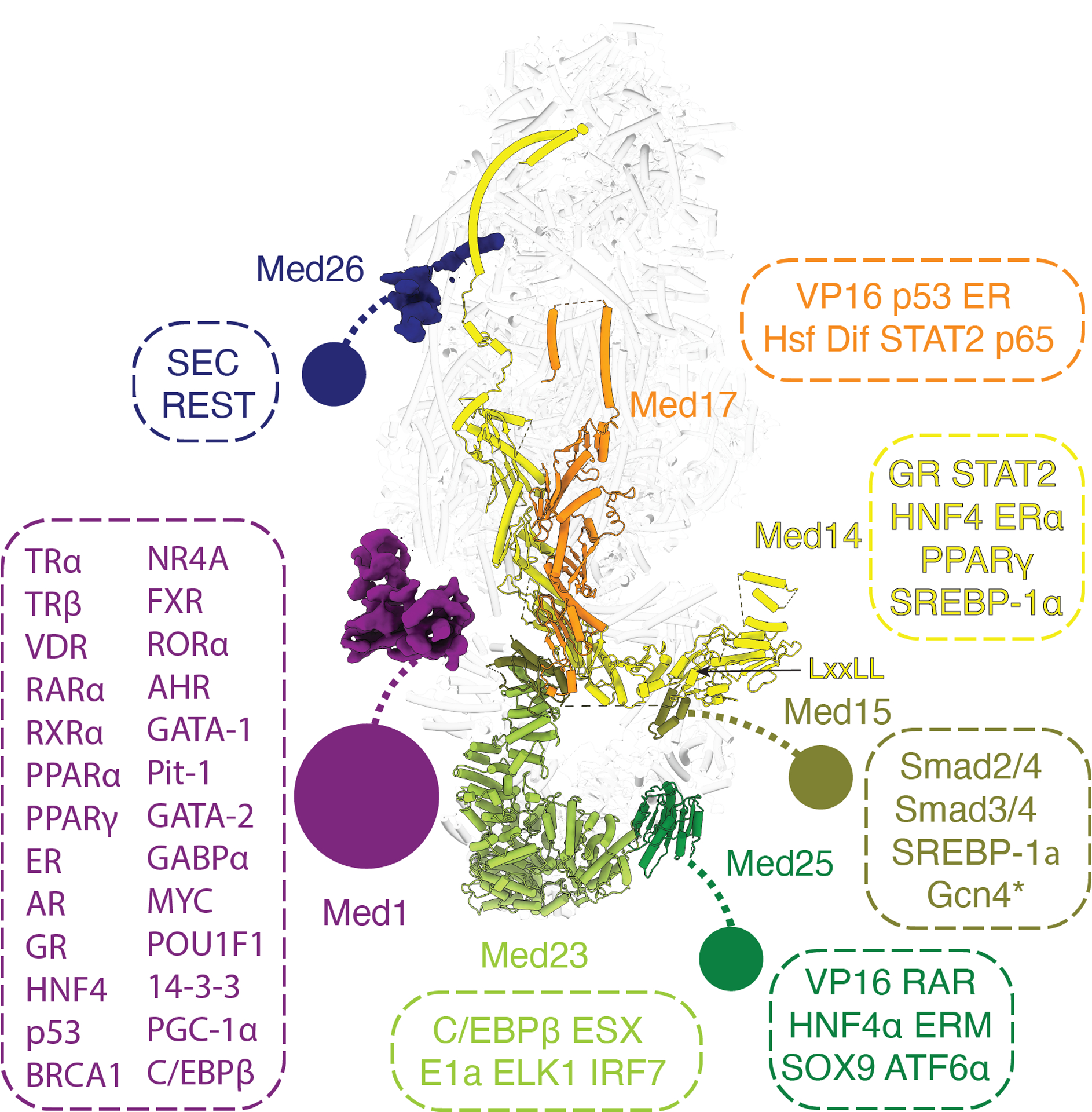
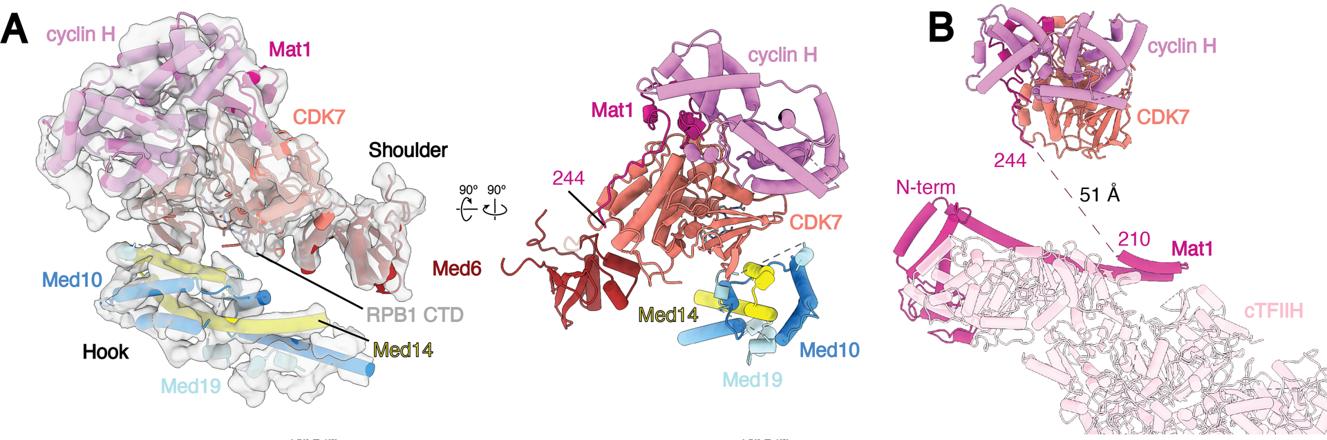
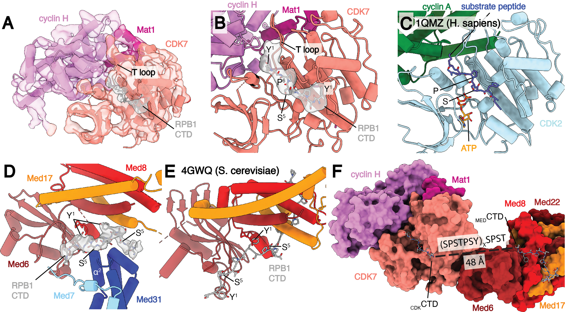
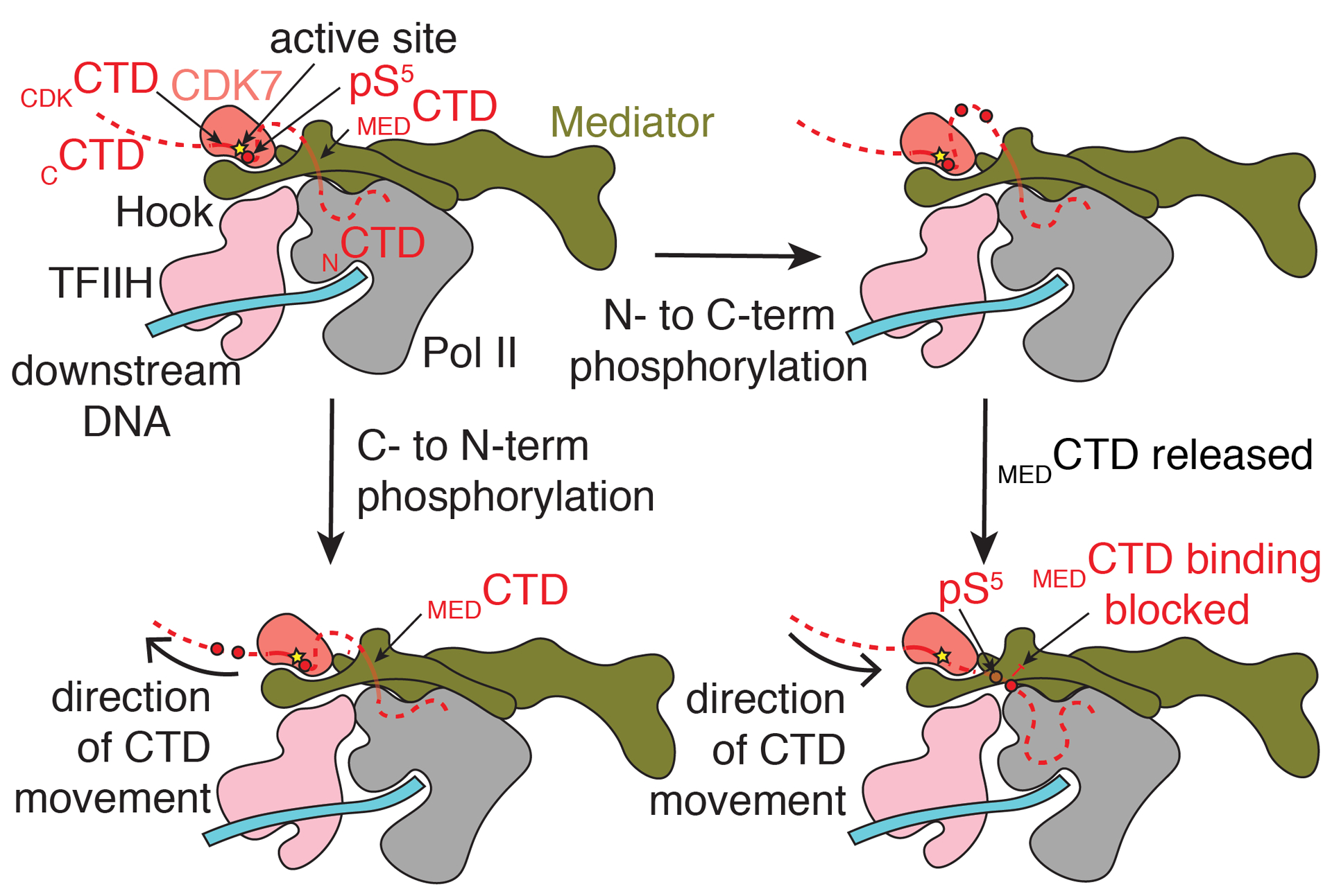
Eukaryotic transcription requires the assembly of a multisubunit preinitiation complex (PIC) composed of RNA polymerase II (Pol II) and the general transcription factors. The coactivator Mediator is recruited by transcription factors, facilitates the assembly of the PIC, and stimulates phosphorylation of the Pol II C-terminal domain (CTD) by the TFIIH subunit CDK7. Here, we present the cryo-electron microscopy structure of the human Mediator-bound PIC at a resolution below 4 angstroms. Transcription factor binding sites within Mediator are primarily flexibly tethered to the tail module. CDK7 is stabilized by multiple contacts with Mediator. Two binding sites exist for the Pol II CTD, one between the head and middle modules of Mediator and the other in the active site of CDK7, providing structural evidence for Pol II CTD phosphorylation within the Mediator-bound PIC.


Conflict of interest statement

Competing interests: Authors declare no competing interests.

Figures

Fig. 1.
Fig. 1.
Structure of the human Mediator-bound pre-initiation complex. A) Composite density map for Med-PIC built from the focused refinement maps for cPIC, cTFIIH, MedHead, MedMiddle-CAK, Med14C, MedTail, and Med1. The colors of the subunits will be repeated throughout the manuscript. B) Model of the human Mediator-bound pre-initiation complex. Gray, Pol II; Dark Gray, general transcription factors; Pink, TFIIH core; Salmon, CDK7; Violet, cyclin H; Medium Violet Red, Mat1; Cyan, DNA, Reds, MedHead; Blues, MedMiddle; Yellow, Med14; Greens, MedTail.
Fig. 2.
Fig. 2.
Models and observed structural interactions for human Mediator. (A-C) Model and observed structural interaction diagram for MedMiddle and the CAK module of TFIIH (A), MedHead (B), and MedTail (C). The N-terminus of the scaffold subunit Med14 extends the length of MedMiddle. Putative density for Med1 and Med26 are shown and colored purple and dark blue, respectively. The C-terminus of Med14 forms extensive interactions with MedHead. MedTail also interacts with the C-terminus of Med14, but on the opposite face. Portions for which models were built are shown in color; unmodeled sections are shown in gray. Known domains are shown with a light-to-dark (top-to-bottom) gradient. Everything else is shown with a dark-to-light gradient. Models colored as in Fig. 1.
Fig. 3.
Fig. 3.
Location of Mediator domains and subunits that interact with transcriptional activators or elongation factors. Flexible tethered domains are indicated by solid circles connected by dashed lines. All interactions shown are between human factors except Gcn4 which is from yeast and indicated by an asterisk.
Fig. 4.
Fig. 4.
Structure of TFIIH within Med-PIC A) Docking of the CAK module (CDK7, cyclin-H, and Mat1) within the MedMiddle-CAK density. The CAK module of TFIIH is stabilized in the Med-PIC by interactions between CDK7 and Med6, the N-terminus of Med14, and a small fragment of Med19. B) The model of the complete human TFIIH complex places the two modeled segments of Mat1 (1–210, 244–308) close to each other. The missing 34 residues can easily span the 51 Å distance between the termini. Models are colored as in Figure 1.
Fig. 5.
Fig. 5.
Location of RPB1 CTD binding in Med-PIC. A) Structure of the TFIIH CAK module. Segmented map of MedMiddle-CAK shows clear density representing an active conformation of the T-loop of CDK7 and density for Pol II CTD in the active site of CDK7. B) Model of the CAK module with density observed for the CDKCTD in the active site. A consensus sequence of the Pol II CTD is modeled due to limited resolution. The T-loop is in the extended, active conformation. C) Model of the CDK2-cyclin A-substrate peptide structure shows high similarity to the CAK module structure with the conserved SP motif that is common to substrates of both enzymes. D) Model and density of MEDCTD with interacting subunits of MedHead and MedMiddle. S5 makes close contacts with α2 of Med31, preventing binding of phosphorylated repeats in this location. E) Model of MEDCTD in the yeast MedHead crystal structure shows a more extensive interface between MEDCTD and MedHead than in the Med-PIC, likely due to the presence of MedMiddle in the Med-PIC. F) View of CDKCTD and MEDCTD within the human Med-PIC structure. Based on the directionality of the CTD, CDKCTD is C-terminal to MEDCTD, and the gap between them would require at least two repeats of the CTD. MedMiddle is hidden for easier visibility. Models are colored as in Figure 1. Annotated domains of Mediator are labeled in black.
Fig. 6.
Fig. 6.
Model for phosphorylation of the Pol II CTD by CDK7. MEDCTD binding positions the rest of the CTD in the CDK7 active site. Following phosphorylation, indicated by a red circle, translocation of the CTD towards the N-terminus (bottom) would place phosphorylated repeats further from the nascent RNA emerging from Pol II. Separation of Mediator and Pol II would be difficult without separation of the CAK module and Mediator. Translocation of the CTD towards the C-terminus would position phosphorylated repeats to block binding of the CTD at MEDCTD, a possible way to favor disassembly of Med-PIC. Phosphorylated repeats would also be significantly closer to the RNA exit tunnel of Pol II to recruit the capping complex properly. CAK = cyclin-activated kinase module; CTD = C-terminal domain of RPB1; pS5 = phosphorylated serine 5 residue (red circle).

References

    1. Thomas MC, Chiang C-M, The general transcription machinery and general cofactors. Critical reviews in biochemistry and molecular biology 41, 105--178 (2006). - PubMed
    1. Soutourina J, Transcription regulation by the Mediator complex. Nature Publishing Group 34, 1--13 (2017).
    1. Nogales E, Louder RK, He Y, Structural Insights into the Eukaryotic Transcription Initiation Machinery. Annual review of biophysics 46, 59--83 (2017). - PMC - PubMed
    1. Bernecky C, Grob P, Ebmeier CC, Nogales E, Taatjes DJ, Molecular architecture of the human Mediator-RNA polymerase II-TFIIF assembly. PLoS Biol 9, e1000603 (2011). - PMC - PubMed
    1. El Khattabi L et al., A Pliable Mediator Acts as a Functional Rather Than an Architectural Bridge between Promoters and Enhancers. Cell 178, 1145--1158.e1120 (2019). - PMC - PubMed

Publication types

MeSH terms