. 2021 Mar 12;371(6534):eabc0956.
doi: 10.1126/science.abc0956.

Lipid presentation by the protein C receptor links coagulation with autoimmunity

Affiliations

Lipid presentation by the protein C receptor links coagulation with autoimmunity

Nadine Müller-Calleja et al. Science. .

Abstract

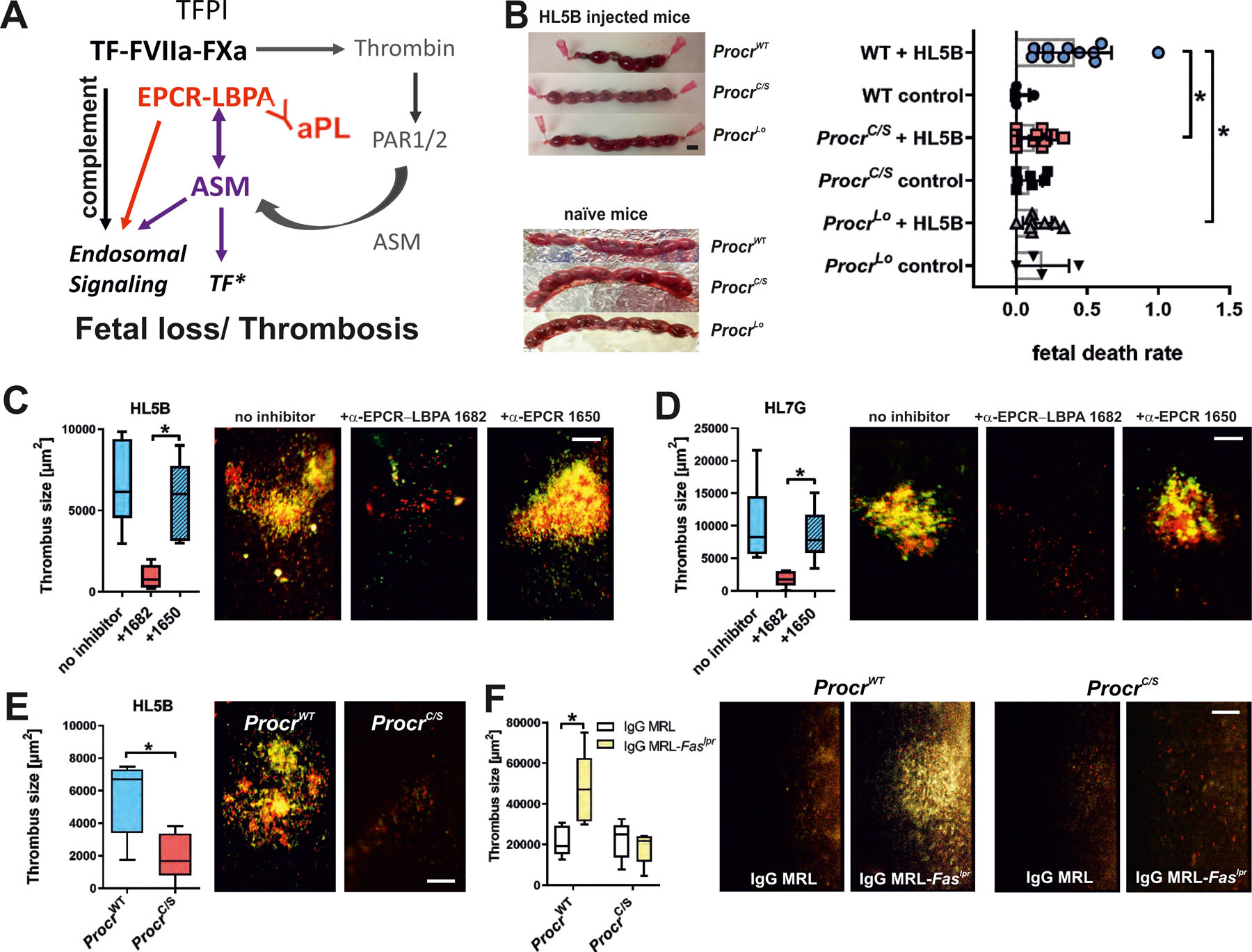
Antiphospholipid antibodies (aPLs) cause severe autoimmune disease characterized by vascular pathologies and pregnancy complications. Here, we identify endosomal lysobisphosphatidic acid (LBPA) presented by the CD1d-like endothelial protein C receptor (EPCR) as a pathogenic cell surface antigen recognized by aPLs for induction of thrombosis and endosomal inflammatory signaling. The engagement of aPLs with EPCR-LBPA expressed on innate immune cells sustains interferon- and toll-like receptor 7-dependent B1a cell expansion and autoantibody production. Specific pharmacological interruption of EPCR-LBPA signaling attenuates major aPL-elicited pathologies and the development of autoimmunity in a mouse model of systemic lupus erythematosus. Thus, aPLs recognize a single cell surface lipid-protein receptor complex to perpetuate a self-amplifying autoimmune signaling loop dependent on the cooperation with the innate immune complement and coagulation pathways.


Conflict of interest statement

Competing interests: A provisional patent application for the discoveries described in the paper has been filed (US 62/955,060).

Figures

Figure 1:
Figure 1:. EPCR is an aPL receptor.
(A) EPCR-dependent induction of IFN-regulated genes and Tnf in mouse monocytes after 1 hour of stimulation with LPS, aPL HL5B, aPL HL7G, or TLR7 agonist R848; relative expression induced by aPLs was normalized to IgG isotype control; n=6, *P<0.05 compared to stimulation without inhibitor; two-way ANOVA, Sidak’s multiple comparisons test. (B) Volcano plot of aPL HL7G-induced transcripts, including Stat1, a known IFN-regulated gene. (C) Induction of Tnf in CD115+ splenic monocytes from ProcrC/S or strain matched ProcrWT mice (upper panel) and human trophoblast cells (lower panel) stimulated for 1 or 3 hours with IgG (100 µg/ml) isolated from APS patients with confirmed reactivity to cardiolipin alone (α-CL), β2GPI alone (α-β2GPI), or dual reactivity (α-CL/β2GP). Human trophoblast cells were pretreated with either non-inhibitory α-EPCR 1489 or inhibitory α-EPCR 1496; *P<0.0001; one-way ANOVA. (D) Live-cell imaging of aPL HL5B IgG or F(ab′)2 colocalization (green) with cholera toxin B (CTB; magenta) or EPCR (blue) using non-inhibitory α-EPCR 1489 in human MM1 cells. Nuclei were stained with Hoechst 33342 (gray). Quantification of colocalization; n=3 ROI (regions of interest) consisting of at least three cells, *P<0.0001; one-way ANOVA. (E) Live-cell imaging of HL5B internalization (green) in monocytes of the indicated mouse strains with CTB or LysoTracker counterstaining (magenta). Nuclei were stained with Hoechst 33342 (blue); bar=5 µm; n=3 ROI, *P<0.0001; one-way ANOVA.
Figure 2:
Figure 2:. EPCR presents late endosomal lysobisphosphatidic acid (LBPA) on the cell surface.
(A) Effect of blocking mouse EPCR with α-EPCR 1682 versus α-EPCR 1650 on Tnf mRNA induction after 3 hours of stimulation of mouse monocytes by aPL HL5B and HL7G; n=6, *P<0.0001; one-way ANOVA. (B) Effect of α-EPCR on aPL HL5B and HL7G (green) internalization in mouse CD115+ spleen monocytes with CTB or LysoTracker counterstaining (magenta). Bar=5 µm. Quantification of colocalization; n=3 ROI consisting of at least three cells, *P<0.0001; one-way ANOVA. (C) Flow cytometric detection of EPCR, LBPA, and FXa as marker for inhibited TF complex formation on CD115+ spleen monocytes isolated from the indicated mouse strains. Effect of pretreatment with 10 µM LBPA for 10 min on surface staining by the indicated antibodies in comparison to isotype control (gray). (D) Competition of α-EPCR 1650 and 1682 for binding of FITC-labeled α-LBPA 6C4 to mouse monocytes; n=3, *P≤0.001; two-way ANOVA, Sidak’s multiple comparisons test. (E) Competition of α-LBPA 6C4 for binding of α-EPCR 1682 to mouse monocytes; n=3, *P≤0.001; two-way ANOVA, Sidak’s multiple comparisons test. (F) Effect of pretreatment with LBPA, cardiolipin (CL), and phosphatidylserine (PS) (10 µM) on aPL HL5B signaling in ProcrC/S monocytes. Induction of Tnf mRNA after 3 hours is shown; n=6, *P<0.0001; one-way ANOVA. (G) LBPA loading of purified mouse or human sEPCR evidenced by faster mobility on native gels. (H) Surface plasmon resonance analysis of aPL HL5B binding to purified human sEPCR or sEPCR–LBPA. The affinity calculation was based on a monovalent binding model because no cooperative binding was evident. Affinity for EPCR with the typical structural lipid phosphatidyl choline (PC) (25) (EPCR-PC) was not measurable (NM).
Figure 3:
Figure 3:. aPLs induce EPCR–LBPA activation of cell surface ASM.
(A) aPL-mediated phosphatidylserine exposure (measured by Annexin V-FITC staining) and aPL-mediated TF activation in MM1 cells (measured as procoagulant activity, PCA) were prevented by desipramine; n=6, *P<0.0001; one-way ANOVA. (B) aPL internalization in MM1 cells is blocked by sphingomyelinase inhibitor desipramine. Bar=5 µm. Quantification of colocalization; n=3 ROI consisting of at least three cells, *P=0.002; t-test. (C) aPL-induced ASM activity in MM1 cells is blocked by inhibitors of FXa (Rivaroxaban, NAP5), thrombin (hirudin), and PAR1 cleavage (αPAR1, ATAP2/WEDE15) but not by inhibitors of complement (compstatin), PDI and ARF6; n=3, *P<0.0001; one-way ANOVA. (D) Surface ASM exposure in MM1 cells after 30 min of stimulation with aPL HL5B F(ab′)2. Scale bar=5 µm. Live-cell imaging of EPCR colocalization (green) with cholera toxin B (CTB; magenta) or ASM (blue). Nuclei were stained with Hoechst 33342 (gray). (E) LBPA (10 µM) loading of mouse ProcrC/S cells enables ASM activation in CD115+ monocytes stimulated with HL5B; n=3, *P<0.0001; one-way ANOVA. (F) ASM activity in unstimulated mouse monocytes lysates after addition of sEPCR–LBPA (2.5 µM) is blocked by α-EPCR–LBPA 1682; n=3, *P<0.0001; one-way ANOVA. (G, H) Proximity ligation assays (PLA) with magenta fluorescence dots for ASM and EPCR on ALIX−/− trophoblast cells after 10 min of stimulation with aPL HL5B F(ab′)2 fragments (G) or thrombin (H) with or without LBPA loading. Nuclei were stained with Hoechst 33342 (blue) Scale bar=25 µm. (I) aPL HL5B signaling in ProcrC/S monocytes was restored by adding 10 µM LBPA (S,R), but not by LBPA (S,S) or hemi LBPA. Stimulation time 3 hours; n=6, *P<0.0001; one-way ANOVA. (J) aPL HL5B internalization in ALIX-deficient JAR cells after pretreatment with the indicated phospholipids at 10 µM. Scale bar=5 µm.
Figure 4:
Figure 4:. aPL–EPCR signaling promotes fetal loss and thrombosis.
(A) Schematic representation of the role of ASM in aPL signaling and thrombosis induction. See also fig. S1 for description of the initial reactions following aPL-induced dissociation of the inhibited TF complex. (B) Representative uteri of pregnant mice of the indicated strains. Pregnancy loss was scored at day 15.5 p.c. after injection of 100 µg aPL HL5B on days 8 and 12; *P<0.02; one-way ANOVA. Scale bar=5 mm. (C and D) HL5B- or HL7G-induced thrombosis in WT mice treated with the indicated α-EPCR antibodies. Quantification of thrombus size in vena cava inferior 3 hours after aPL injection; median, interquartile range, and range; n=6–11, *P<0.004; one-way ANOVA. Platelets are shown in red and leukocytes are shown in green. (E and F) Thrombosis induction by aPL HL5B (E) or IgG isolated from age-matched 16-week-old MRL/MpJ control mice or MRL-Faslpr mice with lupus-like syndrome (F) in the indicated mouse strains. Quantification of thrombus size 3 hours after aPL injection; median, interquartile range, and range; n=5, *P=0.0025; two-way ANOVA with Sidak’s multiple comparisons test. Platelets are shown in red and leukocytes are shown in green.
Figure 5:
Figure 5:. TLR7 is required for IFN signaling-dependent expansion of B cells producing EPCR–LBPA-reactive aPLs.
(A) Mice of the indicated genotypes were immunized with aPL HL5B or isotype-matched control IgG and serum anti-cardiolipin titers were determined at the indicated times; n=10, *P<0.005 for Tlr7−/− or Tlr9−/− versus WT; one-way ANOVA. (B to D) Isolated spleen pDCs and B cells from the indicated mouse strains were co-cultured in the presence of TLR7 agonist R848, aPL HL5B, IFN-α, and LBPA as indicated for 8 days, followed by determination of anti-cardiolipin titers; n=6, *P<0.0001; one-way ANOVA. (E) Gating for CD5+CD19+CD27+CD43+ memory type B1a cells and demonstration of their reactivity with negatively charged fluorescent phospholipid vesicles specifically in mice immunized with aPL HL5B, but not isotype-matched IgG. (F) Competition of sEPCR–LBPA, but not sEPCR with phospholipid vesicle binding to B1a cells; n=5, *P<0.0001; one-way ANOVA.
Figure 6:
Figure 6:. EPCR–LBPA signaling drives aPL expansion and autoimmune pathology in vivo.
(A) Mice of the indicated genotypes were immunized with aPL HL5B and anti-cardiolipin titer determined at the indicated times; n=10, *P<0.0001 for ProcrC/S vs. WT; one-way ANOVA. (B) IgG isolated 12 weeks after the start of aPL immunization were used to stimulate human MM1 cells for 1 hour for induction of the indicated genes; n=5, *P<0.05, **P=0.011, ***P<0.0001; two-way ANOVA, Sidak’s multiple comparisons test. (C) MRL-Faslpr lupus-prone mice were treated with the indicated α-EPCR antibodies at an age of 4 weeks (day 0) and anti-cardiolipin titers were determined in serum at the indicated time points; n=5, *P=0.03; **P<0.0001; two-way ANOVA, Sidak’s multiple comparisons test. (D) Antibodies to double stranded (ds) DNA were measured in α-EPCR 1650- and α-EPCR–LBPA 1682-treated MRL-Faslpr mice 2 weeks after the last dose or in 6-week-old MRL/MpJ control or MRL-Faslpr mice; n=4–5, *P<0.0001; one-way ANOVA. (E) Immune cell infiltration of α-EPCR-treated MRL-Faslpr mice; n=5, *P<0.025. (F) Renal pathology scores of α-EPCR-treated MRL-Faslpr mice; n=5, *P=0.0317; Mann–Whitney U test. Scale bars=80 µm.

Comment in

References

    1. Giannakopoulos B, Krilis SA, The pathogenesis of the antiphospholipid syndrome. N. Engl. J Med 368, 1033–1044 (2013). - PubMed
    1. Ruff WE et al., Pathogenic Autoreactive T and B Cells Cross-React with Mimotopes Expressed by a Common Human Gut Commensal to Trigger Autoimmunity. Cell Host Microbe 26, 100–113 e108 (2019). - PMC - PubMed
    1. Garcia D, Erkan D, Diagnosis and Management of the Antiphospholipid Syndrome. N Engl J Med 378, 2010–2021 (2018). - PubMed
    1. Lieby P et al., The clonal analysis of anticardiolipin antibodies in a single patient with primary antiphospholipid syndrome reveals an extreme antibody heterogeneity. Blood 97, 3820–3828 (2001). - PubMed
    1. Prinz N, Hauser F, Lorenz M, Lackner KJ, von Landenberg P, Structural and functional characterization of a human IgG monoclonal antiphospholipid antibody. Immunobiology 216, 145–151 (2011). - PubMed

Publication types

MeSH terms