. 2021 Mar 11;12(1):1606.
doi: 10.1038/s41467-021-21748-6.

Biofilm formation in the lung contributes to virulence and drug tolerance of Mycobacterium tuberculosis

Affiliations

Biofilm formation in the lung contributes to virulence and drug tolerance of Mycobacterium tuberculosis

Poushali Chakraborty et al. Nat Commun. .

Abstract

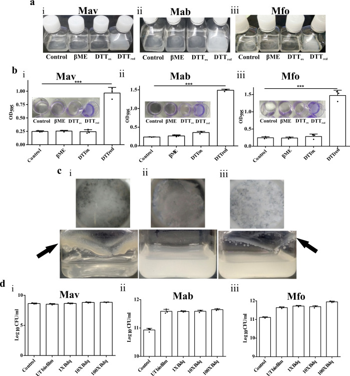
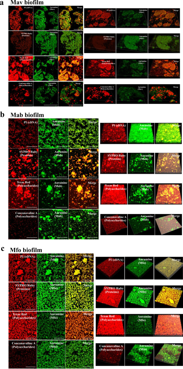
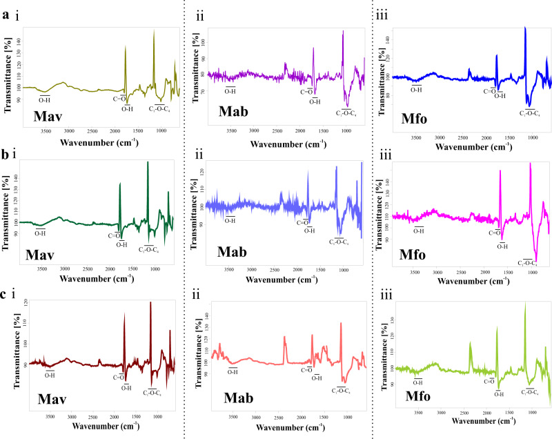
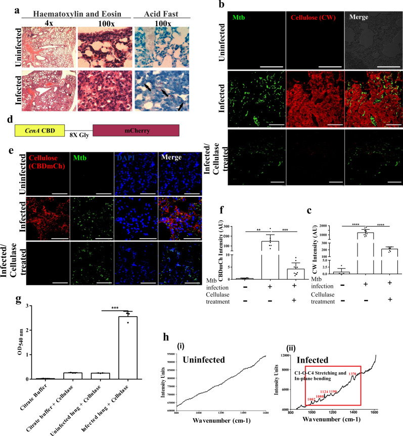
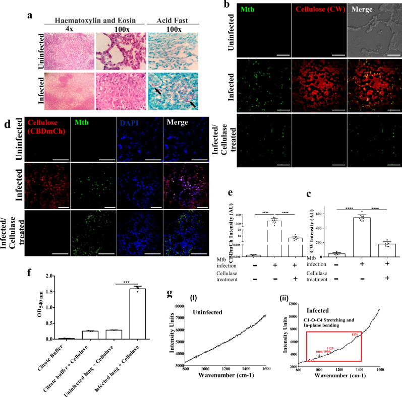
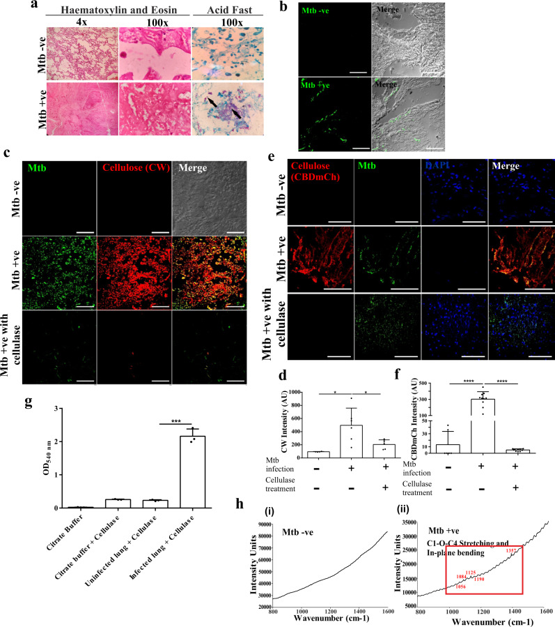
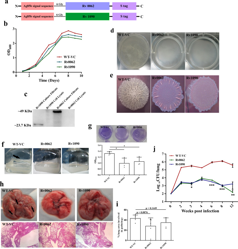
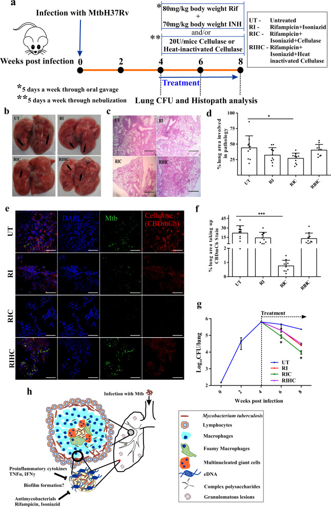
Tuberculosis is a chronic disease that displays several features commonly associated with biofilm-associated infections: immune system evasion, antibiotic treatment failures, and recurrence of infection. However, although Mycobacterium tuberculosis (Mtb) can form cellulose-containing biofilms in vitro, it remains unclear whether biofilms are formed during infection in vivo. Here, we demonstrate the formation of Mtb biofilms in animal models of infection and in patients, and that biofilm formation can contribute to drug tolerance. First, we show that cellulose is also a structural component of the extracellular matrix of in vitro biofilms of fast and slow-growing nontuberculous mycobacteria. Then, we use cellulose as a biomarker to detect Mtb biofilms in the lungs of experimentally infected mice and non-human primates, as well as in lung tissue sections obtained from patients with tuberculosis. Mtb strains defective in biofilm formation are attenuated for survival in mice, suggesting that biofilms protect bacilli from the host immune system. Furthermore, the administration of nebulized cellulase enhances the antimycobacterial activity of isoniazid and rifampicin in infected mice, supporting a role for biofilms in phenotypic drug tolerance. Our findings thus indicate that Mtb biofilms are relevant to human tuberculosis.


Conflict of interest statement

The authors declare no competing interests.

Figures

Fig. 1
Fig. 1. Thiol reductive stress leads to drug-tolerant biofilm formation in NTMs.
a Exponential cultures of Mav (i), Mab (ii) and Mfo (iii) were exposed to 6 mM of βME, oxidized, or reduced DTT individually. Only exposure to reduced DTT resulted in biofilm formation. b Cultures of Mav, Mab, and Mfo were exposed to 6 mM of DTT to induce biofilm formation. CV assays were performed to quantitate Mav (i), Mab (ii), and Mfo (iii) biofilms. c Thiol reductive stress-induced submerged biofilm of Mav (i), Mab (ii) and Mfo (iii) in standing culture (upper panel) and under slow shaking (lower panel). Biofilms formed are indicated with black arrows. d Colony forming units of Mav (i), Mab (ii) and Mfo (iii) were estimated after treatment with 1×, 10× and 100× MIC of Bedaquiline by degrading the biofilms of the NTMs using cellulase and plating them on 7H11 agar after serially diluting them. The column bar graphs were plotted using GraphPad Prism 6 and represented as mean (±s.e.m). Statistical significance was determined using Student’s t-test (two tailed). For b (i), (ii) and (iii), ***P = 0.0003, ***P < 0.0001 and ***P = 0.0002 respectively. All data are representative of three independent biological experiments performed in triplicates. All source data are provided as a Source Data file.
Fig. 2
Fig. 2. Characterization of thiol reductive stress-induced biofilm matrices in NTMs.
ac To characterize the chemical nature of the extracellular matrix of thiol reductive stress-induced biofilms, Mav (a), Mab (b) and Mfo (c) cultures were subjected to 6 mM DTT for 29 h and then stained with PI (for eDNA), SYPRO Ruby (for proteins), Texas red (for polysaccharides) and Concanavalin A (for α-glucopyranosyl and α-mannopyranosyl residues). NTM cells were stained with phenolic Auramine O – Rhodamine B and analyzed using CLSM. All data are representative of three independent biological experiments. Scale bars indicate 10 µm.
Fig. 3
Fig. 3. Characterization of biofilm matrix polysaccharides.
ac Cellulose was purified from submerged biofilms (a), pellicle biofilms (b) and macrocolony biofilms (c) of Mav (i), Mab (ii) and Mfo (iii) and then subjected to FTIR analysis. All the experiments are representatives of at least three biological experiments performed with three technical replicates each.
Fig. 4
Fig. 4. Mtb forms biofilms inside the in vitro granulomas.
a Histopathology staining of 5 μm thick sections of uninfected and Mtb infected PBMCs placed on a collagen based 3D matrix visualized on a compound microscope. b CW and Auramine staining of the granuloma sections suggesting encapsulation of Mtb bacilli within a cellulose-rich extracellular matrix. All data are representative of three independent biological experiments. Scale bars correspond to 10 μm.
Fig. 5
Fig. 5. Mtb forms biofilms inside the mice lungs.
a Histopathology of granulomatous lesions in the lungs of Mtb infected mice (n = 5) in contrast to uninfected ones, as indicated by H & E staining. Clusters of acid-fast bacilli are denoted by arrows. b, c CW staining and corresponding quantitation (using NIS elements) of Mtb infected, uninfected and Cellulysin Cellulase treated lung sections of mice showing Mtb bacilli (stained with Auramine B–Rhodamine O) encased within a polysaccharide-rich matrix. d The schematic construct of the CBD-mCh probe wherein the Cellulose Binding Domain (CBD) of CenA is fused to fluorescent probe mCherry through an octa-glycine linker. e, f CBD-mCh staining and quantification (using NIS elements) for the experiment described above. g DNS assay on uninfected and Mtb infected mice lungs after treatment with cellulase to check the yield of reducing sugar glucose. h Raman microscopy of (i) uninfected and (ii) Mtb infected mice lung sections showing cellulose specific peaks. The column bar graphs presented in figures c, f and g have been plotted in GraphPad Prism 6 and represented as mean (±s.e.m). Statistical significance was determined using Student’s t-test (two tailed). For c, ****P < 0.0001, for f, **P = 0.0022 and ***P < 0.0001, and for g, ***P < 0.0001. All data are representative of three independent biological experiments performed in triplicates. Scale bars correspond to 50 μm. All source data are provided as a Source Data file.
Fig. 6
Fig. 6. Mtb forms biofilms inside the lungs of non-human primates.
a Histopathology of granulomatous lesions in the lungs of Mtb infected rhesus macaques in contrast to uninfected ones, as indicated by H & E staining. Clusters of acid-fast bacilli are indicated by arrows. b, c Cellulose staining with CW and corresponding quantitation (using NIS elements) of Mtb infected, uninfected and Cellulysin cellulase treated lung sections of rhesus macaques showing Mtb bacilli stained with Auramine B–Rhodamine O. d, e CBD-mCh staining for cellulose and subsequent quantification (using NIS elements) of Mtb infected, uninfected and Cellulysin cellulase treated lung sections of rhesus macaques showing Mtb bacilli (stained with Auramine B–Rhodamine O). f DNS assay of cellulase treated lung tissue blocks from uninfected and Mtb infected rhesus macaque. g Raman microscopy of (i) uninfected and (ii) Mtb infected rhesus macaque lung sections showing cellulose specific peaks in the infected lungs. The column bar graphs presented in figures c, e and f have been plotted in GraphPad Prism 6 and represented as mean (±s.e.m). Statistical significance was determined using Student’s t-test (two tailed). For c ****P < 0.0001, for e ****P < 0.0001 and for f ***P < 0.0001. All data are representative of three independent biological experiments performed in triplicates. Scale bars correspond to 50 μm. All source data are provided as a Source Data file.
Fig. 7
Fig. 7. Mtb forms biofilms in the human lungs.
a H & E staining of human lung tissue sections. The clusters of acid-fast bacilli are denoted by arrows. b FISH with Mtb specific 16 S primers was utilized for identifying lung tissues infected with Mtb. c, d Cellulose was visualized using CW staining in Mtb positive and Mtb negative human lung tissue sections. Mtb bacilli were visualized using Auramine B–Rhodamine O staining. Mtb positive tissue sections were also stained with CW after Cellulysin Cellulase treatment. e, f CBD-mCh staining and visualization of Mtb (Auramine B–Rhodamine O) in Mtb positive and Mtb negative lung tissue sections. g DNS assay on Mtb positive and Mtb negative human lung tissue blocks after treatment with Cellulysin Cellulase. h Raman microscopy of (i) Mtb positive and (ii) Mtb negative human lung sections. The column bar graphs presented in figures d, f and g have been plotted in GraphPad Prism 6 and represented as mean (±s.e.m). Statistical significance was determined using Student’s t-test (two tailed). *P < 0.05, **P < 0.001, ***P < 0.0001. For d *P = 0.0169 (column A vs column B) and *P = 0.0401 (column B vs column C), for f ****P < 0.0001 and for g ***P = 0.0001 All data are representative of three independent biological experiments performed in triplicates. Scale bars correspond to 50 μm. All source data are provided as a Source Data file.
Fig. 8
Fig. 8. Mtb biofilms protect the bacilli from the host immune system.
a Construct design for engineering Mtb strains overexpressing endogenous Mtb cellulases (Rv0062 and Rv1090). b Growth profile of planktonic cultures of Mtb strain with vector control or those overexpressing Rv0062 and Rv1090. c The culture supernatant of the above-described cultures was used in Western blot analysis. Antibodies specific to S-tag were utilized to detect the secretion of cellulases outside Mtb cells. Uncropped image of the blot is provided in Source Data. d Pellicle biofilm profile of WT-VC, Rv0062, and Rv1090, suggesting that cellulase overexpressing strains were severely defective in pellicle formation. e Macrocolonies of WT-VC, Rv0062, and Rv1090 on 7H11 agar supplemented with Congo red and Coomassie Brilliant Blue G250 dye. f, g Biofilms of WT-VC, Rv0062, and Rv1090 induced by thiol reductive stress using DTT shows defect in the engineered strains, as indicated by CV assay quantitation. The biofilm formed by WT-VC (f) has been shown by black arrow. h C57BL/6 J mice were independently infected with vector control (WT-VC) or strains overexpressing cellulases Rv0062 and Rv1090 using a low dose of aerosols. Gross lung pathology (upper panel) and histopathology (lower panel) of mice lungs infected with WT-VC, Rv0062, and Rv1090. i Lung pathology post-infection with Mtb strains, as mentioned in h, was quantitated using ImageJ software. j Survival inside mice lungs (n = 5) was estimated using CFU analysis. The data presented in figures g, i and j have been plotted in GraphPad Prism 6 and represented as mean (±s.e.m). Statistical significance of j was determined using two way ANOVA. *P < 0.05, **P < 0.01, ***P < 0.001. And the statistical significance of g and i was determined using Student’s t-test (two tailed). For g *P = 0.0375 (column A vs column B) and *P = 0.0431 (column A and column C). All data are representative of three independent biological experiments performed in triplicates unless otherwise mentioned. Scale bars correspond to 200 μm. All source data are provided as a Source Data file.
Fig. 9
Fig. 9. Mycobacterial biofilms protect the resident bacilli from antimycobacterial agents.
a Schematic representation of Mtb infection followed by drug treatment regime in mice (n = 4, an experiment performed twice). bd Gross lung pathology, histopathology analysis, and pathology scoring of mice lungs infected with Mtb, followed by treatment. e, f CBD-mCh staining and quantification (using NIS elements) in lung sections of Mtb infected mice subsequently treated as described in a. g Survival of Mtb inside mice 2 and 4 weeks post-treatment with Rif and INH orally and/or cellulase or heat-inactivated cellulase through nebulization. h Schematic representation of Mtb biofilms inside the human lungs. Infection with Mtb leads to the formation of granulomatous lesions in the human lungs. A granuloma is a compact immunological structure predominated by macrophages at the center, which, in turn, differentiate into other cell types like foamy macrophages and multinucleate giant cells having lipid droplets, in a specialized manner. The periphery of the granuloma is comprised of T and B lymphocytes. However, in our proposition, the necrotic core of the granuloma, consisting of Mtb bacilli is structured as a biofilm, which is composed of extracellular matrix including cellulose as one of the major components, can act as a barrier to both antimycobacterial agents as well as a host defense mechanism, thus rendering protection to the bacteria. The data presented in figures d, f and g have been plotted in GraphPad Prism 6 and represented as mean (±s.e.m). Statistical significance of g was determined using two way ANOVA, *P < 0.05 and statistical significance of d and f were determined using Student’s t-test (two tailed). *P < 0.05, ***P < 0.0001. For d *P = 0.0205 and for f ***P < 0.0001. All data are representative of three independent biological experiments performed in triplicates unless otherwise mentioned. Scale bars correspond to 200 μm. All source data are provided as a Source Data file.

References

    1. Burmolle M, et al. Biofilms in chronic infections - a matter of opportunity - monospecies biofilms in multispecies infections. FEMS Immunol. Med. Microbiol. 2010;59:324–336. doi: 10.1111/j.1574-695X.2010.00714.x. - DOI - PubMed
    1. Domingue JC, Drewes JL, Merlo CA, Housseau F, Sears CL. Host responses to mucosal biofilms in the lung and gut. Mucosal Immunol. 2020;13:413–422. doi: 10.1038/s41385-020-0270-1. - DOI - PMC - PubMed
    1. Ciofu O, Rojo-Molinero E, Macia MD, Oliver A. Antibiotic treatment of biofilm infections. APMIS. 2017;125:304–319. doi: 10.1111/apm.12673. - DOI - PubMed
    1. Lebeaux D, Ghigo JM, Beloin C. Biofilm-related infections: bridging the gap between clinical management and fundamental aspects of recalcitrance toward antibiotics. Microbiol. Mol. Biol. Rev. 2014;78:510–543. doi: 10.1128/MMBR.00013-14. - DOI - PMC - PubMed
    1. McCune RM, Feldmann FM, Lambert HP, McDermott W. Microbial persistence. I. The capacity of tubercle bacilli to survive sterilization in mouse tissues. J. Exp. Med. 1966;123:445–468. doi: 10.1084/jem.123.3.445. - DOI - PMC - PubMed

Publication types

MeSH terms

LinkOut - more resources