

Development and validation of a CT-based radiomics nomogram for preoperative prediction of tumor histologic grade in gastric adenocarcinoma
- PMID: 33707930
- PMCID: PMC7941693
- DOI: 10.21147/j.issn.1000-9604.2021.01.08
Development and validation of a CT-based radiomics nomogram for preoperative prediction of tumor histologic grade in gastric adenocarcinoma
Abstract
Objectives: To develop and validate a radiomics nomogram for preoperative prediction of tumor histologic grade in gastric adenocarcinoma (GA).
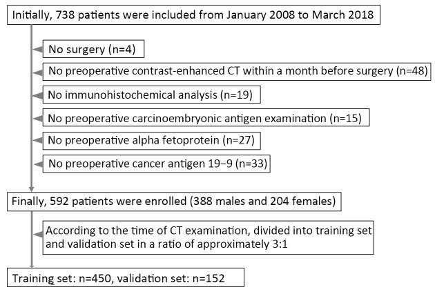
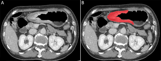
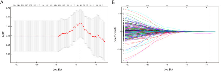
Methods: This retrospective study enrolled 592 patients with clinicopathologically confirmed GA (low-grade: n=154; high-grade: n=438) from January 2008 to March 2018 who were divided into training (n=450) and validation (n=142) sets according to the time of computed tomography (CT) examination. Radiomic features were extracted from the portal venous phase CT images. The Mann-Whitney U test and the least absolute shrinkage and selection operator (LASSO) regression model were used for feature selection, data dimension reduction and radiomics signature construction. Multivariable logistic regression analysis was applied to develop the prediction model. The radiomics signature and independent clinicopathologic risk factors were incorporated and presented as a radiomics nomogram. The performance of the nomogram was assessed with respect to its calibration and discrimination.
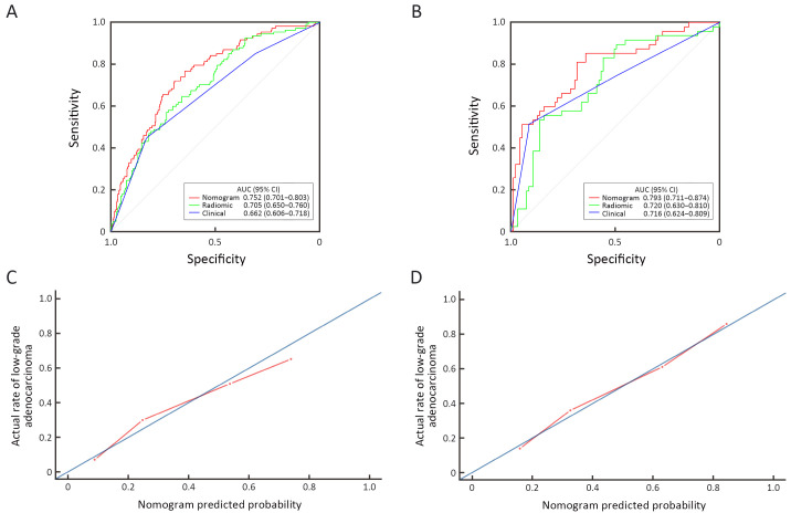
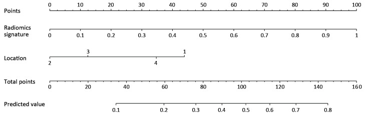
Results: A radiomics signature containing 12 selected features was significantly associated with the histologic grade of GA (P<0.001 for both training and validation sets). A nomogram including the radiomics signature and tumor location as predictors was developed. The model showed both good calibration and good discrimination, in which C-index in the training set, 0.752 [95% confidence interval (95% CI): 0.701-0.803]; C-index in the validation set, 0.793 (95% CI: 0.711-0.874).
Conclusions: This study developed a radiomics nomogram that incorporates tumor location and radiomics signatures, which can be useful in facilitating preoperative individualized prediction of histologic grade of GA.
Keywords: Adenocarcinoma; X-ray computed tomography; histologic grade; nomograms; stomach neoplasm.
Copyright © 2021 Chinese Journal of Cancer Research. All rights reserved.
Figures
Similar articles
-
Development and Validation of a Radiomics Nomogram for Preoperative Prediction of Lymph Node Metastasis in Colorectal Cancer.J Clin Oncol. 2016 Jun 20;34(18):2157-64. doi: 10.1200/JCO.2015.65.9128. Epub 2016 May 2. J Clin Oncol. 2016. PMID: 27138577
-
Development and validation of a radiomics nomogram for identifying invasiveness of pulmonary adenocarcinomas appearing as subcentimeter ground-glass opacity nodules.Eur J Radiol. 2019 Mar;112:161-168. doi: 10.1016/j.ejrad.2019.01.021. Epub 2019 Jan 22. Eur J Radiol. 2019. PMID: 30777206
-
A Radiomics Nomogram Integrated With Clinic-Radiological Features for Preoperative Prediction of DNA Mismatch Repair Deficiency in Gastric Adenocarcinoma.Front Oncol. 2022 Jul 1;12:865548. doi: 10.3389/fonc.2022.865548. eCollection 2022. Front Oncol. 2022. PMID: 35912185 Free PMC article.
-
A radiomics nomogram for preoperative prediction of microvascular invasion risk in hepatitis B virus-related hepatocellular carcinoma.Diagn Interv Radiol. 2018 May-Jun;24(3):121-127. doi: 10.5152/dir.2018.17467. Diagn Interv Radiol. 2018. PMID: 29770763 Free PMC article.
-
Computed Tomography-based Radiomics Nomogram for the Preoperative Prediction of Tumor Deposits and Clinical Outcomes in Colon Cancer: a Multicenter Study.Acad Radiol. 2023 Aug;30(8):1572-1583. doi: 10.1016/j.acra.2022.11.005. Epub 2022 Dec 23. Acad Radiol. 2023. PMID: 36566155
Cited by
-
A CT-Based Radiomics Nomogram Model for Differentiating Primary Malignant Melanoma of the Esophagus from Esophageal Squamous Cell Carcinoma.Biomed Res Int. 2023 Feb 20;2023:6057196. doi: 10.1155/2023/6057196. eCollection 2023. Biomed Res Int. 2023. PMID: 36860814 Free PMC article.
-
Predictive value of magnetic resonance imaging radiomics-based machine learning for disease progression in patients with high-grade glioma.Quant Imaging Med Surg. 2023 Jan 1;13(1):224-236. doi: 10.21037/qims-22-459. Epub 2022 Oct 18. Quant Imaging Med Surg. 2023. PMID: 36620140 Free PMC article.
-
Radiomics-based assessment of HER2 status and prognosis in gastric cancer: a retrospective dual-center CT study.Abdom Radiol (NY). 2025 Apr 8. doi: 10.1007/s00261-025-04912-0. Online ahead of print. Abdom Radiol (NY). 2025. PMID: 40195138
-
Can PD-L1 expression be predicted by contrast-enhanced CT in patients with gastric adenocarcinoma? a preliminary retrospective study.Abdom Radiol (NY). 2023 Jan;48(1):220-228. doi: 10.1007/s00261-022-03709-9. Epub 2022 Oct 21. Abdom Radiol (NY). 2023. PMID: 36271155 Free PMC article.
-
Prognostic and incremental value of computed tomography-based radiomics from tumor and nodal regions in esophageal squamous cell carcinoma.Chin J Cancer Res. 2022 Apr 30;34(2):71-82. doi: 10.21147/j.issn.1000-9604.2022.02.02. Chin J Cancer Res. 2022. PMID: 35685995 Free PMC article.
References
-
-
Stewart BW, Wild CP. World Cancer Report 2014. IARC: Non-Serial Publications. Available online: https://publications.iarc.fr/Non-Series-Publications/World-Cancer-Reports/World-Cancer-Report-2014
-
-
-
Bosman FT, Carneiro F, Hruban RH, et al. WHO classification of tumours of the digestive system. IARC: Lyon, 2010. Available online: https://publications.iarc.fr/Book-And-Report-Series/Who-Classification-Of-Tumours/WHO-Classification-Of-Tumours-Of-The-Digestive-System-2010
-
LinkOut - more resources
Full Text Sources
Other Literature Sources