. 2021 Mar 5:16:1849-1867.
doi: 10.2147/IJN.S295928. eCollection 2021.

The Effects of Luminescent CdSe Quantum Dot-Functionalized Antimicrobial Peptides Nanoparticles on Antibacterial Activity and Molecular Mechanism

Affiliations

The Effects of Luminescent CdSe Quantum Dot-Functionalized Antimicrobial Peptides Nanoparticles on Antibacterial Activity and Molecular Mechanism

Wanzhen Li et al. Int J Nanomedicine. .

Abstract

Background: With the development of bacterial resistance, the range of effective antibiotics is increasingly becoming more limited. The effective use of nanoscale antimicrobial peptides (AP) in therapeutic and diagnostic methods is a strategy for new antibiotics.

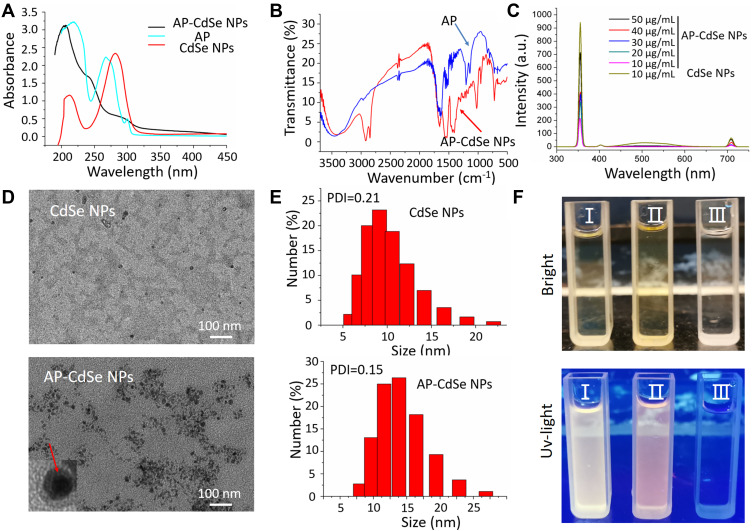
Methods: Combining both AP and cadmium selenide (CdSe) into a composite material may result in a reagent with novel properties, such as enhanced antibacterial activity, fluorescence and favorable stability in aqueous solution.

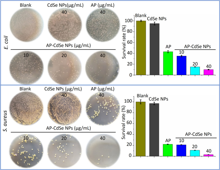
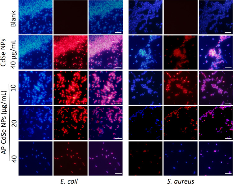
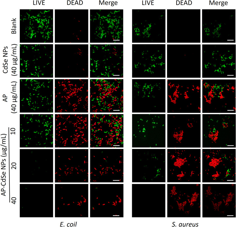
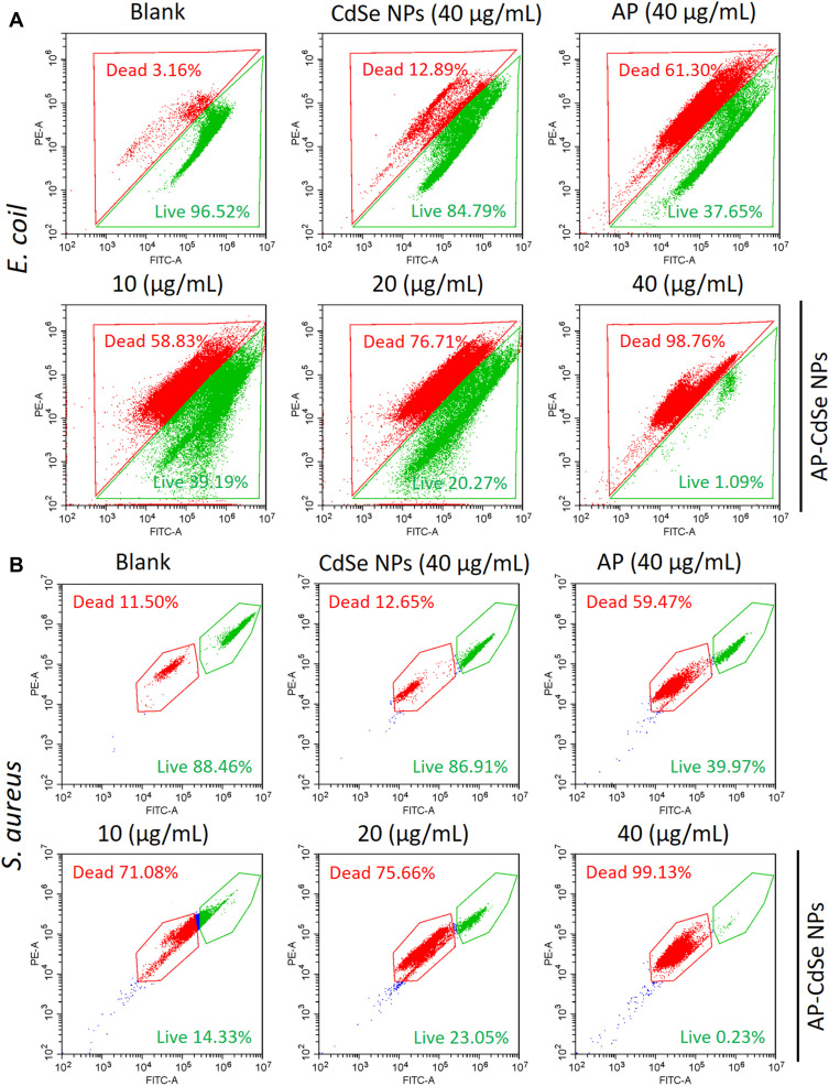
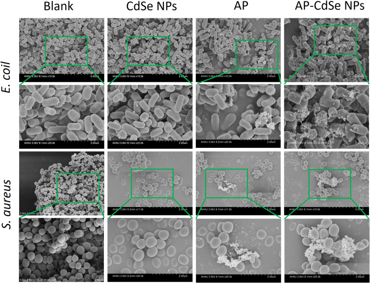
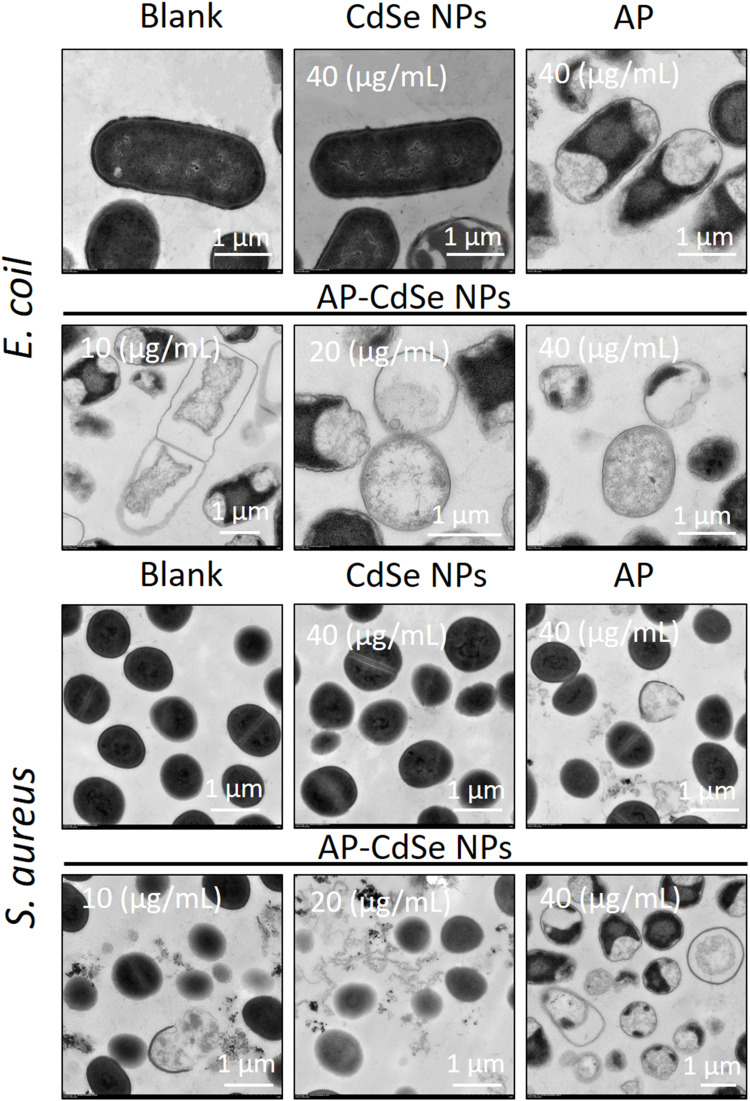
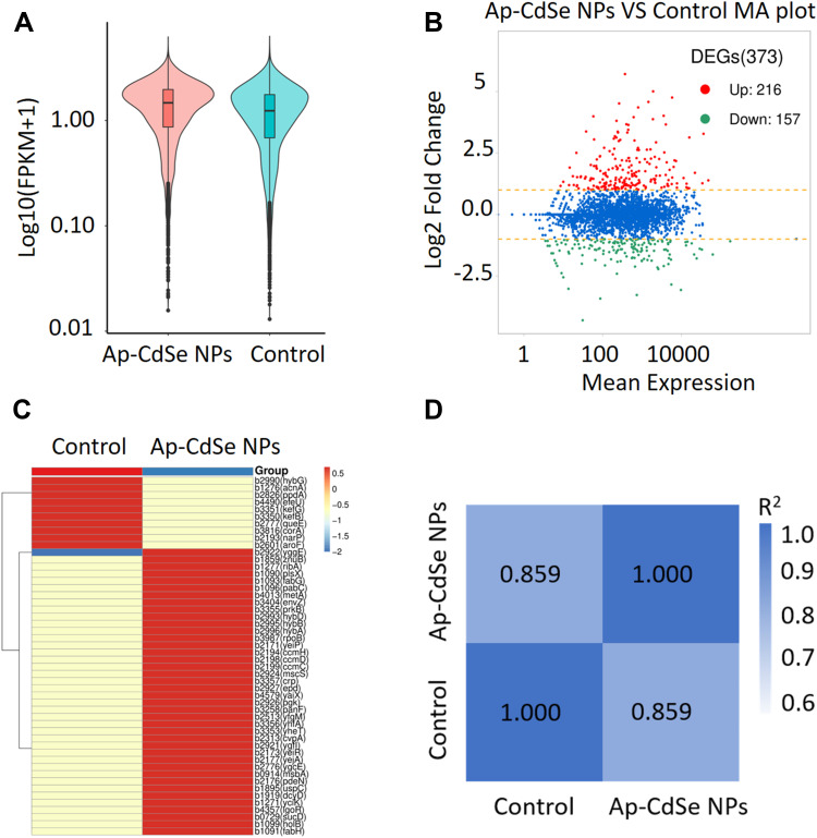
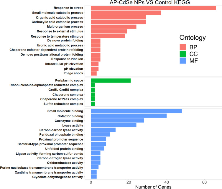
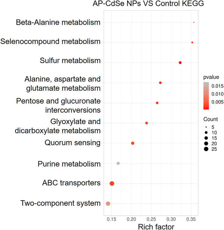
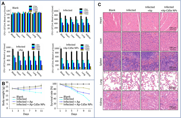
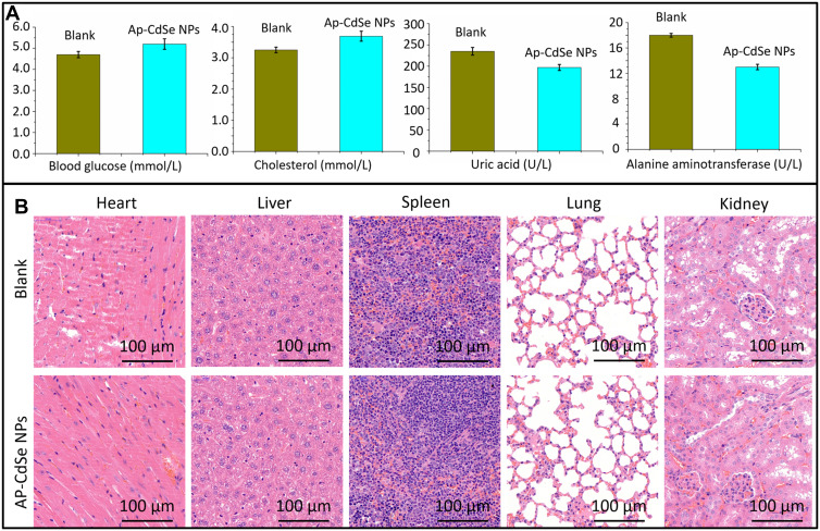
Results: AP-loaded CdSe NPs (AP-CdSe NPs) showed strong antibacterial activity against multidrug-resistant (MDR) Escherichia coli (E. coli) and Staphylococcus aureus (S. aureus) in vitro and in vivo. Colony-forming unit (CFU) and minimum inhibitory concentration (MIC) assays showed that AP-CdSe NPs have highly effective antibacterial activity. The quantitative analysis of apoptosis by flow cytometry analysis further confirmed that MDR E. coli and S. aureus treated with AP-CdSe NPs had death rates of 98.76% and 99.13%, respectively. Also, AP-CdSe NPs was found to inhibit bacterial activity in an in vivo bacteremia model in mice infected with S. aureus. In addition, the antibacterial mechanism of AP-CdSe NPs was determined by RNA sequencing analysis. Gene ontology (GO) analysis and Kyoto encyclopedia of genes and genomes (KEGG) pathway analysis revealed the molecular mechanism of the antibacterial effect of AP-CdSe NPs. Importantly, histopathology analysis, and hematological toxicity analysis indicated that AP-CdSe NPs had few side effects.

Conclusion: These results demonstrate that AP loaded on CdSe NPs had a higher water solubility, bioavailability and antibacterial effect compared with raw AP. This study reports findings that are helpful for the design and development of antibacterial treatment strategies based on AP.

Keywords: CdSe QD; RNASeq; antibacterial mechanism; antibacterial peptides; in vitro and in vivo.


Conflict of interest statement

The authors have declared that there are no competing interests in this work.

Figures

Figure 1
Figure 1
Characterization of AP-CdSe NPs. (A) UV–vis absorption spectra of AP, CdSe NPs and AP-CdSe NPs. (B) Infrared spectra of AP and AP-CdSe NPs. (C) Fluorescence emission spectra of AP-CdSe NPs at various concentrations (10, 20, 30, 40 and 50 µg/mL) and CdSe NPs (10 µg/mL). (D) TEM images of CdSe NPs and AP-CdSe NPs. (E) DLS of CdSe NPs and AP-CdSe NPs. In the inset, the red arrow points to a AP coated CdSe NPs. (F) Digital images of CdSe NPs (I) and AP-CdSe NPs (II) under bright and UV light.
Figure 2
Figure 2
CFU assay of AP-CdSe NPs. Images of bacterial colonies (MDR E. coli and MDR S. aureus) formed on Lysogeny broth-agar plates. PBS was used for the blank group. Corresponding images were graphed using the Origin software. Data are expressed as the mean ± SD (n=9).
Figure 3
Figure 3
Fluorescence microscopy images of MDR E. coli and MDR S. aureus bacterial culture treated with AP-CdSe NPs at various concentrations (10, 20, 40 µg/mL) for 12 h. The PBS was used for the blank group. The CdSe NPs (40 µg/mL) was set as the control group. Cells were stained with DAPI for 30 min are fluorescent blue, and those stained with AP-CdSe NPs are fluorescent red, while those with fluorescent pink color appearance represent merged images of cells. Fluorescence images were captured using an UV illuminator with a 365 nm emission filter for AP-CdSe NPs and a 461 nm emission filters for DAPI. Scale bar = 10 μm.
Figure 4
Figure 4
Confocal fluorescence microscopy assay. MDR E. coli and MDR S. aureus, after treatment with AP-CdSe NPs at various concentrations (10, 20, 40 µg/mL). AP solution (40 µg/mL) and CdSe NPs solution (40 µg/mL) were used as control groups. The PBS-treated group was used as blank group. All cells were stained for 30 min with SYTO 9 (green fluorescence) and PI (red fluorescence). Scale bar = 10 μm.
Figure 5
Figure 5
The results of the apoptotic cell death assay by flow cytometry analysis were statistically analyzed by the CytExpert software. MDR E. coli (A)and MDR S. aureus (B), after incubation with AP-CdSe NPs at various concentrations (10, 20, 40 µg/mL). AP solution (40 µg/mL) and CdSe NPs solution (40 µg/mL) were used as control groups. The PBS-treated group was used as blank group. All cells were subsequently briefly (30 min) stained with SYTO 9 and PI. Data are expressed as the mean ± SD (n=3).
Figure 6
Figure 6
SEM images of MDR E. coli and MDR S. aureus. Bacterial culture treated with AP-CdSe NPs (40 µg/mL). Bacterial culture treated with AP solution (40 µg/mL) and CdSe NPs solution (40 µg/mL) were used as control groups. The PBS-treated group was used as blank group. Green squares indicated the enlarged regions.
Figure 7
Figure 7
TEM images of MDR E. coli and MDR S. aureus. Cell treated with AP-CdSe NPs solutions at various concentrations (10, 20, 40 µg/mL). Bacterial culture treated with AP solution (40 µg/mL) and CdSe NPs solution (40 µg/mL) were used as control groups. The PBS-treated group was used as the blank group.
Figure 8
Figure 8
Differentially expressed genes (DEGs) analyzed in MDR E. coli (AP-CdSe NPs and control). (A) FPKM density distribution of E. coli genes in control and AP-CdSe NPs groups. (B) Number of DEGs comparing the AP-CdSe NPs group with the control group. (C) Gene expression cluster analysis is used to determine the clustering pattern of gene expression in different treatment groups. (D) Pearson correlation coefficient between samples.
Figure 9
Figure 9
Differentially expressed genes (DEGs) GO enrichment analysis results.
Figure 10
Figure 10
Differentially expressed genes (DEGs) KEGG enrichment analysis results.
Figure 11
Figure 11
The antibacterial test in the animal bacteremia model. (A) Bacterial count in blood and major organs (heart, liver, spleen, lungs, and kidneys) in blank, infected, and treatment groups. (B) The survival rate and weight of mice were monitored after tail vein injection. (C) Images of H&E-stained tissues of major organs from mice in the blank, infected, and treatment groups.
Figure 12
Figure 12
Biosafety evaluation of AP-CdSe NPs. (A) Blood biochemistry, liver function markers (alanine aminotransferase), kidney function markers (Uric acid), blood glucose, and cholesterol were all measured. (B) Histopathological changes in major mouse organs examined by H&E staining.

Similar articles

Cited by

References

    1. Almeida JR, Palacios ALV, Patiño RSP, et al. Harnessing snake venom phospholipases A2 to novel approaches for overcoming antibiotic resistance. Drug Develop Res. 2019;80(1):68–85. doi:10.1002/ddr.21456 - DOI - PubMed
    1. Tan P, Lai Z, Zhu Y, Shao C, Shan A. Engineering. multiple strategy optimization of specifically targeted antimicrobial peptide based on structure–activity relationships to enhance bactericidal efficiency. ACS Biomater Sci Eng. 2019;6(1):398–414. doi:10.1021/acsbiomaterials.9b00937 - DOI - PubMed
    1. Chen Y, Gao Y, Chen Y, Liu L, Peng Q, Peng Q. Nanomaterials-based photothermal therapy and its potentials in antibacterial treatment. J Control. Release. 2020;328:251–262. doi:10.1016/j.jconrel.2020.08.055 - DOI - PubMed
    1. He B, Ma S, Peng G, He DJNNB. Medicine. TAT-modified self-assembled cationic peptide nanoparticles as an efficient antibacterial agent. Nanomed Nanotechnol. 2018;14(2):365. doi:10.1016/j.nano.2017.11.002 - DOI - PubMed
    1. Raheem N, Straus S. mechanisms of action for antimicrobial peptides with antibacterial and antibiofilm functions. Front Microbiol. 2019;10:2866. doi:10.3389/fmicb.2019.02866 - DOI - PMC - PubMed

MeSH terms

LinkOut - more resources