

Inter-Laboratory Comparison of Extracellular Vesicle Isolation Based on Ultracentrifugation
- PMID: 33708052
- PMCID: PMC7923922
- DOI: 10.1159/000508712
Inter-Laboratory Comparison of Extracellular Vesicle Isolation Based on Ultracentrifugation
Abstract
Background/aims: Extracellular vesicles (EVs), including microvesicles and exosomes, deliver bioactive cargo mediating intercellular communication in physiological and pathological conditions. EVs are increasingly investigated as therapeutic agents and targets, but also as disease biomarkers. However, a definite consensus regarding EV isolation methods is lacking, which makes it intricate to standardize research practices and eventually reach a desirable level of data comparability. In our study, we performed an inter-laboratory comparison of EV isolation based on a differential ultracentrifugation protocol carried out in 4 laboratories in 2 independent rounds of isolation.
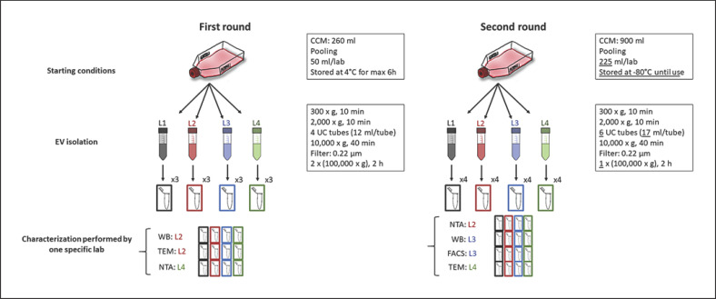
Methods: Conditioned medium of colorectal cancer cells was prepared and pooled by 1 person and distributed to each of the participating laboratories for isolation according to a pre-defined protocol. After EV isolation in each laboratory, quantification and characterization of isolated EVs was collectively done by 1 person having the highest expertise in the respective test method: Western blot, flow cytometry (fluorescence-activated cell sorting [FACS], nanoparticle tracking analysis (NTA), and transmission electron microscopy (TEM).
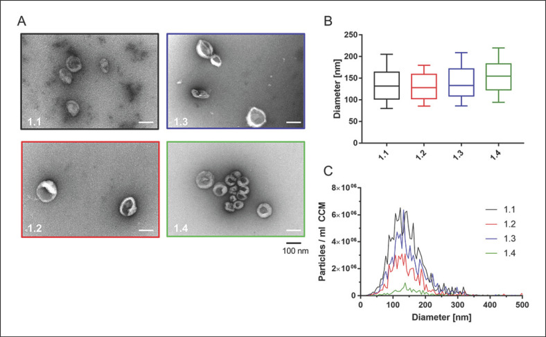
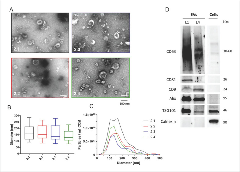
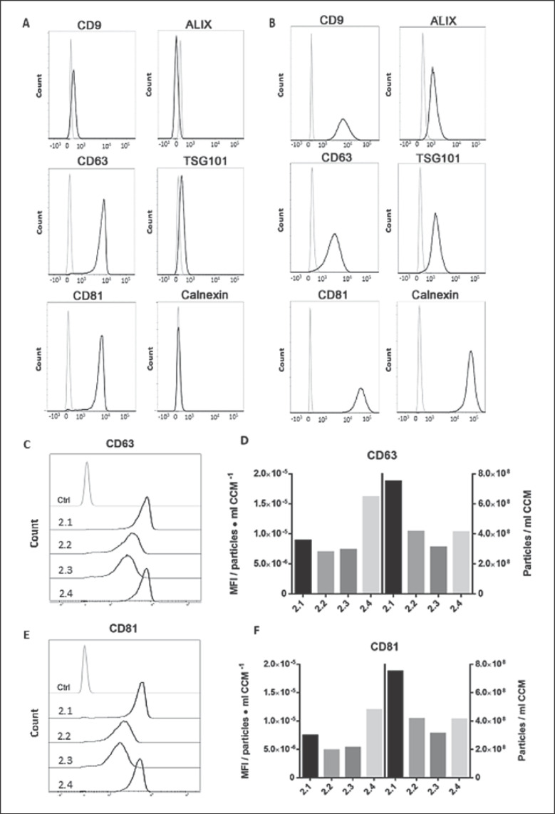
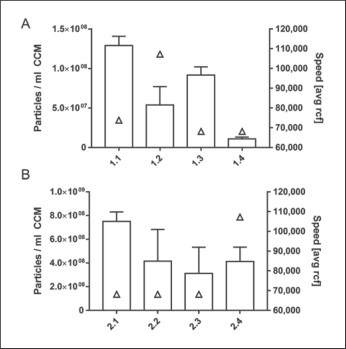
Results: EVs were visualized with TEM, presenting similar cup-shaped and spherical morphology and sizes ranging from 30 to 150 nm. NTA results showed similar size ranges of particles in both isolation rounds. EV preparations showed high purity by the expression of EV marker proteins CD9, CD63, CD81, Alix, and TSG101, and the lack of calnexin. FACS analysis of EVs revealed intense staining for CD63 and CD81 but lower levels for CD9 and TSG101. Preparations from 1 laboratory presented significantly lower particle numbers (p < 0.0001), most probably related to increased processing time. However, even when standardizing processing time, particle yields still differed significantly between groups, indicating inter-laboratory differences in the efficiency of EV isolation. Importantly, no relation was observed between centrifugation speed/k-factor and EV yield.
Conclusions: Our findings demonstrate that quantitative differences in EV yield might be due to equipment- and operator-dependent technical variability in ultracentrifugation-based EV isolation. Furthermore, our study emphasizes the need to standardize technical parameters such as the exact run speed and k-factor in order to transfer protocols between different laboratories. This hints at substantial inter-laboratory biases that should be assessed in multi-centric studies.
Keywords: Extracellular vesicles; Inter-laboratory comparison; Standardization; Ultracentrifugation.
Copyright © 2020 by S. Karger AG, Basel.
Conflict of interest statement
The authors have no conflicts of interest to declare.
Figures
References
-
- Colombo M, Raposo G, Théry C. Biogenesis, secretion, and intercellular interactions of exosomes and other extracellular vesicles. Annu Rev Cell Dev Biol. 2014;30((1)):255–89. - PubMed
-
- Tkach M, Théry C. Communication by Extracellular Vesicles: Where We Are and Where We Need to Go. Cell. 2016 Mar;164((6)):1226–32. - PubMed
LinkOut - more resources
Full Text Sources
Miscellaneous
