. 2021 Feb 23:15:580717.
doi: 10.3389/fncel.2021.580717. eCollection 2021.

Herpes Simplex Virus Type 1 Neuronal Infection Triggers the Disassembly of Key Structural Components of Dendritic Spines

Affiliations

Herpes Simplex Virus Type 1 Neuronal Infection Triggers the Disassembly of Key Structural Components of Dendritic Spines

Francisca Acuña-Hinrichsen et al. Front Cell Neurosci. .

Abstract

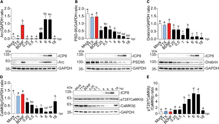
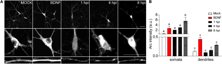
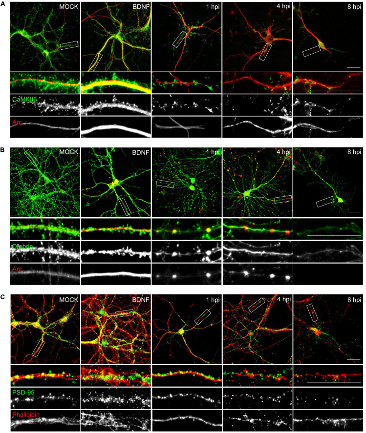
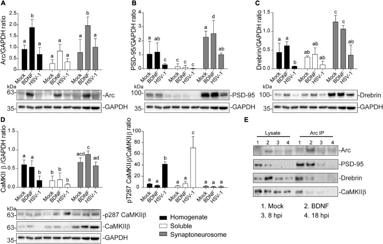
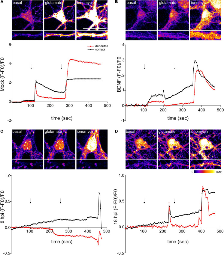
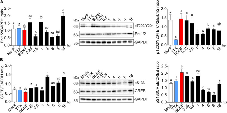
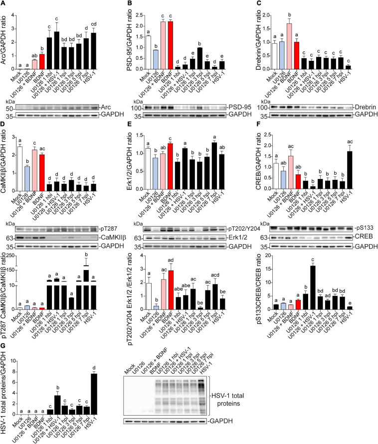
Herpes simplex virus type 1 (HSV-1) is a widespread neurotropic virus. Primary infection of HSV-1 in facial epithelium leads to retrograde axonal transport to the central nervous system (CNS) where it establishes latency. Under stressful conditions, the virus reactivates, and new progeny are transported anterogradely to the primary site of infection. During the late stages of neuronal infection, axonal damage can occur, however, the impact of HSV-1 infection on the morphology and functional integrity of neuronal dendrites during the early stages of infection is unknown. We previously demonstrated that acute HSV-1 infection in neuronal cell lines selectively enhances Arc protein expression - a major regulator of long-term synaptic plasticity and memory consolidation, known for being a protein-interaction hub in the postsynaptic dendritic compartment. Thus, HSV-1 induced Arc expression may alter the functionality of infected neurons and negatively impact dendritic spine dynamics. In this study we demonstrated that HSV-1 infection induces structural disassembly and functional deregulation in cultured cortical neurons, an altered glutamate response, Arc accumulation within the somata, and decreased expression of spine scaffolding-like proteins such as PSD-95, Drebrin and CaMKIIβ. However, whether these alterations are specific to the HSV-1 infection mechanism or reflect a secondary neurodegenerative process remains to be determined.

Keywords: Arc protein; Herpes Simplex Virus Type 1 (HSV-1); dendritic spines; memory consolidation; neurodegeneration; neurotropic virus.


Conflict of interest statement

The authors declare that the research was conducted in the absence of any commercial or financial relationships that could be construed as a potential conflict of interest.

Figures

FIGURE 1
FIGURE 1
Morphological analyses of dendritic complexity in HSV-1 infected neurons. (A) Representative images of cortical neurons infected with HSV-1 MOI 10, BDNF was used as a positive control. Antibodies against PSD-95 and MAP-2 are shown in blue and red, respectively. Scale bar in = 20 μm. (B) Average number of primary neurites (C) average number of secondary neurites. (D) The dendritic arborization was calculated using Sholl analysis and (E) the quantification of spines was represented as the number of spines/10 μm. The quantifications are representative of three independent experiments. Statistical significance was determined by One-Way ANOVA, followed by Tukey’s test. Bars represent the mean ± SD of biological replicates (10 neurons per treatment). Different letters above the mean bars apply to significant differences between groups p < 0.05. Sholl analysis statistical significance was determined by Two-Way ANOVA followed by Bonferroni’s test. ***p < 0.001; **p < 0.01.
FIGURE 2
FIGURE 2
Arc protein expression is increased in cortical neurons infected with HSV-1, while CaMKIIb, Drebrin, and PSD-95 expression are decreased. Quantification of immunoblot analyses showing (A) Arc and its dendritic binding partners: (B) PSD-95, (C) Drebrin, (D) CaMKIIβ (left panel) and pT287 CaMKIIβ (right panel) during infection kinetics with HSV-1 (Mock, 0.25; 0.5; 1, 4, 6, 8, and 18 hpi). BDNF and TTX are used as positive and negative controls of Arc induction, respectively. After gel scanning and densitometry quantitation, the results were expressed as the ratio of protein levels normalized to total GAPDH. Statistical significance was determined by One-Way ANOVA, followed by Tukey’s test. Bars represent the mean ± SD of biological replicates. Different letters above the mean bars apply to significant differences between groups p < 0.05. The blots are representative of three independent experiments.
FIGURE 3
FIGURE 3
HSV-1-induced Arc is enriched in neuronal somata rather than dendrites of infected neurons. (A) Immunocytochemical analyses of Arc protein subcellular distribution during infection kinetics. Middle and low panels show magnifications of somata and dendrites. Scale bars: 20 μm (B) Quantification of Arc fluorescence intensity in HSV-1 infected neurons versus positive (BDNF) and negative (Mock) controls. Statistical significance was determined by One-Way ANOVA, followed by Tukey’s test. Bars represent the mean ± SD of biological replicates (10 neurons per treatment). Different letters above the mean bars apply to significant differences between groups p < 0.05. The images and quantifications are representative of three independent experiments.
FIGURE 4
FIGURE 4
HSV-1 neuronal infection alters the normal distribution of Arc, CaMKIIβ, Drebrin and PSD-95. Immunocytochemistry analyses of infection kinetics. BDNF was used as a positive control for synaptic protein expression. Double staining against (A) Arc and CaMKIIβ; (B) Arc and Drebrin; (C) PSD-95 and cytoskeleton probe: Phalloidin (red). Secondary antibodies coupled to Alexa Fluor 488 and 568. Bars: 20 mm. The results shown are representative of three independent experiments.
FIGURE 5
FIGURE 5
Altered protein content in synaptoneurosomes of HSV-1 infected cortical neurons. Quantification of immunoblot analyses of synaptoneurosomal isolation HSV1-infected and BDNF-stimulated cortical neurons, showing (A) Arc, (B) PSD-95, (C) Drebrin (D) CaMKIIβ and its activation through T287 phosphorylation. GAPDH was used to normalize each blot. (E) Immunoprecipitation of Arc from the synaptoneurosomal fraction from Mock, BDNF-stimulated and HSV-1 -infected neurons at 8 and 18 hpi. Immunoblot against dendritic binding partners: PSD-95, Drebrin CaMKIIβ, and β-actin. After gel scanning and densitometry quantitation, the results were expressed as the ratio of protein levels normalized to total GAPDH. Statistical significance was determined by One-Way ANOVA, followed by Tukey’s test. Bars represent the mean ± SD of biological replicates. Different letters above the mean bars apply to significant differences between groups p < 0.05. The blots are representative of three independent experiments.
FIGURE 6
FIGURE 6
Infected neurons show an impaired response to glutamate stimulation. Top panels show representative microscopy images of somata and dendrites. (A) Mock-infected, (B) BDNF-treated and HSV-1-infected neurons at 8 and 18 hpi, respectively (C and D). Variations of relative cytosolic Ca2+ concentrations were monitored by fluorescence in Fluo4-loaded cortical neurons. The first arrow indicates the addition of glycine plus glutamate. The second arrow indicates the addition of the positive control ionomycin. The data is representative of traces of three independent experiments.
FIGURE 7
FIGURE 7
HSV-1 induces CREB phosphorylation in the early stages of neuronal infection. Immunoblot analyses showing (A) Erk1/2 total protein expression (left panel) and phosphorylated levels in T202/Y204 (right panel) and (B) CREB total protein expression (left panel) and phosphorylated levels in S133 during HSV-1 infection kinetics (Mock, 0.25; 0.5; 1, 4, 6, 8, and 18 hpi), BDNF and TTX were used as positive and negative controls for signaling pathway activation and inhibition, respectively. Statistical significance was determined by One-Way ANOVA, followed by Tukey’s test. Bars represent the mean ± SD of biological replicates. Different letters above the mean bars apply to significant differences between groups p < 0.05. The blots are representative of three independent experiments.
FIGURE 8
FIGURE 8
Dendritic protein levels during HSV-1 neuronal infection is controlled by Erk-independent signaling. Immunoblot analyses showing protein total levels at 8 hpi. (A) Arc, (B) PSD-95, (C) Drebrin, (D) CaMKIIβ, (E) Erk1/2, (F) CREB and, (G) HSV-1 total proteins. BDNF was used as a positive control of Arc expression and the U0126 MEK inhibitor was added at different time points during the infection and together with BDNF to abolish Arc induction. U0126 1 hbi: 1 h before infection; U0126 + HSV-1: added at the same time; U0126 1, 3, 5, 7, hpi, and HSV-1 at 8 h without inhibitor. Statistical significance was determined by One-Way ANOVA, followed by Tukey’s test. Bars represent the mean ± SD of biological replicates. Different letters above the mean bars apply to significant differences between groups p < 0.05. The blots are representative of three independent experiments.
FIGURE 9
FIGURE 9
The impact of HSV-1 neuronal infection at the dendritic spine level. Diagram of the HSV-1-induced phenotype in cortical neurons at 1, 4, 8 and 18 hpi. Changes in shape and structure both in somata and dendrites are shown. Early in infection, 1 hpi CREB is activated (pS133) by p38 MAPK (Hargett et al., 2005) instead of Erk. From 1 hpi there is a progressive decrease of PSD-95, Drebrin and CaMKIIβ along the infection kinetics (upper panel zoom dendrites), most likely due to VHS viral protein in charge of downregulating the host cell mRNAs (Brunella Taddeo et al., 2007). From 4 hpi there is a notorious increase of Arc expression (shown as red color), reaching its highest level at 8 hpi. At this time, CREB activation is downregulated, most likely due to the inhibitory phosphorylation of GSK3 at S129. The viral kinase Us3 phosphorylates GSK3 at inhibitory residue (S9) (Wagner and Smiley, 2009), thus promoting Arc accumulation (Gozdz et al., 2017) (first dashed red arrow). Then, at 18 hpi, the Us3 effect is abolished and GSK3 is activated due to the rising levels of intracellular calcium (Piacentini et al., 2011). The activation of GSK3 promotes Arc degradation at late infection (18 hpi) (second dashed red arrow), this rapid turnover of Arc expression is accompanied by high S206 phosphorylation levels, which could have an impact on protein localization.

References

    1. Acuña-Hinrichsen F., Covarrubias-Pinto A., Ishizuka Y., Stolzenbach M. F., Martin C., Salazar P., et al. (2020). Herpes simplex Virus Type 1 neuronal infection triggers disassembly of key structural components of dendritic spines. bioRxiv [Preprint]. 10.1101/2020.05.30.124958 - DOI - PMC - PubMed
    1. Acuña-Hinrichsen F., Muñoz M., Hott M., Martin C., Mancilla E., Salazar P., et al. (2019). Herpes simplex virus type 1 enhances expression of the synaptic protein arc for its own benefit. Front. Cell. Neurosci. 12:505. 10.3389/fncel.2018.00505 - DOI - PMC - PubMed
    1. Bading H. (2013). Nuclear calcium signalling in the regulation of brain function. Nat. Rev. Neurosci. 14 593–608. 10.1038/nrn3531 - DOI - PubMed
    1. Bloomer W. A. C., VanDongen H. M. A., VanDongen A. M. J. (2007). Activity-regulated cytoskeleton-associated protein Arc/Arg3.1 binds to spectrin and associates with nuclear promyelocytic leukemia (PML) bodies. Brain Res. 1153 20–33. 10.1016/j.brainres.2007.03.079 - DOI - PubMed
    1. Bramham C. R., Alme M. N., Bittins M., Kuipers S. D., Nair R. R., Pai B., et al. (2010). The Arc of synaptic memory. Exp. Brain Res. 200 125–140. 10.1007/s00221-009-1959-2 - DOI - PMC - PubMed

LinkOut - more resources