

Epigenetics, 1-Carbon Metabolism, and Homocysteine During Dysbiosis
- PMID: 33708132
- PMCID: PMC7940193
- DOI: 10.3389/fphys.2020.617953
Epigenetics, 1-Carbon Metabolism, and Homocysteine During Dysbiosis
Abstract
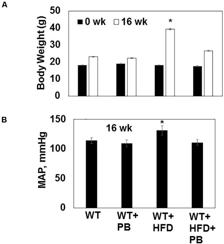
Although a high-fat diet (HFD) induces gut dysbiosis and cardiovascular system remodeling, the precise mechanism is unclear. We hypothesize that HFD instigates dysbiosis and cardiac muscle remodeling by inducing matrix metalloproteinases (MMPs), which leads to an increase in white adipose tissue, and treatment with lactobacillus (a ketone body donor from lactate; the substrate for the mitochondria) reverses dysbiosis-induced cardiac injury, in part, by increasing lipolysis (PGC-1α, and UCP1) and adipose tissue browning and decreasing lipogenesis. To test this hypothesis, we used wild type (WT) mice fed with HFD for 16 weeks with/without a probiotic (PB) in water. Cardiac injury was measured by CKMB activity which was found to be robust in HFD-fed mice. Interestingly, CKMB activity was normalized post PB treatment. Levels of free fatty acids (FFAs) and methylation were increased but butyrate was decreased in HFD mice, suggesting an epigenetically governed 1-carbon metabolism along with dysbiosis. Levels of PGC-1α and UCP1 were measured by Western blot analysis, and MMP activity was scored via zymography. Collagen histology was also performed. Contraction of the isolated myocytes was measured employing the ion-optic system, and functions of the heart were estimated by echocardiography. Our results suggest that mice on HFD gained weight and exhibited an increase in blood pressure. These effects were normalized by PB. Levels of fibrosis and MMP-2 activity were robust in HFD mice, and treatment with PB mitigated the fibrosis. Myocyte calcium-dependent contraction was disrupted by HFD, and treatment with PB could restore its function. We conclude that HFD induces dysbiosis, and treatment with PB creates eubiosis and browning of the adipose tissue.
Keywords: DNMT; MMP; REDD1; butyrate; epigenetics; eubiosis.
Copyright © 2021 Singh, Hardin, George, Eyob, Stanisic, Pushpakumar and Tyagi.
Conflict of interest statement
The authors declare that the research was conducted in the absence of any commercial or financial relationships that could be construed as a potential conflict of interest.
Figures


Similar articles
-
A High-Fat Diet Induces Epigenetic 1-Carbon Metabolism, Homocystinuria, and Renal-Dependent HFpEF.Nutrients. 2025 Jan 8;17(2):216. doi: 10.3390/nu17020216. Nutrients. 2025. PMID: 39861346 Free PMC article.
-
Dysbiotic 1-carbon metabolism in cardiac muscle remodeling.J Cell Physiol. 2020 Mar;235(3):2590-2598. doi: 10.1002/jcp.29163. Epub 2019 Sep 5. J Cell Physiol. 2020. PMID: 31489638 Free PMC article.
-
Propionate alleviates high-fat diet-induced lipid dysmetabolism by modulating gut microbiota in mice.J Appl Microbiol. 2019 Nov;127(5):1546-1555. doi: 10.1111/jam.14389. Epub 2019 Aug 15. J Appl Microbiol. 2019. PMID: 31325215
-
Resveratrol-Induced White Adipose Tissue Browning in Obese Mice by Remodeling Fecal Microbiota.Molecules. 2018 Dec 18;23(12):3356. doi: 10.3390/molecules23123356. Molecules. 2018. PMID: 30567366 Free PMC article.
-
Lactobacillus amylovorus KU4 ameliorates diet-induced obesity in mice by promoting adipose browning through PPARγ signaling.Sci Rep. 2019 Dec 27;9(1):20152. doi: 10.1038/s41598-019-56817-w. Sci Rep. 2019. PMID: 31882939 Free PMC article.
Cited by
-
Remote Hind-Limb Ischemia Mechanism of Preserved Ejection Fraction During Heart Failure.Front Physiol. 2021 Nov 11;12:745328. doi: 10.3389/fphys.2021.745328. eCollection 2021. Front Physiol. 2021. PMID: 34858202 Free PMC article.
-
A High-Fat Diet Induces Epigenetic 1-Carbon Metabolism, Homocystinuria, and Renal-Dependent HFpEF.Nutrients. 2025 Jan 8;17(2):216. doi: 10.3390/nu17020216. Nutrients. 2025. PMID: 39861346 Free PMC article.
-
COVID-19 Mimics Pulmonary Dysfunction in Muscular Dystrophy as a Post-Acute Syndrome in Patients.Int J Mol Sci. 2022 Dec 24;24(1):287. doi: 10.3390/ijms24010287. Int J Mol Sci. 2022. PMID: 36613731 Free PMC article. Review.
-
Mechanism of Blood-Heart-Barrier Leakage: Implications for COVID-19 Induced Cardiovascular Injury.Int J Mol Sci. 2021 Dec 17;22(24):13546. doi: 10.3390/ijms222413546. Int J Mol Sci. 2021. PMID: 34948342 Free PMC article.
-
The role of the mitochondrial trans-sulfuration in cerebro-cardio renal dysfunction during trisomy down syndrome.Mol Cell Biochem. 2024 Apr;479(4):825-829. doi: 10.1007/s11010-023-04761-9. Epub 2023 May 17. Mol Cell Biochem. 2024. PMID: 37198322 Review.
References
-
- Bjørndal B., Ramsvik M. S., Lindquist C., Nordrehaug J. E., Bruheim I., Svardal A., et al. (2015). A phospholipid-protein complex from Antarctic krill reduced plasma homocysteine levels and increased plasma trimethylamine-N-oxide (TMAO) and carnitine levels in male Wistar rats. Mar. Drugs 13 5706–5721. 10.3390/md13095706 - DOI - PMC - PubMed
Grants and funding
LinkOut - more resources
Full Text Sources
Other Literature Sources
Miscellaneous