

Primary adenoid cystic carcinoma of the tracheobronchial tree: report of two cases
- PMID: 33708306
- PMCID: PMC7906545
- DOI: 10.11604/pamj.2019.34.137.14902
Primary adenoid cystic carcinoma of the tracheobronchial tree: report of two cases
Abstract
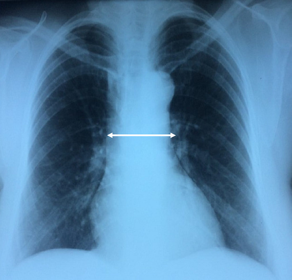
Adenoid cystic carcinoma (ACC) is a rare malignant epithelial tumor that predominantly originates in the salivary glands. Primary ACC of the tracheobronchial tree is extremely rare. We report two new cases of central airways primary ACC: a 58 year-old male with an ACC of the left main bronchus who underwent a pneumonectomy with node dissection, and a 52 year-old female with proximal tracheal ACC presenting as asthma treated by surgical resection and a postoperative radiotherapy. Primary ACC of the tracheobronchial tree is often misdiagnosed given the non-specific clinical presentation. An early diagnosis is essential to ensure good outcomes. An interdisciplinary treatment is required based especially on surgery and radiotherapy.
Keywords: Adenoid cystic carcinoma; bronchogenic carcinoma; lung cancer; salivary gland cancer.
© Ahmed Ben Saad et al.
Conflict of interest statement
The authors declare no competing interests.
Figures


Similar articles
-
[Tracheobronchial adenoid cystic carcinoma: seven case reports].Harefuah. 2012 Apr;151(4):202-4, 255. Harefuah. 2012. PMID: 22616145 Hebrew.
-
Tracheobronchial Adenoid Cystic Carcinoma Treated Successfully With Chemoradiotherapy Followed by Durvalumab: A Case Report.In Vivo. 2024 May-Jun;38(3):1483-1488. doi: 10.21873/invivo.13595. In Vivo. 2024. PMID: 38688619 Free PMC article.
-
Adenoid cystic carcinoma of the trachea: a clinico-pathological analysis.Pan Afr Med J. 2015 Mar 13;20:240. doi: 10.11604/pamj.2015.20.240.3953. eCollection 2015. Pan Afr Med J. 2015. PMID: 27386036 Free PMC article. Review.
-
Treatment outcomes of patients with tracheobronchial mucoepidermoid carcinoma compared with those with adenoid cystic carcinoma.Eur J Surg Oncol. 2020 Oct;46(10 Pt A):1888-1895. doi: 10.1016/j.ejso.2020.04.020. Epub 2020 May 5. Eur J Surg Oncol. 2020. PMID: 32418755
-
Biology of Adenoid Cystic Carcinoma of the Tracheobronchial Tree and Principles of Management.Thorac Surg Clin. 2018 May;28(2):145-148. doi: 10.1016/j.thorsurg.2018.01.002. Thorac Surg Clin. 2018. PMID: 29627047 Review.
Cited by
-
Pulmonary Adenoid Cystic Carcinoma Mimicking Asthma-Like Symptoms: A Case Report and Literature Review.Case Rep Oncol. 2024 Jan 29;17(1):150-160. doi: 10.1159/000535505. eCollection 2024 Jan-Dec. Case Rep Oncol. 2024. PMID: 38288460 Free PMC article.
-
Pulmonary adenoid cystic carcinoma: molecular characteristics and literature review.Diagn Pathol. 2023 May 17;18(1):65. doi: 10.1186/s13000-023-01354-4. Diagn Pathol. 2023. PMID: 37198615 Free PMC article. Review.
References
-
- Chhabra S, Bhutani N, Singh S, Batra A, Sen R. Primary adenoid cystic carcinoma of bronchus: Report of a rare case. Human Pathology. 2017;7:53–56.
-
- Fraire AE, Dail DH. Dail and Hammar's Pulmonary Pathology: Volume II: Neoplastic Lung Disease. New York, NY: Springer New York; 2008. Tracheobronchial Tumors of the Salivary Gland Type; pp. 398–426.
-
- Mukherjee S, Dattachaudhuri A, Bhanja P, Deb J. A rare case of primary adenoid cystic carcinoma of lung. Indian J Chest Dis Allied Sci. 2014;56(3):175–7. - PubMed
-
- zaibi H, ben amar J, Frikha H. Le carcinome adénoide kystique primitif, une tumeur bronchique rare. La tunisie médicale. 2016;94(11):698. - PubMed
Publication types
MeSH terms
LinkOut - more resources
Full Text Sources
