Case Reports
. 2020 Nov 30;14(11):1-15.
doi: 10.3941/jrcr.v14i11.3974. eCollection 2020 Nov.

Involvement of the Mediastinal Subpleural Pulmonary Parenchyma on Chest CT in COVID-19 patients: A Case Series

Affiliations
Case Reports

Involvement of the Mediastinal Subpleural Pulmonary Parenchyma on Chest CT in COVID-19 patients: A Case Series

Luigi Urciuoli et al. J Radiol Case Rep. .

Abstract

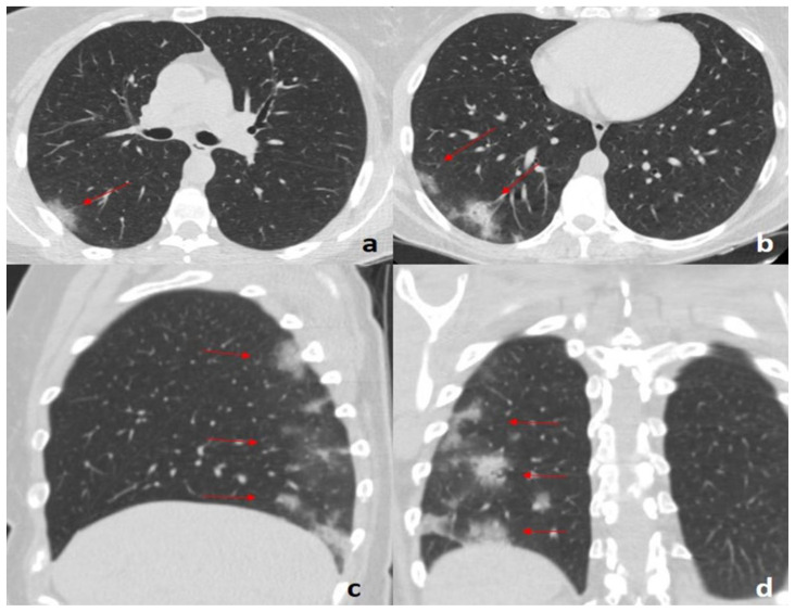
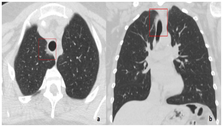
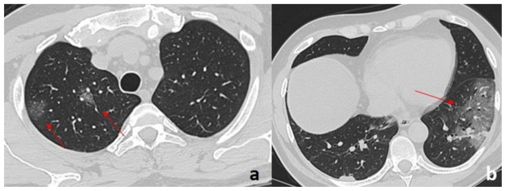
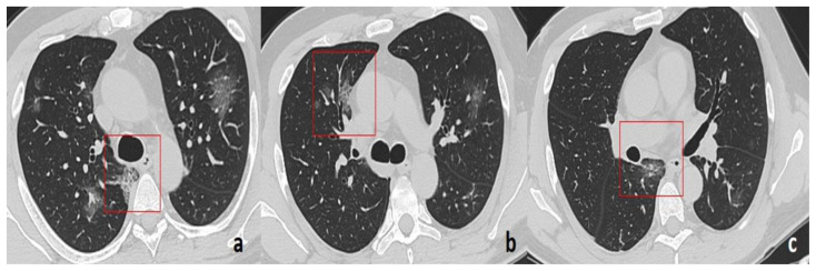
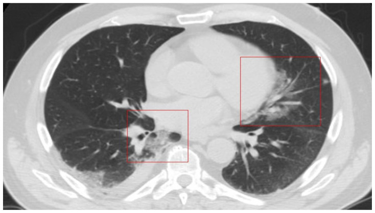
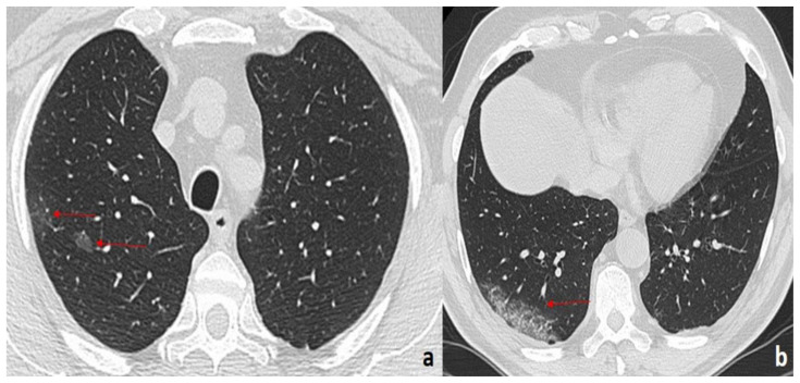
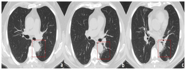
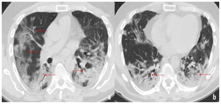
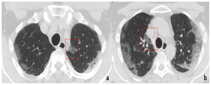
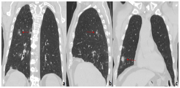
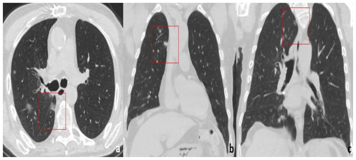
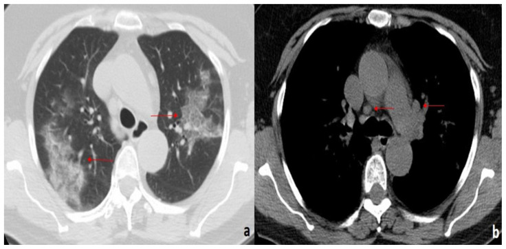
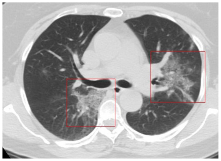
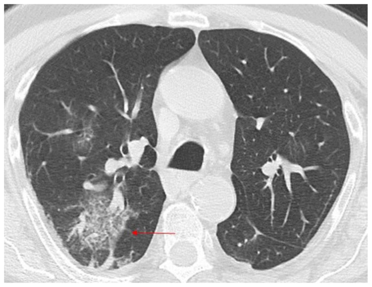
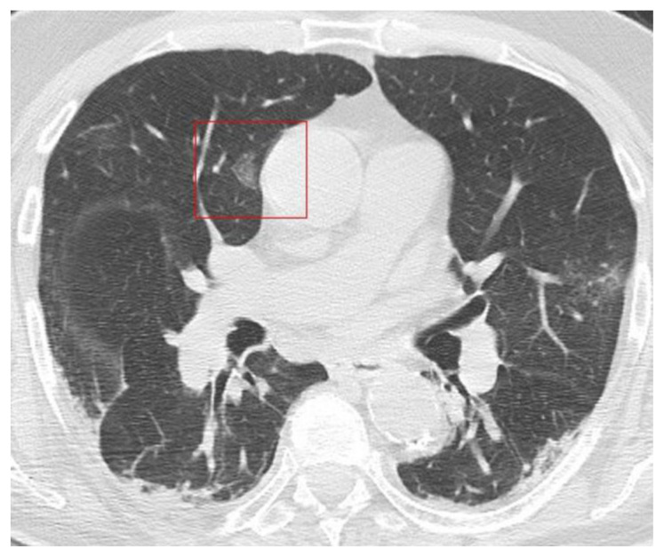
Coronavirus disease 2019 (COVID-19) is an infectious disease caused by the severe acute respiratory syndrome coronavirus 2 (SARS-CoV-2). First identified in December 2019 in Wuhan, China, it has since become a global pandemic. Although the reference standard for SARS-CoV-2 diagnosis is real-time reverse transcription polymerase chain reaction (RT-PCR), computed tomography (CT) is recommended for both initial evaluation and follow-up. The CT findings in COVID-19 are varied, but typical ground-glass opacities are usually reported to occupy a peripheral costal subpleural distribution. Here we report eight confirmed COVID-19 cases who underwent clinical evaluation, laboratory testing, and unenhanced chest CT. In all patients, chest CT showed the presence of ground-glass opacities in the mediastinal subpleural parenchyma. While these cases also showed the typical CT features of COVID-19, involvement of the mediastinal subpleural parenchyma should not lower the index of suspicion for COVID-19.

Keywords: COVID-19; Coronavirus disease 2019; GGOs; chest CT; ground-glass opacities; lung consolidation; mediastinal pleura.


Figures

Figure 1
Figure 1
45-year-old woman with COVID-19 pneumonia. FINDINGS: Axial (a, b), sagittal (c), and coronal (d) unenhanced chest CTs demonstrate multiple, subpleural GGOs with a rounded morphology (arrows) in the right lower lobe. TECHNIQUE: GE 16-slices CT scanner, 80–300 mAs, 120 kV, 1.25 mm slice thickness.
Figure 2
Figure 2
45-year-old woman with COVID-19 pneumonia. FINDINGS: Axial (a) and coronal (b) unenhanced chest CTs show a band-like GGO in the right paratracheal parenchyma (box). TECHNIQUE: GE 16-slices CT scanner, 80–300 mAs, 120 kV, 1.25 mm slice thickness.
Figure 3
Figure 3
49-year-old man with COVID-19 pneumonia. FINDINGS: Axial unenhanced chest CT demonstrates multiple GGOs (arrows) with a rounded morphology in the upper right lobe (arrows in a), and a diffuse area of GGO with a striking peripheral distribution in the left lower lobe (arrow in b). TECHNIQUE: GE 16-slices CT scanner, 80–300 mAs, 120 kV, 1.25 mm slice thickness.
Figure 4
Figure 4
49-year-old man with COVID-19 pneumonia. FINDINGS: Axial unenhanced chest CT demonstrates multiple GGOs with a rounded morphology (boxes) in the right lung involving the mediastinal subpleural parenchyma (posterior costo-mediastinal (a, c) and para-aortic (b)). TECHNIQUE: GE 16-slices CT scanner, 80–300 mAs, 120 kV, 1.25 mm slice thickness.
Figure 5
Figure 5
66-year-old man with COVID-19 pneumonia. FINDINGS: Axial unenhanced chest CT shows bilateral patchy GGOs superimposed on septal thickening (crazy paving pattern) with a pronounced peripheral distribution (arrows in a) and partial consolidation of the right lower lobe (arrows in b). TECHNIQUE: GE 16-slices CT scanner, 80–300 mAs, 120 kV, 1.25 mm slice thickness.
Figure 6
Figure 6
66-year-old man with COVID-19 pneumonia. FINDINGS: Axial unenhanced chest CT shows a right pleural effusion and patchy GGOs in the mediastinal subpleural areas (left paracardiac parenchyma and right posterior costo-mediastinal area (boxes)). TECHNIQUE: GE 16-slices CT scanner, 80–300 mAs, 120 kV, 1.25 mm slice thickness.
Figure 7
Figure 7
61-year-old man with COVID-19 pneumonia. FINDINGS: Axial unenhanced CT shows multiple rounded GGOs in the right upper lobe (arrows in a) and a diffuse GGO with contextual septal thickening (crazy paving pattern) in the periphery of the right lower lobe (arrow in b). TECHNIQUE: GE 16-slices CT scanner, 80–300 mAs, 120 kV, 1.25 mm slice thickness.
Figure 8
Figure 8
61-year-old man with COVID-19 pneumonia. FINDINGS: (a, b, c) Axial unenhanced CT shows area of band-like GGO around the thoracic descending aorta (boxes). TECHNIQUE: GE 16-slices CT scanner, 80–300 mAs, 120 kV, 1.25 mm slice thickness.
Figure 9
Figure 9
58-year-old man with COVID-19 pneumonia. FINDINGS: (a, b) Axial unenhanced CT demonstrates bilateral, diffuse GGOs with a bronchovascular and peripheral distribution. In the posterior segments of the lower lobes, consolidations with air bronchogram are also seen (arrows). TECHNIQUE: GE 16-slices CT scanner, 80–300 mAs, 120 kV, 1.25 mm slice thickness.
Figure 10
Figure 10
58-year-old man with COVID-19 pneumonia. FINDINGS: (a, b) Axial unenhanced CT shows focal rounded GGOs with a mediastinal subpleural distribution (paratracheal area bilaterally (boxes)). TECHNIQUE: GE 16-slices CT scanner, 80–300 mAs, 120 kV, 1.25 mm slice thickness.
Figure 11
Figure 11
75-year-old man with COVID-19 pneumonia. FINDINGS: Coronal (a, c) and sagittal (b) unenhanced CT of the chest demonstrates multiple nodules with faint surrounding ground glass attenuation (halo sign) in the right upper lobe (arrows in a, b) and right lower lobe (arrow in c). TECHNIQUE: GE 16-slices CT scanner, 80–300 mAs, 120 kV, 1.25 mm slice thickness.
Figure 12
Figure 12
75-year-old man with COVID-19 pneumonia. FINDINGS: Axial (a)/coronal (b, c) unenhanced CT shows focal rounded GGOs in the mediastinal subpleural areas (right posterior costo-mediastinal area (box in a), right para-aortic area (box in b), and right paratracheal area (box in c)). TECHNIQUE: GE 16-slices CT scanner, 80–300 mAs, 120 kV, 1.25 mm slice thickness.
Figure 13
Figure 13
60-year-old woman with COVID-19 pneumonia. FINDINGS: Axial unenhanced chest CT shows bilateral, peripheral patchy areas of GGO with superimposed septal thickening (crazy paving pattern) in the upper lobes (arrows in a); evidence of enlargement of some mediastinal lymph nodes (arrows in b). TECHNIQUE: GE 16-slices CT scanner, 80–300 mAs, 120 kV, 1.25 mm slice thickness.
Figure 14
Figure 14
60-year-old woman with COVID-19 pneumonia. FINDNGS: Axial unenhanced chest CT shows areas of GGO with contextual septal thickening (crazy paving pattern) in a mediastinal subpleural distribution (right costo-mediastinal space) and in the left perihilar space (boxes). TECHNIQUE: GE 16-slices CT scanner, 80–300 mAs, 120 kV, 1.25 mm slice thickness
Figure 15
Figure 15
86-year-old man with COVID-19 pneumonia. FINDINGS: Axial unenhanced chest CT shows patchy GGO in the apical segment of the right lower lobe (arrow). TECHNIQUE: GE 16-slices CT scanner, 80–300 mAs, 120 kV, 1.25 mm slice thickness.
Figure 16
Figure 16
86-year-old man with COVID-19 pneumonia. FINDINGS: Axial unenhanced chest CT shows a focal GGO with a mediastinal subpleural distribution in the right periaortic area (box). TECHNIQUE: GE 16-slices CT scanner, 80–300 mAs, 120 kV, 1.25 mm slice thickness.

Similar articles

References

    1. Zhu N, Zhang D, Wang W, et al. A novel coronavirus from patients with pneumonia in China, 2019. N Engl J Med. 2020 Feb 20;382(8):727–733. - PMC - PubMed
    1. Lu H, Stratton CW, Tang YW. Outbreak of pneumonia of unknown etiology in Wuhan, China: The mystery and the miracle. J Med Virol. 2020 Apr;92(4):401–402. - PMC - PubMed
    1. Coronavirus disease (COVID-19) Pandemic. Geneva: World Health Organization; Oct 27, 2020. https://www.who.int/emergencies/diseases/novel-coronavirus-2019.
    1. Guan WJ, Ni ZY, Hu Y, et al. China Medical Treatment Expert Group for Covid-19. Clinical Characteristics of Coronavirus Disease 2019 in China. N Engl J Med. 2020 Apr 30;382(18):1708–1720. - PMC - PubMed
    1. Huang C, Wang Y, Li X, et al. Clinical features of patients infected with 2019 novel coronavirus in Wuhan, China. Lancet. 2020 Feb 15;395(10223):497–506. - PMC - PubMed

Publication types