

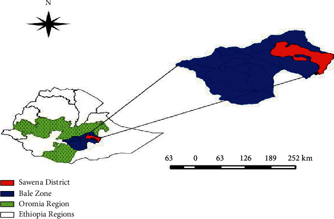
Molecular Characterization of Lumpy Skin Disease Virus Isolates from Outbreak Cases in Cattle from Sawena District of Bale Zone, Oromia, Ethiopia
- PMID: 33708372
- PMCID: PMC7929688
- DOI: 10.1155/2021/8862180
Molecular Characterization of Lumpy Skin Disease Virus Isolates from Outbreak Cases in Cattle from Sawena District of Bale Zone, Oromia, Ethiopia
Abstract
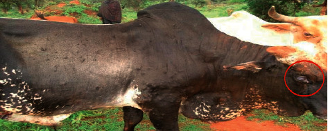
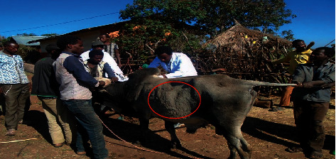
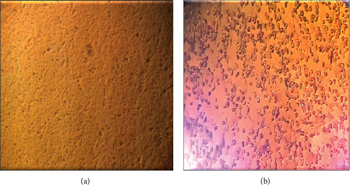
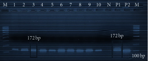
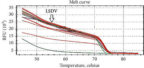
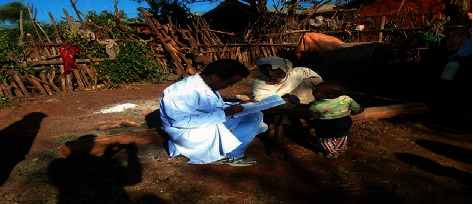
Lumpy skin disease (LSD) is a viral disease caused by LSD virus and is one of the most economically significant transboundary and emerging diseases of cattle. LSD causes considerable economic losses due to emaciation, damage to hides, infertility, and loss of milk production. In Ethiopia, the disease is distributed almost in all regions and is regarded as one of the most economically important livestock diseases in the country. An outbreak investigation of the disease was monitored from October 2016 to April 2017 in southern pastoral areas of Bale Zone, Oromia, Ethiopia. In December 2016, LSD outbreak occurred in Sawena district of Bale Zone, from which necessary biopsy samples were collected from actively infected animals for the purpose of virus isolation, and characterization using different molecular techniques at National Animal Health and Diagnostic Investigation Center (NAHDIC) of Sebeta, Ethiopia. In addition, clinical examination of infected and in-contact animals was carried out together with a questionnaire survey. Based on the clinical manifestations, LSD was recorded in 18% (94/522) of examined cattle, whereas biopsy samples from 20 clinically positive animals were collected for further laboratory process. The morbidity rate was higher in animals less than two years 28.97% (31/107) than other ages and showed a statistically significant difference with P < 0.05. Female animals showed higher morbidity rate of 20.59% (76/369) than male animals (11.76%) (18/153) with a significant difference at P ≤ 0.003. Mortality rate and case fatality were also significantly higher in young animals than other age groups. Viruses were isolated from both skin biopsies and nasal swabs on Vero cell line. From both skin biopsies and nasal swabs, the virus DNA was identified by amplifying the 172 bp DNA fragment using real-time and conventional PCR. Providing adequate diagnostic facilities, establishing strategic policies for effective control and eradication and awareness creations for communities for early identification or reporting were recommendations made to minimize economic losses of the disease.
Copyright © 2021 Shubisa Abera Leliso et al.
Conflict of interest statement
The authors declare that they have no conflicts of interest.
Figures
References
-
- FEWS NET/USAID. Famine early warning system network: food security. Washington, DC, USA: FEWS NET/USAID; 2004.
-
- Mekonnen S., Hussein I., Bedane B. The distribution of ixodid ticks (Acari: ixodidae) in central Ethiopia. Onderstepoort Journal of Veterinary Research. 2001;68:243–251. - PubMed
-
- CSA. Report on Livestock and Livestock Characteristics. Addis Ababa, Ethiopia: 2013. Federal Democratic Republic of Ethiopia, Central Statistical Authority, Agricultural Sample Survey (2016)
-
- MoARD. The Effect of Skin and Hide Quality on Domestic and Export Market and Evaluation of the Campaign against Ectoparasites of Sheep and Goat in Amhara, Tigray and Afar Region, Official Report to Region and Other Sectors. Addis Ababa, Ethiopia: MoARD; 2008.
-
- FAO/WFP. Food and Agriculture Organization and World Food Program; FAO Global Information and Early Warning System on Food and Agriculture. Special Report of FAO/WFP Crop and Food Supply Assessment Mission to Ethiopia. Rome, Italy: FAO/WFP; 2005.
LinkOut - more resources
Full Text Sources
Other Literature Sources
Miscellaneous
