. 2021 Feb;12(1):121-132.
doi: 10.21037/jgo-21-12.

Combined targeting of vascular endothelial growth factor C (VEGFC) and P65 using miR-27b-3p agomir and lipoteichoic acid in the treatment of gastric cancer

Affiliations

Combined targeting of vascular endothelial growth factor C (VEGFC) and P65 using miR-27b-3p agomir and lipoteichoic acid in the treatment of gastric cancer

Yejia Cui et al. J Gastrointest Oncol. 2021 Feb.

Abstract

Background: Gastric cancer is the second leading cancer-related mortality worldwide and more effective treatment strategies are urgently needed to combat the disease. Using lipoteichoic acid (LTA) and miR-27b-3p agomir, we aimed to assess the efficacy of this combination of therapies in treating gastric cancer.

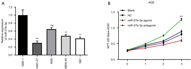
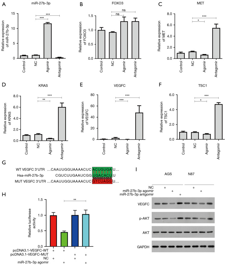
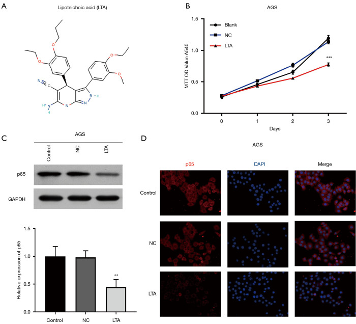
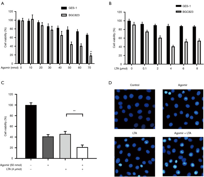
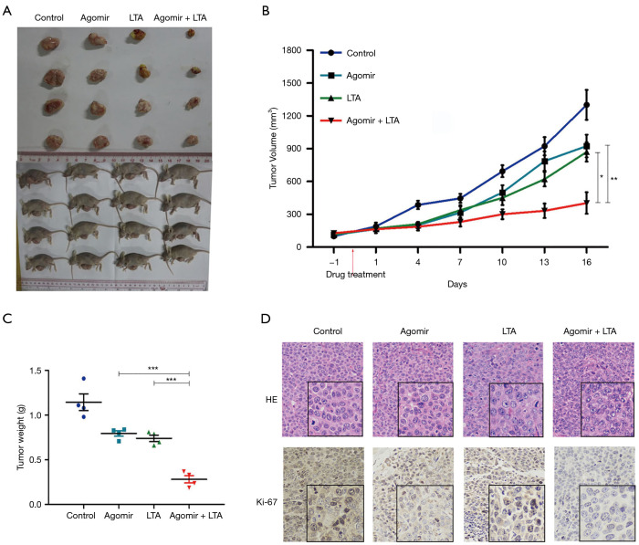
Methods: The RNA levels of miR-27b-3p, FOXO3, MET, KRAS, vascular endothelial growth factor C (VEGFC), TSC1, and P65 were analyzed by quantified-PCR (Q-PCR) and the cell viability of AGS cells was analyzed by MTT. Confirm Luciferase reporter assays were used to explore the putative miR-27b-3p binding sites and Western blot analyzed the protein level of GAPDH, VEGFC, P65, AKT, and phosphorylated-AKT (p-AKT). The level of P65 in both the cytoplasm and nucleus of AGS cells was visualized by immunofluorescence assay. Subcutaneous xenograft models of gastric cancer were established, and mice were treated with miR-27b-3p agomir, LTA, or both. Hematoxylin-eosin staining and Ki-67 immunohistochemistry analysis of tumor tissues were then performed.

Results: The results showed that the decreased expression of miR-27b-3p in gastric cancer cell lines inhibited the viability of AGS cells, and VEGFC was confirmed as the target of miR-27b-3p. In addition, ectopic expression of miR-27b-3p significantly inhibited the AKT pathway in AGS and N87 cells, and LTA suppressed the proliferation of gastric cancer cells by inhibiting the NF-κB pathway. In an established xenograft model, both miR-27b-3p agomir alone and LTA treatment alone inhibited tumor growth and treatment which combined the two showed an even stronger inhibitory effect.

Conclusions: Taken together, the combined use of LTA and miR-27b-3p agomir exhibited a synergistic effect in the treatment of gastric cancer.

Keywords: Akt; NF-κB; apoptosis; gastric cancer; miR-27b-3p; vascular endothelial growth factor C (VEGFC).


Conflict of interest statement

Conflicts of Interest: All authors have completed the ICMJE uniform disclosure form (available at http://dx.doi.org/10.21037/jgo-21-12). The authors have no conflicts of interest to declare.

Figures

Figure 1
Figure 1
Knockdown of miR-27b-3p inhibited cell proliferation in AGS cells. (A) quantified-PCR (Q-PCR) analyzed the expression of miR-27b-3p in GES-1, AGS, BT474, MGC-803, and N87. (B) AGS cells were transfected by negative control (NC), miR-27b-3p agomir, or miR-27b-3p antagomir, and the cell viability was assessed by MTT. All the experiments were carried out in triplicate. *P<0.05, **P<0.01, ***P<0.001.
Figure 2
Figure 2
MiR-27b-3p targets VEGFC and inhibits the AKT pathway in gastric cancer cells. (A) AGS cells were transiently transfected with synthetic miR-27b-3p agomir, or antisense miR-27b-3p antagomir. quantified-PCR (Q-PCR) analyzed the level of miR-27b-3p in AGS cells after transfection. (B,C,D,E,F) Q-PCR analyzed the mRNA level of FOXO3, MET, KRAS, VEGFC, TSC1. (G) Schematic represents putative miR-27b-3p binding sites with VEGFC. (H) Luciferase reporter assays confirming the miR-27b-3p binding sites at 3'-UTRs of VEGFC. (I) AGS and N87 cells were transfected by negative control (NC), miR-27b-3p agomir, or miR-27b-3p antagomir, and the protein level of GAPDH, VEGFC, AKT and p-AKT was assessed by Western blot. All the experiments were carried out in triplicate. *P<0.05, **P<0.01, ***P<0.001.
Figure 3
Figure 3
Lipoteichoic acid (LTA) suppressed the proliferation of gastric cancer cells by inhibiting the NF-kappa-B pathway (A) The molecular structure of LTA. (B) AGS cells were treated with LTA (4 µmol/L) for 1, 2 and 3 days. MTT assessed the cell viability. (C) Western blot analyzed the protein level of GAPDH and P65 at 3 days after LTA stimulation. (D) The level of P65 in both the cytoplasm and nucleus of AGS cells was visualized by immunofluorescence assay at 3 days after LTA stimulation. DAPI was used to stain nucleus (blue). All the experiments were carried out in triplicate. **P<0.01, ***P<0.001.
Figure 4
Figure 4
The combined use of lipoteichoic acid (LTA) and miR-27b-3p agomir has synergistic effect for anti-gastric cancer in vitro. (A) AGS cells and normal gastric mucosa-derived cell line (GES-1) were transfected by increasing concentration of miR-27b-3p agomir (0 nm–70 nmol), and MTT analyzed the cell viability. (B) AGS cells and normal gastric mucosa-derived cell line (GES-1) were transfected by increasing concentration of LTA (0–8 µmol), and cell viability was analyzed by MTT. AGS cells were treated with miR-27b-3p agomir, LTA, or both. The cell viability of AGS cells after indicated treatment was analyzed by MTT (C) and cell apoptosis was analyzed by using Hoechst assays stained with Hoechst (D). All the experiments were carried out in triplicate. *P<0.05, **P<0.01, ***P<0.001.
Figure 5
Figure 5
Combined use of lipoteichoic acid (LTA) and miR-27b-3p agomir has synergistic effect for anti-gastric cancer in vivo. Subcutaneous xenografts were established in nude mice, which were treated with miR-27b-3p agomir, LTA or together as indicated (n=4 per group). (A) The photos of tumor and mice are shown. The growth of tumor (B) was recorded and weight (C) is shown. (D) Hematoxylin-eosin staining of tumor tissues and Ki-67 immunohistochemistry analysis in xenograft tumor tissues were performed. ×400. All the experiments were carried out in triplicate. Magnification, 100×. *P<0.05, **P<0.01, ***P<0.001.

References

    1. Huang J, Yang Y, Yang J, et al. Regenerating gene family member 4 promotes growth and migration of gastric cancer through protein kinase B pathway. Int J Clin Exp Med 2014;7:3037-44. - PMC - PubMed
    1. Wang J, Wang Q, Liu H, et al. MicroRNA expression and its implication for the diagnosis and therapeutic strategies of gastric cancer. Cancer Lett 2010;297:137-43. 10.1016/j.canlet.2010.07.018 - DOI - PubMed
    1. Karimi P, Islami F, Anandasabapathy S, et al. Gastric cancer: descriptive epidemiology, risk factors, screening, and prevention. Cancer Epidemiol Biomarkers Prev 2014;23:700-13. 10.1158/1055-9965.EPI-13-1057 - DOI - PMC - PubMed
    1. Jemal A, Bray F, Center MM, et al. Global cancer statistics. CA Cancer J Clin 2011;61:69-90. 10.3322/caac.20107 - DOI - PubMed
    1. Zhao JJ, Pan K, Wang W, et al. The prognostic value of tumor-infiltrating neutrophils in gastric adenocarcinoma after resection. PLoS One 2012;7:e33655. 10.1371/journal.pone.0033655 - DOI - PMC - PubMed

LinkOut - more resources