

Actinomycotic Osteomyelitis of the Mandible - A Rare Case Report
- PMID: 33708610
- PMCID: PMC7944017
- DOI: 10.4103/ams.ams_99_20
Actinomycotic Osteomyelitis of the Mandible - A Rare Case Report
Abstract
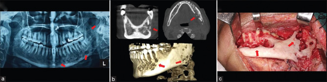
Actinomycetes are a relatively sporadic cause of infection of the head-and-neck region and their appearance is usually uncharacteristic, and hence pose a challenge for the diagnosis. The present article intends to exhibit this rarity afflicting mandible and highlight its management. The present report describes a case of a 55-year-old countryside female who presented with pain and swelling affecting the left side of the mandible. Orthopantomograph and cone-beam computed tomography imaging showed multiple ill-defined radiolucencies and perforations of the buccal and lingual cortical plates. Fine-needle aspiration microbiology was used to ascertain the microbial organism and the patient was treated with amoxicillin + clavulanic acid with curettage of the infected site. The patient responded well to prompt systemic antibiotics and local surgical measures with complete resolution of the infection and spontaneous bone regeneration. Although rare actinomycosis of the mandible is curable and should be included in the differential diagnosis of osteomyelitis of the jaw. Early and accurate diagnosis and prompt intervention confirm better outcomes.
Keywords: Actinomycosis; filamentous; jaw infection; osteomyelitis; ray fungus; suppuration.
Copyright: © 2020 Annals of Maxillofacial Surgery.
Conflict of interest statement
There are no conflicts of interest.
Figures


References
-
- Sharkawy AA. Cervicofacial actinomycosis and mandibular osteomyelitis. Infect Dis Clin North Am. 2007;21:543–56. viii. - PubMed
-
- Stewart MG, Sulek M. Pediatric actinomycosis of the head and neck. Ear Nose Throat J. 1993;72:614. - PubMed
-
- Belmont MJ, Behar PM, Wax MK. Atypical presentations of actinomycosis. Head Neck. 1999;21:264–8. - PubMed
-
- Rankow RM, Abraham DM. Actinomycosis: Masquerader in the head and neck. Ann Otol Rhinol Laryngol. 1978;87:230–7. - PubMed
-
- Mandell G, Douglas RG, Bennett JE. Principles and Practice of Infectious Diseases. New York: John Wiley & Sons, Inc; 1979.
Publication types
LinkOut - more resources
Full Text Sources