. 2021 Feb 23:11:601957.
doi: 10.3389/fonc.2021.601957. eCollection 2021.

Defining the Extracellular Matrix of Rhabdomyosarcoma

Affiliations

Defining the Extracellular Matrix of Rhabdomyosarcoma

Xiaolei Lian et al. Front Oncol. .

Abstract

Rhabdomyosarcoma (RMS) is the most common soft-tissue sarcoma of childhood with a propensity to metastasize. Current treatment for patients with RMS includes conventional systemic chemotherapy, radiation therapy, and surgical resection; nevertheless, little to no improvement in long term survival has been achieved in decades-underlining the need for target discovery and new therapeutic approaches to targeting tumor cells or the tumor microenvironment. To evaluate cross-species sarcoma extracellular matrix production, we have used murine models which feature knowledge of the myogenic cell-of-origin. With focus on the RMS/undifferentiated pleomorphic sarcoma (UPS) continuum, we have constructed tissue microarrays of 48 murine and four human sarcomas to analyze expression of seven different collagens, fibrillins, and collagen-modifying proteins, with cross-correlation to RNA deep sequencing. We have uncovered that RMS produces increased expression of type XVIII collagen alpha 1 (COL18A1), which is clinically associated with decreased long-term survival. We have also identified significantly increased RNA expression of COL4A1, FBN2, PLOD1, and PLOD2 in human RMS relative to normal skeletal muscle. These results complement recent studies investigating whether soft tissue sarcomas utilize collagens, fibrillins, and collagen-modifying enzymes to alter the structural integrity of surrounding host extracellular matrix/collagen quaternary structure resulting in improved ability to improve the ability to invade regionally and metastasize, for which therapeutic targeting is possible.

Keywords: COL18A1; PLOD1/2; matrix; rhabdomyosarcoma; survival.


Conflict of interest statement

Authors ML and GS are employed by the company Omics Data Automation. The remaining authors declare that the research was conducted in the absence of any commercial or financial relationships that could be construed as a potential conflict of interest.

Figures

Figure 1
Figure 1
Conditional (Cre/LoxP) Mouse Models of Rhabdomyosarcoma (RMS) and Non-Rhabdomyosarcoma Soft Tissue Sarcomas, including like undifferentiated pleomorphic sarcoma (UPS). Phenotype of the sarcoma depends not only on mutational profile, but also on cell (lineage) of origin and timing of the initiation before or after birth (–8). Surprisingly, eRMS, and UPS exist in a continuum (7). Also, to our surprise postnatal muscle progenitors almost never gave rise to aRMS.
Figure 2
Figure 2
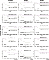
Extracellular Matrix Gene Expression (ECM) gene expression in mouse and human aRMS. Boxplots are given for biopsies versus cell lines versus normal muscle (n = 35 human aRMS biopsies, n = 4 human aRMS cell lines, n = 564 human normal skeletal muscle, n = 13 mouse necropsy aRMS tumors, n = 12 mouse normal skeletal muscle).
Figure 3
Figure 3
ECM gene expression in mouse and human eRMS. Boxplots are given for biopsies versus cell lines versus normal muscle (n = 57 human eRMS biopsies, n = 5 human eRMS cell lines, n = 564 human normal skeletal muscle, n = 4 mouse necropsy eRMS tumors, n = 12 mouse normal skeletal muscle).
Figure 4
Figure 4
ECM gene expression in mouse NRSTS. Boxplots are given for biopsies versus normal muscle (n = 6 mouse necropsy NRSTS tumors, n = 12 mouse normal skeletal muscle).
Figure 5
Figure 5
Protein expression of collagens, fibrillins and collagen modifying enzymes in murine aRMS and eRMS. Representative images showing tissue microarray negative control immunohistochemical staining and positive tumoral immunohistochemical staining of COL18A1, COL4A1, COL4A2, FBN1, FBN2, PLOD1, and PLOD2. Note that the representative aRMS IHC image for FBN1 shown is nearly negative (scoring is 1). Antibody specificity is with tumor specimen ID in parentheses listed on the figure. Models used for IHC include murine aRMS models (U21459, U24988, U48484, U20745, and U23674), a human aRMS model (pcb00380), murine eRMS models (U37125, U34278, U24085, and U24055), and a human eRMS model (pcb00232).
Figure 6
Figure 6
Immunocytochemical detection of ECM-related proteins in murine sarcoma models. Expression of ECM proteins in primary cell cultures derived from murine RMS. Immunocytochemistry using five independent antibodies against fibrillin-1, collagen XVI, collagen IV and two antibodies recognizing NC1 and NC11 domains of collagen XVIII. For each antibody, staining was intracellular and/or extracellular. Murine cell cultures used for ICC included U23674 (murine aRMS), U48484 (murine metastatic aRMS), U33915 (murine spindle cell eRMS), U57810 (murine eRMS), and U34279 (murine pleomorphic sarcoma).
Figure 7
Figure 7
Biochemical analysis of secreted collagen XVIII in murine sarcoma models. Secretion of soluble type XVIII collagen molecules in cell cultures derived from murine RMS. Western-blot analysis of conditioned media from four cultures with two antibodies against type XVIII collagen. Antibody anti-NC1 recognizes the C-terminal non-collagenous domain (left panel), whereas antibody anti-NC11 recognizes the N-terminal non-collagenous domain (right panel). The bands depicted by arrow α are intact collagen XVIII. The bands depicted by arrow β are presumed collagen XVIII without heparan-sulphate. The bands depicted by arrow γ are presumed fragments of collagen XVIII without NC11 domain. The bands depicted by arrow δ are presumed endostatin/endostatin-containing fragments.
Figure 8
Figure 8
Overexpression of COL18A1, COL4A1 and COL4A2 at the RNA level is associated with worsened outcome. Decreased overall survival at 9 years for human RMS patients with elevated expression of COL18A1, COL4A1 and COL4A2, even when adjusted for other known clinical covariates [as per analysis in (27)].

References

    1. Svalina MN, Keller C. YAPping about differentiation therapy in muscle cancer. Cancer Cell (2014) 26:154–5. 10.1016/j.ccr.2014.07.011 - DOI - PMC - PubMed
    1. Arndt CA, Rose PS, Folpe AL, Laack NN. Common musculoskeletal tumors of childhood and adolescence. Mayo Clin Proc (2012) 87:475–87. 10.1016/j.mayocp.2012.01.015 - DOI - PMC - PubMed
    1. Abraham J, Prajapati SI, Nishijo K, Schaffer BS, Taniguchi E, Kilcoyne A, et al. . Evasion mechanisms to Igf1r inhibition in rhabdomyosarcoma. Mol Cancer Ther (2011) 10:697–707. 10.1158/1535-7163.MCT-10-0695 - DOI - PMC - PubMed
    1. Nishijo K, Chen QR, Zhang L, McCleish AT, Rodriguez A, Cho MJ, et al. . Credentialing a preclinical mouse model of alveolar rhabdomyosarcoma. Cancer Res (2009) 69:2902–11. 10.1158/0008-5472.CAN-08-3723 - DOI - PMC - PubMed
    1. Abraham J, Nuñez-Álvarez Y, Hettmer S, Carrió E, Chen HI, Nishijo K, et al. . Lineage of origin in rhabdomyosarcoma informs pharmacological response. Genes Dev (2014) 28:1578–91. 10.1101/gad.238733.114 - DOI - PMC - PubMed