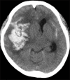
Ruptured Mycotic Cerebral Aneurysm Secondary to Disseminated Nocardiosis
- PMID: 33708692
- PMCID: PMC7869307
- DOI: 10.4103/ajns.AJNS_283_20
Ruptured Mycotic Cerebral Aneurysm Secondary to Disseminated Nocardiosis
Abstract
We report a case of a ruptured mycotic cerebral aneurysm caused by Nocardia infection. A 22-year-old immunocompromised woman with adult-onset Still's disease developed a subarachnoid hemorrhage (SAH). Digital subtraction angiography revealed a small aneurysm at the M2-3 bifurcation of the right middle cerebral artery. Cardiac ultrasonography showed vegetation at the posterior cardiac wall, suspecting infective endocarditis (IE). Gram-positive filamentous bacteria were observed in the necrotic tissue surrounding the aneurysm obtained during trapping surgery. Long-term blood culture showed that the cause of her cerebral mycotic aneurysm was nocardiosis. A mycotic ruptured cerebral aneurysm is an important cause of SAH in immunocompromised patients. Early diagnosis of IE, detection of gram-positive rods by Gram staining, and long-term culture to identify the bacteria is crucial in diagnosing nocardiosis.
Keywords: Immunosuppressed host; nocardiosis; ruptured mycotic cerebral aneurysm.
Copyright: © 2020 Asian Journal of Neurosurgery.
Conflict of interest statement
There are no conflicts of interest.
Figures


References
-
- Long PF. A retrospective study of Nocardia infections associated with the acquired immune deficiency syndrome (AIDS) Infection. 1994;22:362–4. - PubMed
-
- Tremblay J, Thibert L, Alarie I, Valiquette L, Pépin J. Nocardiosis in Quebec, Canada, 1988-2008. Clin Microbiol Infect. 2011;17:690–6. - PubMed
-
- Farran Y, Antony S. Nocardia abscessus-related intracranial aneurysm of the internal carotid artery with associated brain abscess: A case report and review of the literature. J Infect Public Health. 2016;9:358–61. - PubMed

