. 2021 Feb 25:2021:6685605.
doi: 10.1155/2021/6685605. eCollection 2021.

Human Umbilical Cord Mesenchymal Stem Cells Ameliorate Hepatic Stellate Cell Activation and Liver Fibrosis by Upregulating MicroRNA-455-3p through Suppression of p21-Activated Kinase-2

Affiliations

Human Umbilical Cord Mesenchymal Stem Cells Ameliorate Hepatic Stellate Cell Activation and Liver Fibrosis by Upregulating MicroRNA-455-3p through Suppression of p21-Activated Kinase-2

Qing Zhou et al. Biomed Res Int. .

Abstract

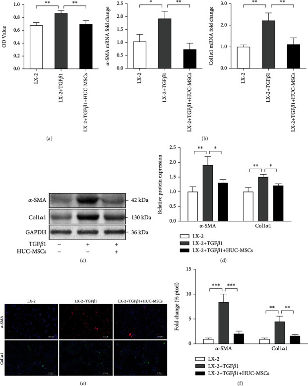
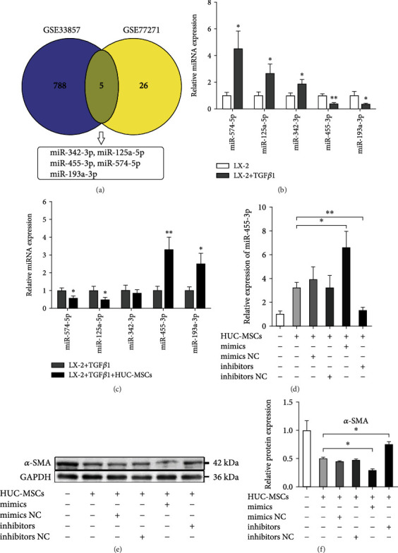
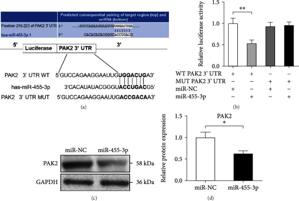
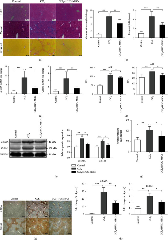
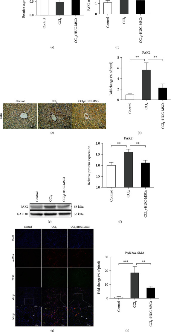
Mesenchymal stem cells (MSCs) were shown to have potential therapeutic effects for treatment of liver fibrosis, and dysregulated expression of microRNAs (miRNAs) played a pivotal role in the pathogenesis of liver fibrosis by regulating their downstream target genes. However, the mechanism by which MSCs affect the progression of liver fibrosis by regulating miRNA expression remains unclear. Here, we investigated whether human umbilical cord MSCs (HUC-MSCs) attenuated hepatic fibrosis by regulating miR-455-3p and its target gene. Significantly upregulated miRNA (miR-455-3p) was screened out by GEO datasets analysis and coculture HUC-MSCs with hepatic stellate cell (HSC) LX-2 cells. p21-activated kinase-2 (PAK2) was forecasted to be the target gene of miR-455-3p by bioinformatics analyses and confirmed by luciferase reporter assay. HUC-MSCs were transplanted into mice with carbon tetrachloride- (CCl4-) induced liver fibrosis, the result showed that HUC-MSC transplantation significantly ameliorated the severity of CCl4-induced liver fibrosis, attenuated collagen deposition, improved liver function by reducing the expression of alanine aminotransferase (ALT) and aspartate aminotransferase (AST) in serum, upregulated miR-455-3p, and suppressed PAK2 expression of liver tissue in mice. Taken together, our study suggests that HUC-MSCs inhibit the activation of HSCs and mouse CCl4-induced liver fibrosis by upregulation of miR-455-3p through targeting PAK2.


Conflict of interest statement

The authors declare that they have no conflicts of interest.

Figures

Figure 1
Figure 1
HUC-MSCs inhibit proliferation and activation of HSCs. (a) CCK8 assay analysis of HUC-MSCs on inhibition of TGFβ1-mediated proliferation in LX-2 cells. (b) The mRNA levels of α-SMA and Col1α1 were measured by qRT-PCR. (c) The protein levels of α-SMA and Col1α1 were measured by Western blot. (d) Quantitation of α-SMA and Col1α1 levels from three independent Western blot analyses. (e) Immunofluorescence images of LX-2 cells, the red fluorescence represents α-SMA, and the green fluorescence represents Col1α1. Scale bars, 200 μm. (f) Quantification of α-SMA and Col1α1 positive areas. Percentages of immunoreactive areas were expressed as relative values to those in the LX-2 group. The quantitative data are represented as the mean ± SD. Each experiment was repeated three times. P < 0.05, ∗∗P < 0.01, and ∗∗∗P < 0.001.
Figure 2
Figure 2
HUC-MSCs promote inactivation of LX-2 cells by upregulating miR-455-3p. (a) Venn diagram of DEMs about liver fibrosis from two miRNA profile data. (b) Comparison of qRT-PCR results of five selected DEMs in LX-2 cells treated with or without TGFβ1. (c) Comparison of the expression levels of five selected DEMs in TGFβ1-activated LX-2 cells cultured alone or cocultured with HUC-MSCs. (d) The expression levels of miR-455-3p were examined in TGFβ1-treated LX-2 cells cocultured with HUC-MSCs and then were transfected with miR-455-3p mimics, mimics NC, inhibitors, or inhibitors NC, respectively. (e) The protein levels of α-SMA measured by Western blot in each group. (f) Quantitation of α-SMA levels from three independent Western blot analyses. The quantitative data are represented as the mean ± SD. Each experiment was repeated three times. P < 0.05 and ∗∗P < 0.01.
Figure 3
Figure 3
miR-455-3p regulates PAK2 expression by directly targeting the 3′ UTR of its mRNA. (a) Predicted miR-455-3p targeting sequence in 3′ UTR of WT or MUT PAK2 mRNA. (b) Dual-luciferase reporter assay of LX-2 cells cotransfected with WT or MUT PAK2 3' UTR reporter and miR-455-3p mimics or mimics NC. (c) PAK2 protein expression analysis in LX-2 cells transfected with miR-455-3p mimics using Western blotting. (d) Quantitation of PAK2 protein levels from three independent Western blot analyses. The quantitative data are represented as the mean ± SD. Each experiment was repeated three times. P < 0.05 and ∗∗P < 0.01.
Figure 4
Figure 4
HUC-MSCs significantly attenuate severity of liver fibrosis in mice. (a) Representative images of H&E, Masson, and Sirius red staining of liver sections in olive oil-treated mice and mice of CCl4-induced liver fibrosis treated with PBS or HUC-MSCs. Scale bars, 200 μm. (b) Relative fold change of Masson and Sirius red positive fibrosis areas compared with the olive oil-treated group. (c) qRT-PCR shows the expression of α-SMA and Col1α1 in the three groups. (d) Serum levels of ALT and AST were determined to indicate the extent of liver damage in the different groups. (e) The protein levels of α-SMA and Col1α1 in liver tissues were examined by Western blotting. (f) The amount of liver hydroxyproline was detected in the different groups. (g) Immunohistochemical analysis of mouse liver tissues for the expression of α-SMA and Col1α1. Scale bars, 200 μm. (h) Quantification of immunohistochemical analysis. Percentages of immunoreactive (α-SMA and Col1α1) areas were expressed as relative values to those in oil-treated mouse livers. The quantitative data are represented as the mean ± SD. Each experiment was repeated three times. P < 0.05, ∗∗∗P < 0.01, and ∗∗∗P < 0.001.
Figure 5
Figure 5
HUC-MSCs upregulate the expression of miR-455-3p of CCl4-induced liver fibrosis in mice by suppressing PAK2 expression. (a) The expression levels of miR-455-3p were examined in mouse hepatic tissues of the olive oil-treated group and CCl4-induced liver fibrosis groups treated with PBS or HUC-MSCs. (b) The expression levels of PAK2 mRNA in mouse hepatic tissues were examined by qRT-PCR. (c) Immunohistochemical staining of PAK2 in liver tissues of different groups. Scale bars, 50 μm. (d) Quantification of PAK2-positive area in mouse hepatic tissues. (e) The protein levels of PAK2 in mice hepatic tissue were examined by Western blotting. (f) Quantitation of PAK2 protein levels from three independent Western blot analyses. (g) Immunofluorescence images of liver sections for the presence of DAPI (blue) or α-SMA (red) or PAK2 (green). The yellow areas indicated by the arrow are the coexpression regions of α-SMA and PAK2. Scale bars, 200 μm. (h) Quantification of coexpression region of α-SMA and PAK2 in mouse hepatic tissues. The quantitative data are represented as the mean ± SD. Each experiment was repeated three times. P < 0.05 and ∗∗P < 0.01.

Similar articles

Cited by

References

    1. Higashi T., Friedman S. L., Hoshida Y. Hepatic stellate cells as key target in liver fibrosis. Advanced Drug Delivery Reviews. 2017;121:27–42. doi: 10.1016/j.addr.2017.05.007. - DOI - PMC - PubMed
    1. Tsuchida T., Friedman S. L. Mechanisms of hepatic stellate cell activation. Nature Reviews Gastroenterology & Hepatology. 2017;14(7):397–411. doi: 10.1038/nrgastro.2017.38. - DOI - PubMed
    1. Mederacke I., Hsu C. C., Troeger J. S., et al. Fate tracing reveals hepatic stellate cells as dominant contributors to liver fibrosis independent of its aetiology. Nature Communications. 2013;4(1) doi: 10.1038/ncomms3823. - DOI - PMC - PubMed
    1. Kadyk L. C., Collins L. R., Littman N. J., Millan M. T. Proceedings: moving toward cell-based therapies for liver disease. STEM CELLS Translational Medicine. 2015;4(3):207–210. doi: 10.5966/sctm.2014-0276. - DOI - PMC - PubMed
    1. Haldar D., Henderson N. C., Hirschfield G., Newsome P. N. Mesenchymal stromal cells and liver fibrosis: a complicated relationship. FASEB J. 2016;30(12):3905–3928. doi: 10.1096/fj.201600433R. - DOI - PubMed

LinkOut - more resources