

Expression of E-Cadherin in Pig-Tailed Monkey (Macaca nemestrina) Endometrium after Controlled Ovarian Hyperstimulation
- PMID: 33708995
- PMCID: PMC7932768
- DOI: 10.1155/2021/8824614
Expression of E-Cadherin in Pig-Tailed Monkey (Macaca nemestrina) Endometrium after Controlled Ovarian Hyperstimulation
Abstract
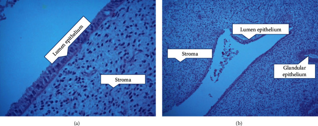
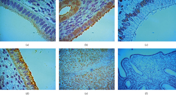
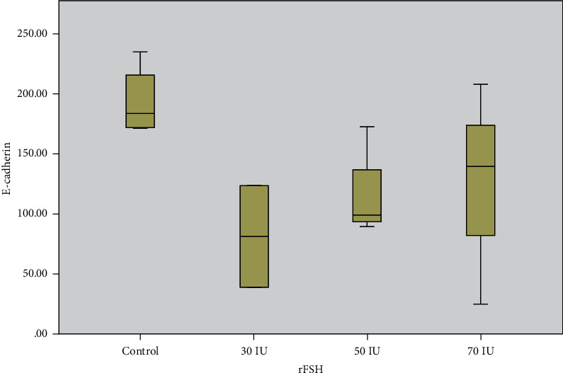
An increase of steroid hormones in controlled ovarian hyperstimulation (COH) procedures is reducing the success rate in assisted reproductive technology (ART), and this includes the pregnancy rate and/or implantation rate. Research has found that the decrease in the success rate occurred due to the decreased expression of the protein that is needed to prepare the endometrium so that the embryo could attach. The aim of the study was to analyse the changes in E-chaderin expression due to COH and its relations with increased level of steroid hormones as one of the proteins in the endometrium. There were 13 samples of stored biological tissue from Macaca nemestrina endometrial tissue; came from one group of natural cycles as the control group (n = 4) and three groups of stimulated cycles. The first stimulated cycle group was injected by a 30 IU dose of rFSH (n = 2). The second stimulated cycle group was injected by a 50 IU dose of rFSH (n = 4). The third stimulated cycle group was injected by a 70 IU dose of rFSH (n = 3). The expression of E-cadherin was measured by the immunohistochemistry (IHC) technique. Estradiol (E2) and progesterone (P4) levels were assessed using ELISA and have already been done. The IHC staining expression of E-cadherin was found in the cytoplasm of glandular epithelium. Immunostaining measurement used the H_SCORE. We found that the expression of E-cadherin within the group was not significantly different (p value: 0.178). Similarly, both the correlation between the estradiol level with E-cadherin and the correlation between the progesterone level with E-cadherin were not significantly different (p value: 0.872 and p value: 0.836). The conclusion is that the level of E-Cadherin expression in the endometrium that were taken in themiddle secretion phase not affected by the dose regimen that given. In addition, the level of expression is not influenced by the increase of serum E2 and P4 levels.
Copyright © 2021 Nurhuda Sahar et al.
Conflict of interest statement
The authors declare that they have no conflicts of interest.
Figures

Similar articles
-
Histochemical localization of endometrial insulin-like growth factor binding protein-1 and -3 during the luteal phase in controlled ovarian hyperstimulation cycles: a controlled study.Fertil Steril. 2000 Aug;74(2):338-42. doi: 10.1016/s0015-0282(00)00596-3. Fertil Steril. 2000. PMID: 10927055
-
Endometrial glycodelin-A expression in the luteal phase of stimulated ovarian cycles.Fertil Steril. 2000 Jul;74(1):130-3. doi: 10.1016/s0015-0282(00)00586-0. Fertil Steril. 2000. PMID: 10899509
-
Increased Progesterone on the Day of Administration of hCG in Controlled Ovarian Hyperstimulation Affects the Expression of HOXA10 in Primates' Endometrial Receptivity.Biomedicines. 2019 Oct 21;7(4):83. doi: 10.3390/biomedicines7040083. Biomedicines. 2019. PMID: 31640230 Free PMC article.
-
[Endometrial changes caused by induction of ovarian hyperstimulation which affect the process of embryo implantation].Ginecol Obstet Mex. 1994 Dec;62:415-8. Ginecol Obstet Mex. 1994. PMID: 7835742 Review. Spanish.
-
The endometrium during and after ovarian hyperstimulation and the role of segmentation of infertility treatment.Best Pract Res Clin Endocrinol Metab. 2019 Feb;33(1):61-75. doi: 10.1016/j.beem.2018.09.003. Epub 2018 Sep 13. Best Pract Res Clin Endocrinol Metab. 2019. PMID: 30770200 Review.
Cited by
-
Shoutai Pill Enhances Endometrial Receptivity in Controlled Ovarian Hyperstimulation Mice by Improving the In-Vivo Immune Environment.Comb Chem High Throughput Screen. 2025;28(4):711-723. doi: 10.2174/0113862073274708231028185333. Comb Chem High Throughput Screen. 2025. PMID: 37929727
-
MicroRNA participates in embryo implantation by modulating endometrial tolerance in sows during peri-implantation period.Front Endocrinol (Lausanne). 2025 Aug 5;16:1555636. doi: 10.3389/fendo.2025.1555636. eCollection 2025. Front Endocrinol (Lausanne). 2025. PMID: 40838210 Free PMC article. Review.
References
-
- Simón C., Cano F., Valbuena D., Remohí J., Pellicer A. Implantation: clinical evidence for a detrimental effect on uterine receptivity of high serum oestradiol concentrations in high and normal responder patients. Human Reproduction. 1995;10(9):2432–2437. doi: 10.1093/oxfordjournals.humrep.a136313. - DOI - PubMed
MeSH terms
Substances
LinkOut - more resources
Full Text Sources
Other Literature Sources
