

Non-sebaceous Lymphadenoma of the Parotid Gland Mimicking a Pleomorphic Adenoma
- PMID: 33709077
- PMCID: PMC7907658
- DOI: 10.1177/2632010X21996338
Non-sebaceous Lymphadenoma of the Parotid Gland Mimicking a Pleomorphic Adenoma
Abstract
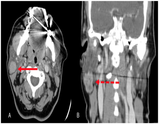
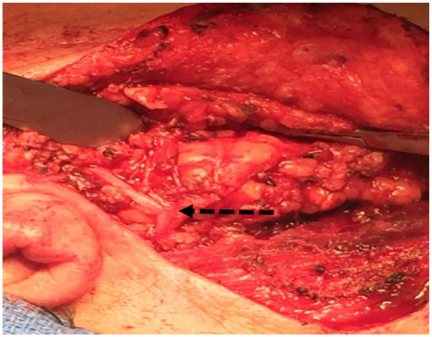
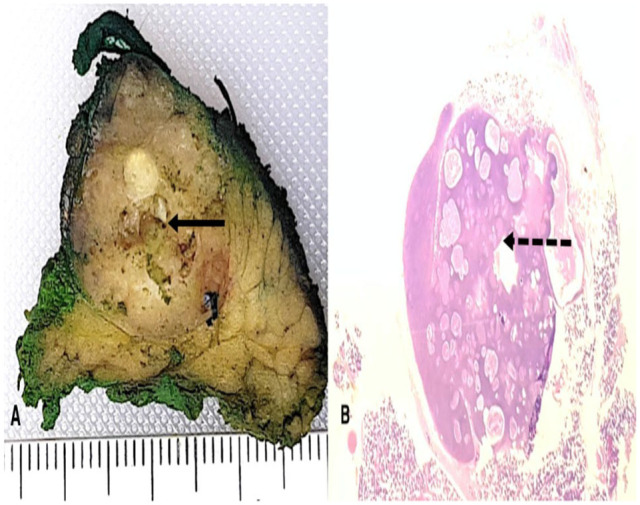
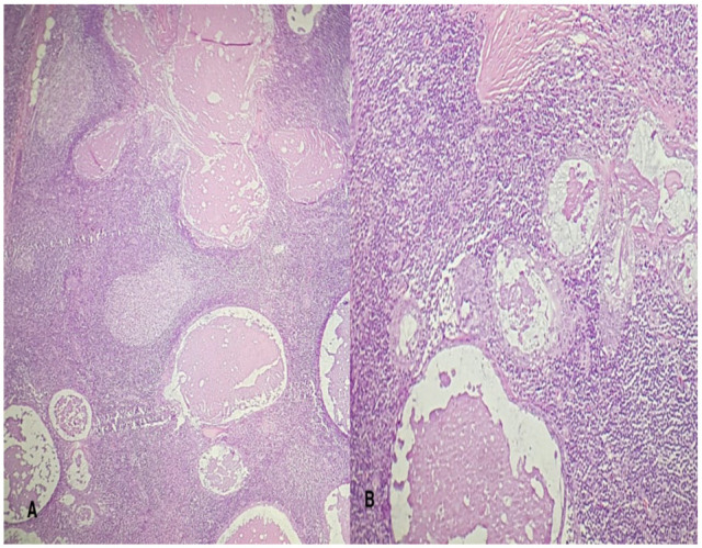
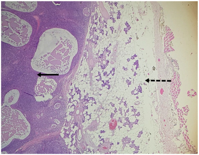
Non-sebaceous lymphadenoma of the salivary glands is a rare benign lesion, first described in 1991. We present the case of a 54-year-old woman, with a right parotid mass. She underwent right superficial parotidectomy, and histopathology reported a non-sebaceous lymphadenoma due to an encapsulated lesion and multiple non-atypical epithelial inclusions without sebaceous differentiation. The etiology of non-sebaceous lymphadenoma is not yet understood, but it can arise predominantly from the parotid gland. Surgical excision is the treatment of choice.
Keywords: Parotid tumor; non-sebaceous lymphadenoma; parotid.
© The Author(s) 2021.
Conflict of interest statement
Declaration of conflicting interests:The author(s) declared no potential conflicts of interest with respect to the research, authorship, and/or publication of this article.
Figures
Similar articles
-
Sebaceous lymphadenoma of the parotid: a rare case report of an entity mimicking other salivary tumors.J Surg Case Rep. 2025 Jun 5;2025(6):rjaf382. doi: 10.1093/jscr/rjaf382. eCollection 2025 Jun. J Surg Case Rep. 2025. PMID: 40476025 Free PMC article.
-
Sebaceous lymphadenoma of parotid gland in a child.Oral Surg Oral Med Oral Pathol Oral Radiol Endod. 2009 Feb;107(2):253-5. doi: 10.1016/j.tripleo.2008.09.009. Oral Surg Oral Med Oral Pathol Oral Radiol Endod. 2009. PMID: 19138644
-
Sebaceous lymphadenoma of parotid gland: A case report of a unique presentation in an immunocompromised patient.J Family Med Prim Care. 2020 Feb 28;9(2):1202-1205. doi: 10.4103/jfmpc.jfmpc_1115_19. eCollection 2020 Feb. J Family Med Prim Care. 2020. PMID: 32318494 Free PMC article.
-
Malignant Transformation of Parotid Gland Non-sebaceous Lymphadenoma: Case Report and Review of Literature.Head Neck Pathol. 2020 Dec;14(4):1123-1128. doi: 10.1007/s12105-020-01133-3. Epub 2020 Jan 29. Head Neck Pathol. 2020. PMID: 31997132 Free PMC article. Review.
-
Myeloid Sarcoma That Infiltrated a Preexisting Sebaceous Lymphadenoma in the Parotid Gland: Diagnostic Challenges and Literature Review.Biomed Res Int. 2019 Nov 22;2019:9869406. doi: 10.1155/2019/9869406. eCollection 2019. Biomed Res Int. 2019. PMID: 31886274 Free PMC article. Review.
References
-
- Yang S, Chen X, Wang L, Zhang J. Non-sebaceous lymphadenoma of the salivary gland: case report with immunohistochemical investigation. Virchows Arch. 2007;450:595-599. - PubMed
-
- Weiler C, Agaimy A, Zengel P, Zenk J, Kirchner T, Ihrler S. Nonsebaceous lymphadenoma of salivary glands: proposed development from intraparotid lymph nodes and risk of misdiagnosis. Virchows Arch. 2012;460:467-472. - PubMed
-
- Castelino-Prabhu S, Li QK, Ali SZ. Nonsebaceous lymphadenoma of the parotid gland: cytopathologic findings and differential diagnosis. Diagn Cytopathol. 2010;38:137-140. - PubMed
Publication types
LinkOut - more resources
Full Text Sources
Other Literature Sources
