Review
. 2022 Dec;29(6):3044-3056.
doi: 10.1007/s12350-021-02570-5. Epub 2021 Mar 11.

Cardiac lymphoma with early response to chemotherapy: A case report and review of the literature

Affiliations
Review

Cardiac lymphoma with early response to chemotherapy: A case report and review of the literature

Andrea Bonelli et al. J Nucl Cardiol. 2022 Dec.

Abstract

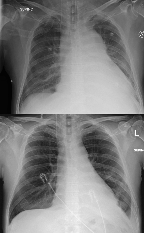
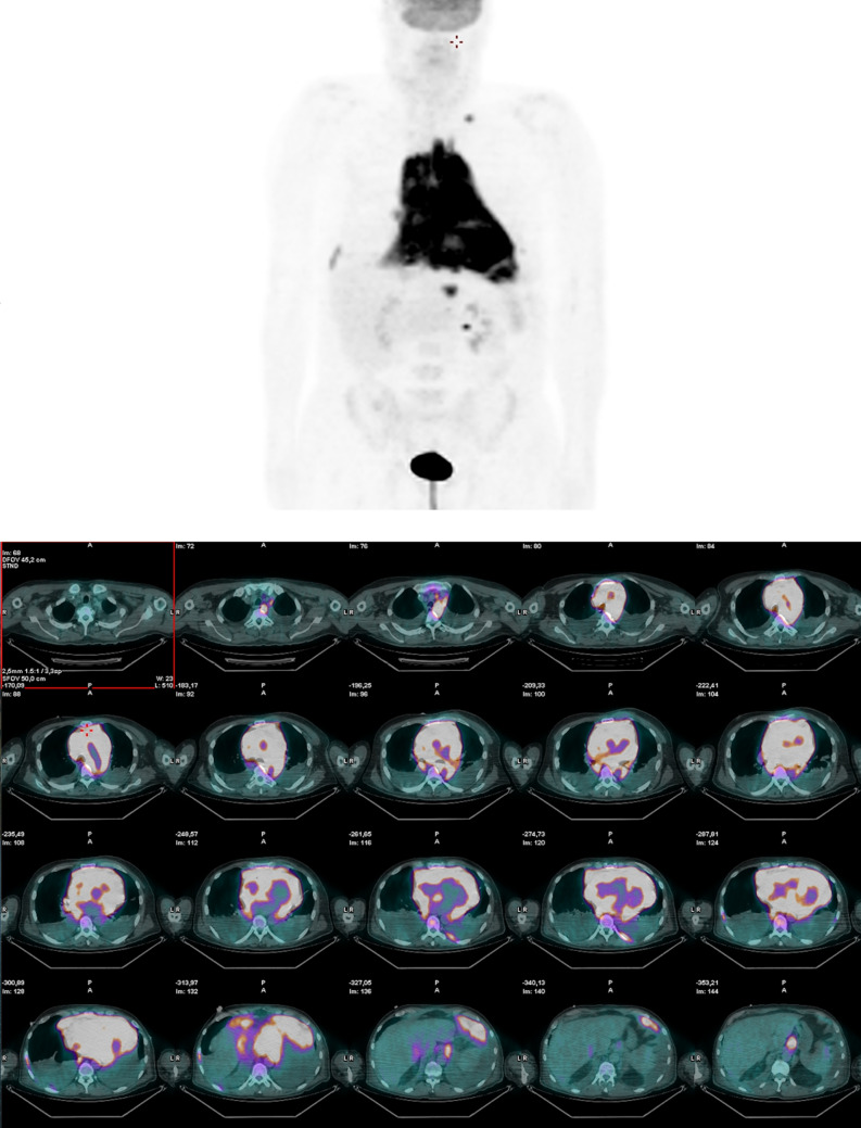
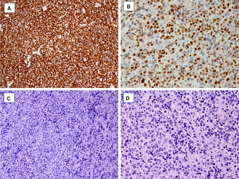
Cardiac tumors are rare and benign masses account for the most part of the diagnosis. When malignant cancer is detected, primary or secondary cardiac lymphoma are quite frequent. Cardiac lymphoma may present as an intra or peri-cardiac mass or, rarely, it may diffusely infiltrate the myocardium. Although often asymptomatic, patients can have non-specific symptoms. Acute presentations with cardiogenic shock, unstable angina, or acute myocardial infarction are also described. Modern imaging techniques can help the clinicians not only in the diagnostic phase but also during administration of chemotherapy. A multidisciplinary counseling and serial multi-parametric assessment (echocardiography, cardiac troponin) seem to be the most effective approach to prevent possible fatal complications (i.e., cardiac rupture). Currently, only chemo- and radiotherapy are available options for treatment, but the prognosis remains poor. This is a case of secondary cardiac lymphoma presenting as a mediastinal mass with large infiltration of the heart and the great vessels with a good improvement after only one cycle of chemotherapy. It demonstrates the importance of an early diagnosis to modify the natural history of the disease.

Keywords: Basic science; diseases/processes; modalities; technical.


Figures

Figure 1
Figure 1
Trans-thoracic echocardiography - parasternal long-axis view. On the left, it is evident the marked and diffused thickening of the myocardium, especially of the interventricular septum, the posterior, the interatrial septum and the right ventricular free wall, which appear hypokinetic. Also, the large iso-echogenic mass takes up space around the ascending aorta and in the anterior mediastinum (yellow narrow). On the right, the view after the first chemotherapy regimen
Figure 2
Figure 2
Chest X-ray - The upper picture shows the parenchymal thickening of the left inferior pulmonary lobe with associated pleural effusion. Note the enlarged mediastinum and the increased dimension of the heart. The inferior picture shows the chest X-ray after the first cycle of chemotherapy, with net reduction of the pleural effusion and the mediastinal mass
Figure 3
Figure 3
[18F]-Fluorodeoxyglucose positron emission tomography. Global and diffused accumulation of glucose in the mediastinum and in the heart, with the involvement of both ventricles, interventricular septum, and atria. Subdiaphragmatic lymph nodes were described too
Figure 4
Figure 4
Cardiac magnetic resonance. A 3-chamber view and mid-ventricular short-axis view before chemotherapeutic treatment. Note the massive invasion of the heart by the mediastinal mass, whose cleavage borders are not evaluable; the myocardium appears thickened. Diffuse edema in T2-weighted sequences was detected, with a similar diffuse LGE pattern. B After the first regimen of chemotherapy, no more myocardial edema and late gadolinium enhancement were noticed, with near-normalization of left ventricle thickness
Figure 5
Figure 5
Tumor histology. High-grade diffuse large B-cell lymphoma with expression of CD20 (A) and BCL-6 (B). The hematoxylin–eosin stain (C and D) shows medium to large cells with irregular nucleus
Figure 6
Figure 6
Electrocardiogram before and after the first cycle of chemotherapy showing atrial fibrillation and subsequent spontaneous restoration of sinus rhythm. Note the negative T waves on the precordial leads

References

    1. Grebenc ML, Rosado De Christenson ML, Burke AP, Green CE, Galvin JR. Primary cardiac and pericardial neoplasms: Radiologic-pathologic correlation. Radiographics. 2000 doi: 10.1148/radiographics.20.4.g00jl081073. - DOI - PubMed
    1. Gordon MJ, Danilova O, Spurgeon S, Danilov AV. Cardiac non-Hodgkin’s lymphoma: Clinical characteristics and trends in survival. Eur J Haematol. 2016 doi: 10.1111/ejh.12751. - DOI - PubMed
    1. Jeudy J, Burke AP, Frazier AA. Cardiac lymphoma. Radiol Clin N Am. 2016 doi: 10.1016/j.rcl.2016.03.006. - DOI - PubMed
    1. O’Mahony D, Peikarz RL, Bandettini WP, Arai AE, Wilson WH, Bates SE. Cardiac involvement with lymphoma: A review of the literature. Clin Lymphoma Myeloma. 2008 doi: 10.3816/CLM.2008.n.034. - DOI - PMC - PubMed
    1. Vinicki JP. Complete regression of myocardial involvement associated with lymphoma following chemotherapy. World J Cardiol. 2013 doi: 10.4330/wjc.v5.i9.364. - DOI - PMC - PubMed